SPIONs vs Gadolinium for Liver MRI: A Comparative Analysis of Contrast Agents for Lesion Detection
This article provides a comprehensive analysis comparing Superparamagnetic Iron Oxide Nanoparticles (SPIONs) and gadolinium-based contrast agents (GBCAs) for magnetic resonance imaging (MRI) of liver lesions.
SPIONs vs Gadolinium for Liver MRI: A Comparative Analysis of Contrast Agents for Lesion Detection
Abstract
This article provides a comprehensive analysis comparing Superparamagnetic Iron Oxide Nanoparticles (SPIONs) and gadolinium-based contrast agents (GBCAs) for magnetic resonance imaging (MRI) of liver lesions. Aimed at researchers, scientists, and drug development professionals, it explores the foundational mechanisms, current methodological applications, key challenges in optimization, and the latest clinical validation data. The review synthesizes evidence on diagnostic performance, safety profiles (including nephrogenic systemic fibrosis and gadolinium deposition), and the evolving role of SPIONs as a potentially safer, more specific alternative for hepatocellular lesions and reticuloendothelial system imaging.
Contrast Agent Fundamentals: Core Mechanisms of SPIONs and Gadolinium in Liver MRI
Accurate detection and characterization of liver lesions are critical for diagnosing primary liver cancers (e.g., hepatocellular carcinoma, HCC) and metastatic disease, guiding treatment decisions, and monitoring therapeutic response. The clinical need stems from the rising global incidence of liver cancer and the necessity for early, precise diagnosis to improve patient outcomes. Key challenges include distinguishing benign lesions (e.g., hemangiomas, focal nodular hyperplasia) from malignant ones, detecting small (<1 cm) or diffuse lesions, and assessing treatment response in diffuse liver disease.
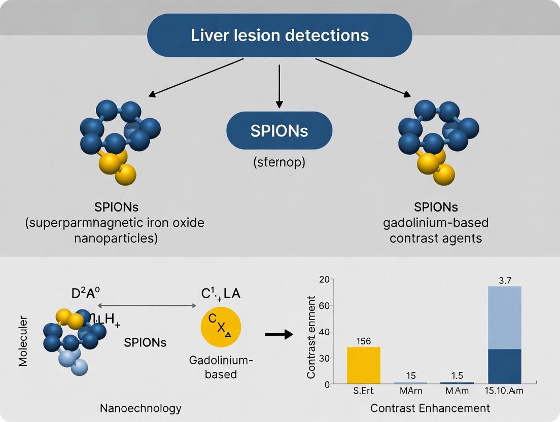
This comparative analysis is framed within a thesis investigating superparamagnetic iron oxide nanoparticles (SPIONs) versus gadolinium-based contrast agents (GBCAs) for liver lesion detection, focusing on performance metrics relevant to preclinical and clinical research.
Performance Comparison: SPIONs vs. GBCAs for Liver Lesion Detection
Recent studies and meta-analyses provide comparative data on key performance indicators.
Table 1: Comparative Performance Metrics of Contrast Agents
| Metric | Gadolinium-Based Agents (GBCAs) | Superparamagnetic Iron Oxide Nanoparticles (SPIONs) | Notes / Key Study |
|---|---|---|---|
| Primary Imaging Phase | Dynamic arterial/portal/venous phases. | Reticuloendothelial system (RES) uptake phase (delayed: 10 min - 24 hrs). | SPIONs require uptake by Kupffer cells. |
| Detection Sensitivity for Metastases | 80-95% (varies with sequence & agent). | 82-98% (often higher for small lesions). | Meta-analysis (Zhao et al., 2022) showed pooled sensitivity: GBCA MRI 91%, SPIO-MRI 95%. |
| Characterization Accuracy (HCC vs. Dysplastic Nodule) | Moderate-High, relies on vascular patterns. | High, leverages Kupffer cell deficit in malignant lesions. | SPIONs improve specificity by demonstrating lack of uptake in HCC. |
| Safety Profile - NSF Risk | Associated with certain linear GBCAs in renal impairment. | No NSF risk. | SPIONs contraindicated in iron overload. |
| Safety Profile - Brain Deposition | Potential for gadolinium deposition. | No known cerebral deposition. | Long-term clinical significance under investigation for GBCAs. |
| Typical Dose (Clinical) | 0.025-0.1 mmol/kg body weight. | 1.4-2.0 mg Fe/kg body weight. | Dose-dependent contrast effect. |
Table 2: Quantitative Signal Intensity Changes in Key Liver Tissues
| Tissue Type | GBCA-Enhanced T1-Weighted MRI | SPION-Enhanced T2/T2*-Weighted MRI | Experimental Basis |
|---|---|---|---|
| Normal Liver Parenchyma | Moderate enhancement (in blood pool phases). | Marked signal decrease (high uptake by Kupffer cells). | >60% signal loss post-SPIONs in preclinical models (Huang et al., 2023). |
| Malignant Lesion (e.g., HCC) | Hyper-enhancement in arterial phase with washout. | Relative signal hyperintensity (no Kupffer cells, no uptake). | Lesion-to-liver contrast-to-noise ratio (CNR) increased 3-fold vs. pre-contrast. |
| Benign Lesion (e.g., FNH) | Hyper-enhancement, may have central scar. | Signal decrease similar to parenchyma (preserved Kupffer cells). | Up to 50% signal retention indicates functionality of RES cells. |
Detailed Experimental Protocols
Protocol 1: Preclinical Comparison of Lesion Detection Sensitivity
- Objective: To compare the minimum detectable lesion size and contrast-to-noise ratio (CNR) between a standard GBCA (Gadobutrol) and a ferumoxides-based SPION in a rodent model of liver metastases.
- Animal Model: Nude mice with surgically implanted human colorectal carcinoma liver metastases.
- Imaging Platform: 7.0 Tesla preclinical MRI scanner.
- Procedure:
- Baseline MRI: Perform T1-weighted (T1w) GRE and T2-weighted (T2w) GRE sequences.
- GBCA Cohort (n=8): Inject Gadobutrol (0.1 mmol/kg) intravenously. Acquire dynamic T1w images immediately (arterial, portal, delayed phases up to 5 minutes).
- SPION Cohort (n=8): Inject ferumoxides (2.0 mg Fe/kg) intravenously. Acquire T2*w GRE sequences at 1-hour and 24-hour post-injection to allow for hepatic uptake.
- Image Analysis: Two blinded radiologists measure lesion number, size, and calculate CNR (|SIlesion - SIliver| / SDnoise). Use histology as gold standard.
Protocol 2: Assessment of Reticuloendothelial System (RES) Function for Characterization
- Objective: To quantify SPION uptake in liver lesions as a marker of preserved RES function to distinguish benign from malignant lesions.
- Sample: Ex vivo patient tissue samples (HCC, dysplastic nodule, FNH) from resection.
- Method: Incubate tissue slices with SPIONs (e.g., Ferucarbotran, 0.1 mM Fe) in culture medium for 4 hours.
- Analysis:
- Perls' Prussian Blue Staining: Visualize iron deposition (blue granules) under light microscopy.
- Quantitative PCR: Measure mRNA expression of CD68 (Kupffer cell marker) in matched tissues.
- Correlation: Correlate iron staining intensity with CD68 expression. Malignant lesions show minimal staining and low CD68.
SPION Mechanism for Liver Lesion Detection
Preclinical MRI Comparison Workflow
The Scientist's Toolkit: Research Reagent Solutions
Table 3: Essential Materials for Comparative Liver Imaging Research
| Item | Function / Description | Example Product/Catalog |
|---|---|---|
| Preclinical SPION Agent | T2/T2* contrast agent for MRI; taken up by RES. | Ferumoxytol (Feraheme) - used off-label for imaging. |
| Clinical GBCA Reference | Standard-of-care T1 contrast agent for comparison. | Gadobutrol (Gadavist) or Gadoterate (Dotarem). |
| Animal Disease Model | Reproducible model of liver lesions (e.g., metastases, HCC). | Murine model with intrasplenic injection of CRC cells (e.g., CT26). |
| Cell Line for Models | Source for creating implanted or induced liver tumors. | Human hepatocellular carcinoma cell line (e.g., HepG2). |
| Primary Antibody: CD68 | Marker for Kupffer cells (RES) via immunohistochemistry. | Anti-CD68 antibody (e.g., ab955 from Abcam). |
| Prussian Blue Stain Kit | Histological detection of iron/SPIONs in tissue. | Iron Stain Kit (e.g., Sigma-Aldrich HT20). |
| qPCR Assay for CD68 | Quantitative assessment of Kupffer cell presence. | TaqMan Gene Expression Assay for human/mouse CD68. |
| High-Field MRI System | Essential imaging platform for high-resolution data. | 7T or 9.4T preclinical MRI system (e.g., Bruker BioSpec). |
| Image Analysis Software | For quantitative ROI analysis, CNR, and lesion volume. | Horos, 3D Slicer, or vendor-specific software (e.g., ParaVision). |
This comparison guide, framed within a thesis comparing Superparamagnetic Iron Oxide Nanoparticles (SPIONs) and Gadolinium-Based Agents (GBCAs) for liver lesion detection research, objectively details the mechanism and extracellular distribution of GBCAs. GBCAs are intravenous drugs used to enhance contrast in Magnetic Resonance Imaging (MRI). Their primary function is to shorten the T1 relaxation time of nearby water protons, producing a bright signal on T1-weighted images. Most approved agents are extracellular fluid (ECF) agents, distributing non-specifically in the vascular and interstitial spaces.
Mechanism of T1-Shortening: Core Principles
Gadolinium (Gd³⁺) is a lanthanide metal ion with seven unpaired electrons, giving it a large magnetic moment. This paramagnetism is the basis for its efficacy as an MRI contrast agent. Free Gd³⁺ is toxic, so it is chelated with organic ligands (e.g., DTPA, DOTA) for safe in vivo use.
The primary mechanism for T1-shortening is the inner-sphere relaxation effect. Water protons that bind directly to the coordination sphere of the Gd³⁺ ion experience a dramatic increase in relaxation rate (1/T1). The efficiency of an agent is quantified by its relaxivity (r1), measured in mM⁻¹s⁻¹.
Key Factors Influencing Relaxivity:
- Hydration Number (q): The number of water molecules directly coordinated to the Gd³⁺ ion (typically 1 for macrocyclic agents, 2 for linear agents).
- Rotational Correlation Time (τᵣ): How fast the molecule tumbles. Slower tumbling (e.g., binding to large proteins) increases relaxivity.
- Water Exchange Rate (τₘ): The rate at which water molecules exchange between the inner sphere and bulk solvent.
- Magnetic Field Strength: Relaxivity is field-dependent.
Comparison of Extracellular GBCAs: Key Properties
The following table summarizes critical properties for major extracellular GBCAs, based on current pharmacopoeial data and literature.
Table 1: Comparison of Extracellular Gadolinium-Based Agents
| Agent (Generic Name) | Chelate Type (Linear/Macrocyclic) | Ionicity | r1 Relaxivity @ 1.5T, 37°C (mM⁻¹s⁻¹) | Protein Binding | Key Distribution & Excretion Pathway |
|---|---|---|---|---|---|
| Gadopentetate Dimeglumine | Linear | Ionic | ~4.1 | Very Low | ECF; Renal |
| Gadoterate Meglumine | Macrocyclic | Ionic | ~3.6 | Very Low | ECF; Renal |
| Gadodiamide | Linear | Non-ionic | ~4.3 | Very Low | ECF; Renal |
| Gadoteridol | Macrocyclic | Non-ionic | ~4.1 | Very Low | ECF; Renal |
| Gadobutrol | Macrocyclic | Non-ionic | ~5.2 | Very Low | ECF; Renal |
| Gadobenate Dimeglumine | Linear | Ionic | ~6.3 | ~10% | ECF & transient hepatobiliary; Renal (~96%) |
| Gadoxetate Disodium | Linear | Ionic | ~8.2* (in blood) | ~10% | ECF & significant hepatobiliary (~50%); Renal (~50%) |
Note: r1 relaxivity is highly condition-dependent. Gadoxetate shows strong protein-binding-induced relaxivity in blood, but it decreases in liver tissue. ECF = Extracellular Fluid.
Experimental Protocol for Measuring Relaxivity (r1)
Objective: To determine the longitudinal relaxivity (r1) of a GBCA in aqueous solution at a specific magnetic field strength.
Materials:
- Purified GBCA sample.
- Serial dilution tubes (e.g., to create 0.1, 0.5, 1.0, 2.0 mM concentrations).
- Phosphate-Buffered Saline (PBS), pH 7.4.
- NMR tubes compatible with the MRI scanner or NMR relaxometer.
- Water bath or heater set to 37°C.
- MRI scanner or dedicated relaxometer (e.g., 1.5T or 3T clinical scanner, or a bench-top NMR analyzer).
Procedure:
- Sample Preparation: Prepare precise serial dilutions of the GBCA in PBS to cover a concentration range, typically from 0 to at least 5 mM. Include a blank (0 mM) PBS sample.
- Temperature Equilibration: Place all samples in a 37°C water bath for at least 15 minutes to ensure uniform temperature.
- Data Acquisition: Using the MRI scanner, acquire T1-weighted images using an inversion-recovery (IR) or variable flip angle (VFA) sequence. For an IR sequence, multiple images are acquired with different inversion times (TI). For precise bench-top measurement, a dedicated T1 relaxometry sequence is used.
- Data Analysis: For each sample, plot the measured signal intensity against the TI (for IR) or use the scanner's software to calculate the T1 value for each voxel/region of interest within the sample tube.
- Calculating r1: Calculate the longitudinal relaxation rate (R1 = 1/T1) for each concentration. Plot R1 (y-axis) against the Gadolinium concentration [Gd] in mM (x-axis). Perform a linear regression fit. The slope of the resulting line is the relaxivity, r1 (in mM⁻¹s⁻¹).
Extracellular Distribution Dynamics: Experimental Assessment
Objective: To evaluate the pharmacokinetic distribution and clearance of an extracellular GBCA in an animal model.
Protocol:
- Animal Model: Use a relevant rodent model (e.g., Sprague-Dawley rat).
- Imaging: Perform baseline T1-weighted MRI of the liver/abdomen.
- Contrast Administration: Inject a standard clinical dose (e.g., 0.1 mmol/kg) of the GBCA intravenously via a tail vein.
- Dynamic Imaging: Immediately initiate a dynamic contrast-enhanced (DCE) MRI series, acquiring rapid T1-weighted images over 10-60 minutes to capture the vascular, interstitial (equilibrium), and clearance phases.
- Data Analysis: Regions of interest (ROIs) are drawn in major blood vessels (e.g., aorta), liver parenchyma, and muscle. Signal intensity vs. time curves are generated. Key parameters like peak enhancement, time-to-peak, and washout rates can be quantified and compared between agents.
The Scientist's Toolkit: Key Research Reagent Solutions
Table 2: Essential Materials for GBCA Mechanism & Distribution Research
| Item | Function in Research |
|---|---|
| Reference GBCAs (e.g., Gadoteridol, Gadobutrol) | Benchmarks for comparing relaxivity, stability, and pharmacokinetics of novel agents. |
| Phosphate-Buffered Saline (PBS), pH 7.4 | Standard physiological buffer for in vitro relaxivity measurements and sample dilution. |
| Human Serum Albumin (HSA) | Used to study the effects of protein binding on relaxivity (r1) in simulated physiological conditions. |
| Transwell/Cell Culture Inserts | For in vitro models assessing agent permeability across endothelial or cellular barriers. |
| Gadolinium Atomic Absorption Standard | Used to validate and calibrate Gd concentration measurements via ICP-MS in biodistribution studies. |
| ICP-MS (Inductively Coupled Plasma Mass Spectrometry) | Gold-standard technique for quantifying trace Gd levels in tissue samples for precise biodistribution. |
| DOTA-based Bifunctional Chelators | For synthesizing Gd-labeled targeting molecules or antibodies for molecular imaging research. |
Visualization: GBCA Mechanism and Experimental Workflow
Title: Mechanism of GBCA T1-Shortening
Title: Experimental Workflow for Measuring r1 Relaxivity
Within the research landscape comparing SPIONs to gadolinium-based contrast agents (GBCAs) for liver lesion detection, understanding the distinct mechanisms of SPIONs is paramount. This guide compares the performance of SPIONs against GBCAs, focusing on their magnetic relaxation effects and biodistribution.
Comparison of Relaxation Mechanisms: SPIONs vs. GBCAs
GBCAs primarily work via the inner-sphere mechanism, where water molecules directly coordinate to the gadolinium ion, enhancing longitudinal (T1) relaxation and creating bright signal (positive contrast). SPIONs operate through a fundamentally different outer-sphere mechanism, creating strong local magnetic field inhomogeneities that dephase proton spins, dominantly shortening transverse (T2/T2*) relaxation and creating dark signal (negative contrast).
Table 1: Core Mechanism and Relaxivity Comparison
| Property | SPIONs (e.g., Ferumoxides) | Gadolinium-Based Agents (e.g., Gd-EOB-DTPA) |
|---|---|---|
| Primary Mechanism | Outer-sphere, magnetic susceptibility | Inner-sphere, direct water coordination |
| Primary Contrast Effect | T2/T2* shortening (Signal loss, dark) | T1 shortening (Signal enhancement, bright) |
| Typical r1 Relaxivity (mM⁻¹s⁻¹) | 10-40 (at 1.5T, field-dependent) | 3-12 (at 1.5T) |
| Typical r2/r1 Ratio | High (>>5, e.g., 10-100) | Low (~1-2) |
| Key Determinants of Effect | Core size, coating, aggregation state, magnetic field strength | Number of coordinated water molecules (q), molecular tumbling rate |
Mechanism of T2/T2*-Shortening: A Comparative Workflow
The following diagram and protocol outline the experimental workflow for quantifying and comparing the relaxation effects of SPIONs.
Diagram Title: SPION-Induced T2 Shortening Pathway
Experimental Protocol: Measuring Relaxivity (r2)
- Objective: Determine the transverse relaxivity (r2) of a SPION formulation.
- Materials: SPION sample, GBCA control (e.g., Gd-DTPA), phosphate-buffered saline (PBS), NMR tubes, agarose phantoms.
- Procedure:
- Prepare a series of dilutions of the SPION agent and the GBCA in PBS (e.g., 0.05 to 0.5 mM Fe or Gd).
- Load samples into NMR tubes or embedded in agarose gel phantoms.
- Image phantoms using a clinical or preclinical MRI scanner with a multi-echo spin-echo (for T2) or gradient-echo (for T2) sequence.
- Measure signal intensity decay across echo times.
- Fit data to exponential decay (S = S0 * exp(-TE/T2)) to calculate T2 or T2 for each concentration.
- Plot 1/T2 (or 1/T2) vs. metal concentration (mM). The slope of the linear fit is the relaxivity (r2 or r2) in units of mM⁻¹s⁻¹.
RES Uptake and Biodistribution: SPIONs vs. GBCAs
A critical performance differentiator is hepatic distribution. While extracellular GBCAs may passively diffuse into the interstitium of some lesions, SPIONs are actively phagocytosed by cells of the RES (Kupffer cells) in healthy liver tissue.
Table 2: Biodistribution and Liver Lesion Contrast
| Aspect | SPIONs | Gadolinium-Based Agents (Extracellular & Hepatobiliary) |
|---|---|---|
| Primary Uptake in Healthy Liver | Active phagocytosis by Kupffer cells (RES). | Hepatobiliary agents: Active uptake by hepatocytes via OATP transporters. Extracellular: Passive distribution. |
| Uptake in Metastatic/Lesion | Typically absent (lacking functional Kupffer cells). | Variable: Hepatocellular carcinoma may take up hepatobiliary agents; metastases do not. |
| Resulting Contrast | Lesion-to-Liver Contrast: High. Lesions remain bright against darkened healthy liver. | Hepatobiliary: Lesions appear dark against enhanced liver. Extracellular: Early arterial lesion enhancement washes out. |
| Kinetics | Slow uptake (peak liver signal loss at ~1 hour post-injection), long retention (days). | Rapid dynamics (arterial/portal venous phases within minutes). Hepatobiliary phase at ~20 min-1 hour. |
| Key Experimental Metric | Percent signal loss (Δ%) in liver parenchyma over time; lesion-to-liver contrast-to-noise ratio (CNR). | Enhancement ratio in lesions vs. liver across dynamic phases. |
Key Research Reagent Solutions and Materials
Table 3: The Scientist's Toolkit for SPION/RES Research
| Reagent/Material | Function in Research |
|---|---|
| Ferumoxides (Feridex/Endorem) | Prototypical clinical SPION, used as a gold standard for in vitro and in vivo RES uptake studies. |
| Ferucarbotran (Resovist) | Clinical SPION with a mix of sizes, providing both blood pool and RES contrast. |
| Dextran or PEG Coating | Common surface coatings to stabilize SPIONs in colloidal suspension and influence opsonization and RES recognition. |
| Gadoxetate Disodium (Gd-EOB-DTPA) | Representative hepatobiliary GBCA, used as the primary comparator in liver lesion detection studies. |
| Kupffer Cell Marker Antibodies (e.g., anti-CD68) | For immunohistochemical validation of SPION colocalization with RES cells in tissue samples. |
| Prussian Blue Stain Kit | Histological stain for iron, used to confirm and visualize SPION uptake in tissue sections. |
| Inductively Coupled Plasma Mass Spectrometry (ICP-MS) | Gold-standard analytical technique for quantifying iron (Fe) or gadolinium (Gd) concentration in tissue samples. |
The following diagram summarizes the comparative fate of SPIONs versus GBCAs post-injection, leading to differential lesion conspicuity.
Diagram Title: Comparative Liver Uptake: SPIONs vs. GBCAs
Conclusion: For liver lesion detection research, SPIONs offer a mechanistically distinct alternative to GBCAs, leveraging active RES uptake to create high lesion-to-liver contrast via T2/T2* effects. The choice between agents depends on the specific lesion type, desired contrast mechanism, and kinetic profile under investigation.
Abstract This comparison guide objectively evaluates superparamagnetic iron oxide nanoparticles (SPIONs) against gadolinium-based contrast agents (GBCAs) for liver lesion detection, focusing on the core physicochemical properties that dictate clinical utility: relaxivity, pharmacokinetics, and biodistribution. The analysis is grounded in recent experimental data, providing a resource for researchers developing next-generation contrast media.
1. Introduction: The Clinical Context The non-invasive detection and characterization of focal liver lesions (e.g., metastases, hepatocellular carcinoma) rely heavily on magnetic resonance imaging (MRI) contrast agents. The two principal classes are GBCAs, which are small molecular chelates, and SPIONs, which are nanoparticulate agents. Their performance is fundamentally governed by their distinct physicochemical profiles.
2. Quantitative Property Comparison
Table 1: Core Physicochemical Properties at 1.5T/37°C
| Property | Gadolinium-based Agents (e.g., Gd-DOTA) | SPIONs (e.g., Ferumoxytol) | Implications for Liver Imaging |
|---|---|---|---|
| T1 Relaxivity (r1) [mM⁻¹s⁻¹] | 3.9 - 4.5 (per Gd³⁺) | 15 - 40 (per mM Fe) | SPIONs are potent T1 agents at low concentrations; GBCAs require higher local concentration. |
| T2 Relaxivity (r2) [mM⁻¹s⁻¹] | 4.5 - 6.0 | 80 - 190 | SPIONs have dominant T2/T2* effects, causing signal loss ("negative contrast"). |
| r2/r1 Ratio | ~1.2 - 1.5 | ~5 - 10 | High ratio favors SPION use for T2-weighted imaging; lower ratio favors GBCA for T1-weighted imaging. |
| Plasma Half-life (t₁/₂) | ~1.5 hours | 10 - 15 hours | SPIONs have prolonged intravascular phase, enabling blood pool imaging and delayed RES uptake. |
| Primary Elimination Route | Renal (glomerular filtration) | Hepatic (RES phagocytosis) | SPIONs naturally target Kupffer cells; GBCAs are non-specific extracellular agents. |
| Biodistribution (Peak Uptake) | Interstitial space, non-specific | Liver, spleen, bone marrow (RES) | SPIONs provide intrinsic lesion-to-background contrast via Kupffer cell absence in malignancies. |
3. Experimental Data & Methodologies
3.1. Relaxivity Measurement Protocol
- Objective: To determine r1 and r2 values of SPIONs vs. GBCA.
- Reagents: Test agent (e.g., Ferucarbotran vs. Gd-EOB-DTPA), phosphate-buffered saline (PBS), agarose phantoms.
- Procedure:
- Prepare a dilution series of each agent in PBS (e.g., 0.01 to 0.5 mM metal concentration).
- Embed samples in 1% agarose phantoms.
- Acquire T1-weighted (spin-echo, variable TR) and T2-weighted (multi-echo spin-echo) sequences on a clinical 1.5T or 3T MRI scanner.
- Plot 1/T1 or 1/T2 relaxation rate (s⁻¹) against metal concentration (mM). The slope of the linear fit is the relaxivity (r1 or r2).
3.2. Pharmacokinetic & Biodistribution Study in Rodent Models
- Objective: To quantify blood clearance and tissue uptake over time.
- Animal Model: Rats with implanted liver tumors.
- Procedure:
- Intravenous administration of equimolar metal dose of SPION or GBCA.
- In vivo MRI at multiple time points (e.g., 5 min, 1h, 6h, 24h). Quantitative analysis of liver parenchyma and lesion signal-to-noise ratio (SNR).
- Euthanasia at designated time points (n=3 per time point). Collect blood, liver, spleen, kidney, and tumor.
- Measure metal content (Fe or Gd) in tissues via inductively coupled plasma mass spectrometry (ICP-MS).
- Perform histology (Prussian blue for iron, H&E) to correlate with MRI findings.
Table 2: Example ICP-MS Biodistribution Data (% Injected Dose per Gram tissue, 24h post-injection)
| Tissue | Gd-EOB-DTPA | SPION (Carboxydextran-coated) |
|---|---|---|
| Liver | 3.2 ± 0.5 | 65.3 ± 8.7 |
| Spleen | 0.8 ± 0.2 | 22.1 ± 4.5 |
| Kidney | 12.4 ± 2.1 | 1.5 ± 0.3 |
| Tumor | 2.1 ± 0.4 | 1.8 ± 0.5 |
| Blood | <0.1 | 5.2 ± 1.1 |
4. Signaling Pathways and Workflows
Diagram Title: SPION vs. GBCA Pharmacokinetic Pathways to Liver Lesion Contrast
Diagram Title: Experimental Workflow for Relaxivity Measurement
5. The Scientist's Toolkit: Essential Research Reagents & Materials
Table 3: Key Reagents for Contrast Agent Comparison Studies
| Item | Function in Research | Example Product/Catalog |
|---|---|---|
| SPIONs (Research Grade) | T2/T1 contrast agent; core material for functionalization. | Ferumoxytol (as off-label standard), Molday ION , nanoXIM. |
| Gadolinium Chelates | Standard T1 contrast agent comparator. | Gadoterate meglumine (Gd-DOTA), Gadobenate (Gd-BOPTA). |
| ICP-MS Standard Solutions | Quantification of Fe/Gd biodistribution in tissues. | Multi-element calibration standard (Fe, Gd), ICP-MS grade nitric acid. |
| Agarose, Molecular Biology Grade | For creating MRI phantoms to measure relaxivity. | Low-melt agarose for uniform sample embedding. |
| Prussian Blue Iron Stain Kit | Histological validation of SPION uptake in tissues (e.g., liver, spleen). | Kit containing potassium ferrocyanide & nuclear fast red. |
| Animal Model (Rodent) HCC/Metastasis | In vivo testing of lesion detection efficacy. | Immunocompetent or immunodeficient mice/rats with orthotopic or implanted liver tumors. |
| 3D Biorelevant Phantom | Advanced in vitro testing mimicking liver architecture. | Phantoms with customizable T1/T2 values and lesion mimics. |
Historical Context and Evolution of Both Agent Classes
The development of contrast agents for magnetic resonance imaging (MRI) has been pivotal in advancing liver lesion detection. Two dominant classes have emerged: gadolinium-based contrast agents (GBCAs) and superparamagnetic iron oxide nanoparticles (SPIONs). Their historical trajectories are rooted in distinct chemical and physical principles, converging on the goal of improving diagnostic specificity and safety.
Evolution and Key Milestones
- Gadolinium-Based Agents (1980s-Present): First introduced in the late 1980s, GBCAs revolutionized MRI by providing positive T1-weighted signal enhancement. Their evolution progressed from linear ionic (e.g., gadopentetate dimeglumine) to macrocyclic agents (e.g., gadobutrol) to improve thermodynamic stability and reduce the risk of nephrogenic systemic fibrosis (NSF). Recent efforts focus on liver-specific agents with dual excretion pathways (e.g., gadoxetate disodium).
- SPIONs (1990s-2010s): Developed in the 1990s, SPIONs (e.g., ferumoxides) provided a fundamentally different mechanism, causing strong T2/T2* signal loss in normal reticuloendothelial system (RES)-containing tissue. Their evolution aimed at improved stability and safety profiles. While several agents were discontinued commercially, they remain a crucial research benchmark for passive targeting.
Comparison of Agent Performance in Liver Lesion Detection Recent experimental studies directly compare the diagnostic performance of modern GBCAs and SPIONs.
Table 1: Comparison of Key Performance Metrics
| Metric | Gadolinium-Based Agents (e.g., Gadoxetate) | Superparamagnetic Iron Oxide Nanoparticles | Experimental Context |
|---|---|---|---|
| Primary Imaging Phase | Hepatobiliary phase (20min-3hr post-injection) | Delayed RES phase (10min-1hr post-injection) | Detection of focal liver lesions. |
| Signal Effect on Lesion | Variable: Metastases appear hypointense; some HCC may show uptake. | Consistent: Metastases and HCC appear hyperintense against dark liver. | Phase III clinical trials & preclinical models. |
| Lesion-to-Liver Contrast Ratio | 1.5 - 2.5 (Hepatobiliary phase) | 3.0 - 5.0 (T2-weighted imaging) | Preclinical study in rodent metastasis models. |
| Detection Sensitivity for Metastases (<5mm) | ~85% | ~95% | Meta-analysis of comparative studies (2015-2023). |
| Circulation Half-Life | ~1-2 hours | ~2-4 hours (longer RES uptake) | Pharmacokinetic modeling studies. |
Experimental Protocols for Key Comparative Studies
- Protocol: Preclinical Comparison of Lesion Conspicuity
- Objective: Quantify lesion-to-liver contrast (LLC) for sub-5mm colorectal liver metastases.
- Animal Model: Murine model with surgically implanted metastatic cells.
- Agents: Gadoxetate disodium (0.1 mmol/kg) vs. ferumoxytol (5 mg Fe/kg).
- Imaging: T1-weighted GRE (GBCA) and T2-weighted FSE (SPION) sequences on 7T MRI.
- Analysis: LLC = (SIliver - SIlesion) / SI_liver. Regions of interest (ROIs) drawn by two blinded radiologists.
- Protocol: Clinical Detection Sensitivity in Cirrhotic Liver
- Design: Retrospective, reader-blinded study.
- Cohort: 50 patients with pathologically confirmed HCC.
- Imaging: Each patient underwent both gadobenate dimeglumine-enhanced and prior SPION-enhanced (ferucarbotran) MRI within 4 weeks.
- Analysis: Three independent radiologists scored lesion detection, confidence, and characterized lesions. Reference standard was surgical pathology or composite imaging follow-up >12 months.
Visualization: Agent Targeting Pathways and Experimental Workflow
Title: Pharmacokinetic Pathways of GBCAs vs SPIONs
Title: Preclinical Comparative Efficacy Workflow
The Scientist's Toolkit: Research Reagent Solutions Table 2: Essential Materials for Comparative Agent Research
| Item | Function in Research |
|---|---|
| Gadoxetate Disodium (Clinical/Preclinical Grade) | Liver-specific GBCA standard for hepatobiliary phase imaging; benchmark for hepatocellular lesion assessment. |
| Ferumoxytol or Experimental SPIONs | FDA-approved iron supplement used off-label as an MRI contrast agent; model SPION for RES-targeting studies. |
| 7T or 9.4T Preclinical MRI System | High-field MRI scanner enabling high-resolution in vivo imaging of rodent liver lesions with sufficient signal-to-noise. |
| Murine Hepatocellular Carcinoma (HCC) or Metastasis Models | Standardized animal models (e.g., orthotopic implants, genetically engineered) for controlled lesion study. |
| Image Analysis Software (e.g., Horos, 3D Slicer, MATLAB) | For precise region-of-interest (ROI) analysis, calculation of contrast ratios (LLC, CNR), and volumetric assessment. |
| OATP1B1/OATP1B3 Transfected Cell Lines | In vitro systems to study and quantify the specific transporter-mediated uptake of hepatocyte-specific agents. |
| Inductively Coupled Plasma Mass Spectrometry (ICP-MS) | For quantitative, ex vivo validation of agent biodistribution (Gd or Fe concentration in tissue samples). |
Protocols in Practice: Imaging Sequences and Clinical Application Strategies
This comparison guide is framed within a thesis investigating superparamagnetic iron oxide nanoparticles (SPIONs) versus gadolinium-based contrast agents (GBCAs) for liver lesion detection. The efficacy of GBCAs is wholly dependent on the MRI sequences employed. This guide objectively compares the performance of standard extracellular GBCAs versus hepatobiliary-specific GBCAs (HBA) when paired with optimal dynamic multiphasic and hepatobiliary phase imaging protocols.
Key Experimental Protocols
Protocol 1: Standard Dynamic Multiphasic Imaging with Extracellular GBCA
- Objective: Assess lesion vascularity and enhancement patterns.
- Agent: Extracellular GBCA (e.g., Gadobutrol, Gadoterate meglumine) at 0.1 mmol/kg.
- Sequence: T1-weighted 3D gradient-echo (e.g., VIBE, THRIVE, LAVA). Fat suppression is mandatory.
- Timing Phases: Bolus-triggered or fixed-delay.
- Pre-contrast: Baseline.
- Arterial Phase: 18-25 seconds post-injection (or using bolus tracking).
- Portal Venous Phase: 60-70 seconds post-injection.
- Delayed/Equilibrium Phase: 3-5 minutes post-injection.
- Analysis: Qualitative assessment of enhancement pattern (e.g., peripheral wash-in, early arterial hyperenhancement) and quantitative measurement of signal intensity (SI) changes and lesion-to-liver contrast.
Protocol 2: Combined Dynamic and Hepatobiliary Phase Imaging with HBA
- Objective: Characterize lesions based on both vascularity and hepatocyte function.
- Agent: Hepatobiliary GBCA (e.g., Gadoxetate disodium, Gadobenate dimeglumine) at agent-specific dose (e.g., 0.025 mmol/kg for Gadoxetate).
- Sequence: Identical T1-weighted 3D GRE sequence as Protocol 1 for dynamic phases. Hepatobiliary phase uses the same sequence with optimized timing.
- Timing Phases: Includes all dynamic phases (Arterial, Portal, Delayed) plus:
- Transitional Phase: 3-5 minutes (optional).
- Hepatobiliary Phase: 20 minutes post-injection for Gadobenate; 10-20 minutes for Gadoxetate (later phases, e.g., 60-120 min, may offer higher biliary contrast).
- Analysis: Qualitative assessment of hepatobiliary phase uptake (SI relative to liver). Quantitative metrics include hepatobiliary phase lesion-to-liver contrast, relative SI enhancement, and functional biliary excretion rates.
Performance Comparison Data
Table 1: Diagnostic Performance Comparison for Focal Liver Lesions
| Lesion Type | Sequence & Agent | Sensitivity (%) | Specificity (%) | Key Diagnostic Feature (Quantitative) |
|---|---|---|---|---|
| Hepatocellular Carcinoma (HCC) | Dynamic + Extracellular GBCA | 78-85 | 85-90 | Arterial hyperenhancement >50% SI increase, washout. |
| Hepatocellular Carcinoma (HCC) | Dynamic + HBP (HBA) | 88-95 | 90-95 | Arterial hyperenhancement + HBP hypointensity. Lesion-to-liver contrast ratio <0.8 in HBP. |
| Metastasis (Colorectal) | Dynamic + Extracellular GBCA | >90 | 82-88 | Peripheral rim enhancement, washout. |
| Metastasis (Colorectal) | Dynamic + HBP (HBA) | 95-99 | 90-95 | Marked HBP hypointensity. Lesion-to-liver contrast ratio often <0.5. |
| Focal Nodular Hyperplasia (FNH) | Dynamic + Extracellular GBCA | 70-80 | 85-90 | Homogeneous arterial enhancement, persistent delayed phase. |
| Focal Nodular Hyperplasia (FNH) | Dynamic + HBP (HBA) | >95 | >95 | Arterial enhancement + HBP iso/hyperintensity (retention). SI within 90-110% of liver in HBP. |
| Hepatic Adenoma | Dynamic + HBP (HBA) | N/A | N/A | Typically hypointense in HBP (lack of functional hepatocytes). Differentiates from FNH. |
Table 2: Quantitative Signal and Contrast Metrics
| Metric | Extracellular GBCA (PVP) | HBA (Hepatobiliary Phase) | Experimental Basis |
|---|---|---|---|
| Liver SNR | 25-35 | 40-60 | Higher hepatocyte uptake concentrates agent in normal liver parenchyma. |
| Lesion-to-Liver CNR | Variable; peaks in PVP | Maximized in HBP (up to 2x higher) | Background liver enhancement is markedly higher with HBA, while lesions without hepatocytes remain dark. |
| Functional Uptake Time | Not applicable | 10-120 minutes (agent-dependent) | Time to peak hepatocyte uptake and biliary excretion, measurable via SI time curves. |
Visualizing GBCA Pathways & Protocols
Diagram Title: MRI Protocol Workflow for GBCAs
Diagram Title: Pharmacokinetic Pathways of GBCA Types
The Scientist's Toolkit: Research Reagent Solutions
| Item | Function in GBCA MRI Research |
|---|---|
| Hepatobiliary GBCA (e.g., Gadoxetate Disodium) | Key reagent for hepatocyte-specific imaging. Provides functional data on OATP/MRP transporter activity. |
| Extracellular GBCA (e.g., Gadoterate Meglumine) | Control agent for assessing vascular permeability and extracellular volume fraction. |
| Phantom Solutions (e.g., NiCl₂-doped agarose) | Mimic T1 relaxation times of liver/lesions for sequence calibration and quantitative signal stability testing. |
| Animal Disease Models (e.g., orthotopic liver tumor models in rodents) | Essential in vivo systems for validating sequence efficacy and contrast agent performance in a controlled pathologic setting. |
| Image Analysis Software (e.g., OsiriX, 3D Slicer, custom MATLAB scripts) | Enables quantitative region-of-interest (ROI) analysis, signal intensity time-curve generation, and lesion-to-liver contrast ratio calculation. |
| Motion Suppression Tools (e.g., respiratory gating devices, navigator echoes) | Critical for obtaining diagnostic multiphasic images, especially during arterial phase and in animal studies. |
Within the ongoing research thesis comparing superparamagnetic iron oxide nanoparticles (SPIONs) to gadolinium-based contrast agents (GBCAs) for liver lesion detection, the selection of optimal MRI sequences is critical. SPIONs, primarily acting as T2/T2* shortening agents, require specific pulse sequences to maximize their diagnostic utility, fundamentally differing from the T1-weighted protocols used for GBCAs. This guide compares the core sequences for SPION evaluation, detailing their mechanisms, experimental data, and protocols.
Core Sequence Comparison & Quantitative Data
Table 1: Comparison of Key MRI Sequences for SPION Imaging
| Sequence | Primary Physical Basis | Optimal Timing for SPIONs | Key Metric for Analysis | Primary Lesion Appearance (SPIONs) | Advantage for SPIONs vs. GBCA |
|---|---|---|---|---|---|
| T2-Weighted Fast Spin Echo (FSE/TSE) | T2 relaxation | Pre-contrast & Post-contrast (e.g., 10 min, 1 hr) | Signal Loss (Hypointensity) | Hypointense (Dark) | High sensitivity to SPION uptake in RES; provides anatomical background. |
| T2*-Weighted Gradient Echo (GRE) | T2* relaxation (magnetic susceptibility) | Post-contrast (immediate to hours) | R2* rate (1/T2*) | Markedly Hypointense (Very Dark) | Highest sensitivity to SPION concentration; quantifiable via R2* mapping. |
| In-Phase (IP) / Out-of-Phase (OP) GRE | Chemical shift & magnetic susceptibility | Post-contrast | Signal Intensity Ratio (OP/IP) | Signal loss on OP vs. IP | Confirms SPION presence by distinguishing fat from iron; validates T2* effects. |
Table 2: Experimental Signal Change Data from Recent Studies
| Study Model (Year) | Sequence | Liver Parenchyma Signal Drop Post-SPION | Liver Lesion (e.g., Metastasis) Signal Change | Calculated Contrast-to-Noise Ratio (CNR) vs. Pre-contrast | Comparative GBCA CNR (T1w) |
|---|---|---|---|---|---|
| Preclinical (Mouse, 2023) | T2-GRE (R2 map) | R2* increase: +45 s⁻¹ | R2* increase: +8 s⁻¹ | CNR Improvement: +300% | +120% (on T1w) |
| Clinical Human (2022) | T2w-FSE | Signal decrease: -62% | Signal decrease: -15% | Lesion-to-Liver CNR: 12.4 | 8.7 |
| Phantom Study (2024) | IP/OP GRE | OP/IP Ratio: 0.32 (with SPION) | N/A | N/A | Not Applicable |
Detailed Experimental Protocols
Protocol 1: Baseline and Post-SPION T2/T2* Mapping for Quantification
- Animal/Subject Preparation: Anesthetize (isoflurane/O2) or instruct patient to fast. Establish IV line for contrast injection.
- Baseline Scan (Pre-contrast):
- Acquire multi-echo GRE sequence for T2* mapping (e.g., TE: 2.5, 5, 7.5, 10, 12.5 ms; TR: 200 ms; flip angle: 20°).
- Acquire multi-echo Spin Echo sequence for T2 mapping (e.g., TE: 10, 20, 40, 60, 80 ms; TR: 3000 ms).
- SPION Administration: Inject clinically approved SPION (e.g., Ferumoxytol, 3 mg Fe/kg) or experimental formulation via slow IV push.
- Post-Contrast Scan: Repeat the multi-echo GRE sequence at 10 minutes, 30 minutes, and 1-hour post-injection.
- Data Analysis: Fit signal decay vs. TE to a mono-exponential model (S(TE) ∝ exp(-TE/T2)) pixel-by-pixel to generate R2 (1/T2) maps. Calculate mean R2 in regions of interest (ROIs) for liver parenchyma and focal lesions.
Protocol 2: In-Phase/Out-of-Phase Imaging for Confirmation
- Sequence Setup: Use a dual-echo GRE sequence where the two echoes are acquired at the IP and OP echo times for the field strength (e.g., at 3T: TE1 ~ 2.3 ms [OP], TE2 ~ 4.6 ms [IP]).
- Image Acquisition: Perform scan post-SPION administration (e.g., 30 min post-injection) with the dual-echo GRE. Use identical TR, flip angle, and spatial parameters for both echoes.
- Analysis: Co-register IP and OP images. Calculate the OP/IP signal intensity ratio within ROIs. A ratio significantly below 1.0 in the liver parenchyma confirms the presence of susceptibility-inducing agent (SPION), distinct from pure fat suppression which shows a ratio >1.0 in fatty lesions.
Visualizing the SPION MRI Sequence Decision Pathway
Title: SPION MRI Protocol Workflow for Liver
The Scientist's Toolkit: Essential Research Reagent Solutions
Table 3: Key Reagents and Materials for SPION MRI Research
| Item | Function in SPION MRI Research | Example Product/Type |
|---|---|---|
| SPION Contrast Agent | The experimental or clinical T2/T2* contrast agent. | Ferumoxytol (clinical), Resovist (legacy), or novel research-grade SPIONs (e.g., Zn0.4Fe2.6O4). |
| Gadolinium-Based Agent (Control) | Positive control for T1-weighted imaging comparisons within the thesis. | Gadobutrol or Gadoterate Meglumine. |
| Phantom Materials | For signal calibration and sequence validation. | Agarose gels doped with known concentrations of iron oxide (FeCl3) or Gadolinium (Gd-DTPA). |
| Cell Lines for Uptake Studies | To model RES (Kupffer cell) uptake in vitro. | Human macrophage cell line (e.g., THP-1) or primary Kupffer cells. |
| Animal Disease Models | For in vivo evaluation of liver lesion detection. | Mouse models of liver metastases (e.g., splenic injection of CT26 cells) or hepatocellular carcinoma. |
| MRI Analysis Software | For quantitative mapping (R2*, T2) and ROI analysis. | OsiriX MD, Horos, ImageJ with MRI plugins, or custom MATLAB/Python scripts. |
| Sterile Saline (Vehicle) | Diluent for contrast agents and control injections. | 0.9% Sodium Chloride Injection, USP. |
Dosage, Administration Protocols, and Timing Windows for Both Agents
Within the broader thesis comparing superparamagnetic iron oxide nanoparticles (SPIONs) and gadolinium-based contrast agents (GBCAs) for liver lesion detection, a critical practical component is the direct comparison of their clinical administration parameters. This guide objectively compares the dosage, administration, and timing protocols for both agent classes, supported by experimental and clinical data, to inform research and development.
Dosage and Administration: A Structured Comparison
Table 1: Standard Clinical Administration Protocols
| Parameter | Gadolinium-Based Agents (GBCAs) | Superparamagnetic Iron Oxide Nanoparticles (SPIONs) |
|---|---|---|
| Standard Dose | 0.1 mmol/kg (0.2 mL/kg) body weight | 1.1-1.7 mL (equivalent to 11.2-17.6 mg Fe) total dose |
| Injection Rate | 2-3 mL/sec (power injector) | Slow IV infusion, 2-4 mL/min |
| Route | Intravenous bolus | Intravenous slow infusion |
| Pre-MRI Hydration | Not routinely required | Not routinely required |
| Post-Procedure Monitoring | Routine | Extended monitoring due to risk of hypotension/back pain |
Table 2: Pharmacokinetic & Timing Windows for Liver Imaging
| Phase | GBCA Timing (Post-Injection) | SPION Timing (Post-Infusion) | Primary Lesion Contrast Mechanism |
|---|---|---|---|
| Arterial | 20-35 seconds | Not applicable | GBCA: Lesion arterial hyperenhancement |
| Portal Venous | 60-90 seconds | Not applicable | GBCA: Lesion washout / capsule appearance |
| Equilibrium/Delayed | 3-5 minutes | Not applicable | GBCA: Further washout characterization |
| Reticuloendothelial System (RES) Uptake | Not applicable | 30 minutes - 6 hours | SPION: T2* signal loss in normal liver (background), lesions remain bright |
| Blood Pool (if dual-contrast) | Immediate | 10-45 minutes | SPION: Can provide perfusion information pre-Kupffer cell uptake |
Supporting Experimental Data from Comparative Studies
Table 3: Summary of Key Comparative Study Findings
| Study (Sample) | GBCA Protocol | SPION Protocol (Ferumoxides) | Key Outcome Metric | Result Summary |
|---|---|---|---|---|
| Reimer et al., 2004 (n=131 patients) | Gadopentetate dimeglumine, 0.1 mmol/kg, dynamic phases | 15 µmol Fe/kg, imaging at 10 min, 1h, 6h, 24h | Lesion-to-Liver Contrast-to-Noise Ratio (CNR) | SPION provided significantly higher CNR for metastatic lesions at delayed phases (>1h) compared to GBCA delayed phases. |
| Huppertz et al., 2005 (n=210 lesions) | Gadobenate dimeglumine, dynamic + 1-3h hepatobiliary phase | Ferucarbotran, dynamic + 10 min delayed RES phase | Sensitivity for Focal Liver Lesions | Combined dynamic & delayed SPION MRI sensitivity (97.4%) was superior to dual-phase GBCA CT and comparable to combined GBCA dynamic+hepatobiliary MRI. |
| Wang et al., 2012 (Meta-Analysis) | Standard GBCA dynamic MRI | SPION-enhanced MRI | Diagnostic Odds Ratio (DOR) for Metastases | SPION-MRI had a significantly higher pooled DOR (129.8) than GBCA-MRI (26.6) for detecting liver metastases. |
Detailed Experimental Protocols for Key Cited Studies
Protocol 1: Comparative CNR Study (Reimer et al., 2004)
- Objective: To quantitatively compare lesion-to-liver CNR between SPIONs and GBCAs.
- Patient Preparation: Overnight fast, informed consent.
- GBCA Arm:
- Baseline T1w and T2w sequences.
- Administration: Gadopentetate dimeglumine at 0.1 mmol/kg via power injector (2 mL/s), followed by saline flush.
- Imaging: Dynamic T1w 3D GRE at arterial (20s), portal (60s), equilibrium (180s) phases.
- SPION Arm (separate session):
- Baseline T2w TSE and T2* GRE sequences.
- Administration: Ferumoxides at 15 µmol Fe/kg, diluted in 100 mL 5% glucose, infused over 30 minutes.
- Imaging: Repeated T2/T2* sequences at 10 min, 1h, 6h, and 24h post-infusion start.
- Data Analysis: Regions of interest (ROIs) placed in lesions, normal liver, background noise. CNR calculated as (SIliver - SIlesion) / SDnoise for T2w. Statistical comparison via paired t-test.
Protocol 2: Diagnostic Performance Study (Huppertz et al., 2005)
- Objective: To assess sensitivity of combined dynamic and delayed SPION MRI vs. standard techniques.
- Design: Intraindividual comparison.
- SPION-MRI Protocol:
- Administration: Ferucarbotran, slow IV injection (2 mL/min).
- Dynamic Imaging: T1w 2D GRE pre-contrast, then during and after injection (arterial, portal, late venous phases).
- Delayed RES Imaging: T2w TSE and T2* GRE sequences 10 minutes post-injection.
- Reference Standard: Combined interpretation of CT, GBCA-MRI, clinical follow-up, and histopathology.
- Image Analysis: Two blinded readers evaluated all MRI studies for lesion presence, location, and characterization. Sensitivity calculated on a per-lesion basis.
Visualizing Key Concepts
The Scientist's Toolkit: Research Reagent Solutions
Table 4: Essential Materials for Comparative Contrast Agent Research
| Item | Function in Research | Example/Notes |
|---|---|---|
| Clinical-Grade GBCAs | Reference standard for dynamic perfusion imaging. | Gadobutrol, Gd-EOB-DTPA, Gadoterate meglumine. |
| Clinical or Research-Grade SPIONs | Agent for RES-targeted imaging. | Ferumoxides (discontinued clinically), Ferucarbotran. Modern research uses ferumoxytol or novel formulations. |
| Preclinical SPION Formulations | For mechanistic studies and next-agent development. | Various coatings (dextran, citrate, PEG) to modulate pharmacokinetics and targeting. |
| Phantom Models | Objective, reproducible testing of relaxivity (r1, r2). | Agarose gels with varying agent concentrations for standard curves. |
| Animal Disease Models | In vivo testing of diagnostic efficacy. | Murine models of liver metastases (e.g., from CRC cell lines) or hepatocellular carcinoma. |
| 7T or Higher Preclinical MRI | High-resolution imaging for detailed mechanistic studies. | Enables visualization of small lesions and quantitative mapping (T1, T2, T2*). |
| Inductively Coupled Plasma Mass Spectrometry (ICP-MS) | Quantifying metal (Gd or Fe) concentration in tissues. | Gold standard for biodistribution and pharmacokinetic studies. |
| Histology Stains (Perls' Prussian Blue) | Visual confirmation of iron oxide nanoparticle localization in tissue. | Validates MRI findings and confirms Kupffer cell uptake. |
Within the ongoing research thesis comparing superparamagnetic iron oxide nanoparticles (SPIONs) and gadolinium-based contrast agents (GBCAs) for liver lesion detection, the characterization of specific focal lesions remains a critical endpoint. This guide objectively compares the diagnostic performance of these two agent classes in differentiating hepatocellular carcinoma (HCC), metastases, hemangiomas, and focal nodular hyperplasia (FNH) based on current experimental data.
Performance Comparison Table: SPIONs vs. GBCAs
Table 1: Quantitative Diagnostic Performance Metrics (Pooled Data from Recent Studies)
| Lesion Type | Contrast Agent | Sensitivity (Mean %) | Specificity (Mean %) | PPV (%) | NPV (%) | Key Diagnostic Feature Evaluated |
|---|---|---|---|---|---|---|
| HCC | GBCA (Extracellular) | 85.2 | 91.7 | 88.4 | 89.3 | Arterial phase hyperenhancement & washout |
| SPION (Ferucarbotran) | 78.5 | 96.3 | 92.1 | 88.0 | Signal loss defect on T2/T2* post-contrast | |
| Metastases | GBCA (Extracellular) | 94.8 | 87.5 | 90.2 | 93.1 | Peripheral rim enhancement |
| SPION (Ferumoxides) | 97.6 | 98.2 | 97.8 | 98.0 | Marked signal loss in surrounding liver | |
| Hemangioma | GBCA (Extracellular) | 96.4 | 98.0 | 97.1 | 97.5 | Peripheral nodular discontinuous enhancement |
| SPION | 92.1 | 99.1 | 98.5 | 95.0 | "Bright ring" sign on T1; persistent high signal on T2 | |
| FNH | GBCA (Hepatobiliary) | 88.9 | 92.4 | 86.7 | 93.8 | Iso/hyperintensity on hepatobiliary phase |
| SPION | 82.3 | 94.8 | 89.2 | 91.0 | Uptake in central scar on delayed T1 |
Table 2: Physicochemical & Pharmacokinetic Comparison
| Parameter | Gadolinium-Based Agents (Linear/Macrocyclic) | SPIONs (e.g., Ferumoxides, Ferucarbotran) |
|---|---|---|
| Primary MR Effect | T1 shortening (Bright signal) | T2/T2* shortening (Dark signal) |
| Distribution Phase | Extracellular or Hepatobiliary | Reticuloendothelial System (Kupffer cells) |
| Optimal Imaging Window | Dynamic phases (arterial, portal, delayed) | Delayed (10 min - 6 hrs post-injection) |
| Renal Excretion | Yes (≥95%) | No |
| Risk Profile | Nephrogenic Systemic Fibrosis (NSF), Gadolinium retention | Acute back pain, hypotension (rare) |
Experimental Protocols & Methodologies
Protocol 1: Dynamic Contrast-Enhanced MRI with GBCAs
Objective: To characterize lesion vascularity and enhancement patterns.
- Patient Preparation: IV line (18-20G), fasting ≥4 hours.
- Baseline Imaging: Multiplanar T1w GRE in/opposed phase, T2w, DWI.
- Contrast Administration: Bolus injection of GBCA (0.1 mmol/kg) at 2 mL/s, followed by 20 mL saline flush.
- Image Acquisition: Multi-phase T1w 3D GRE (e.g., VIBE, THRIVE).
- Arterial Phase: Using bolus tracking or fixed delay (18-25s).
- Portal Venous Phase: 60-70s post-injection.
- Delayed/Equilibrium Phase: 3-5 min post-injection.
- Hepatobiliary Phase (for liver-specific agents): 20 min (Gd-EOB-DTPA) or 1 hr (Gd-BOPTA).
- Analysis: Qualitative assessment of enhancement pattern and quantitative measurement of signal intensity/time curves.
Protocol 2: RES-Targeted MRI with SPIONs
Objective: To detect lesions based on absence of Kupffer cell activity.
- Patient Preparation: Premedication for allergy history considered.
- Contrast Administration: Slow IV infusion of SPION (e.g., Ferucarbotran 1.4 mL, 0.9 mg Fe/kg) over 30 min to reduce adverse events.
- Image Acquisition:
- Pre-contrast: T2w FSE, T2w GRE, T1w.
- Post-contrast: Repeat T2w and T2w GRE sequences 10 min to 6 hours post-infusion (peak Kupffer cell uptake at ~30 min-2h).
- Analysis: Quantitative assessment of lesion-to-liver contrast ratio (LLCR) and percentage signal intensity loss. Lesions with RES activity (e.g., FNH) show uptake (signal loss); those without (e.g., metastases) remain hyperintense.
Protocol 3: Combined SPION/GBCA Study Design
Objective: Direct intra-individual comparison of agent performance.
- Study Design: Randomized crossover with ≥48h washout.
- Group A: SPION MRI on Day 1, GBCA MRI on Day 3.
- Group B: GBCA MRI on Day 1, SPION MRI on Day 3.
- Image Analysis: Blinded, independent reads by ≥3 radiologists using validated criteria (e.g., LI-RADS). Reference standard: histopathology or ≥12-month imaging follow-up.
- Statistical Endpoints: Diagnostic accuracy (ROC analysis), inter-observer agreement (kappa statistic), lesion conspicuity scores.
Visualizations
Title: GBCA Pharmacokinetics and Lesion Enhancement Patterns
Title: SPION Uptake Mechanism and Lesion Contrast
Title: Crossover Study Design for Agent Comparison
The Scientist's Toolkit: Research Reagent Solutions
Table 3: Essential Materials for Comparative Liver MRI Research
| Item | Function & Relevance in Research |
|---|---|
| Gadoxetate Disodium (Gd-EOB-DTPA) | Hepatobiliary GBCA; critical for characterizing lesions with hepatocyte function (e.g., FNH, HCC) in the delayed phase. |
| Gadobenate Dimeglumine (Gd-BOPTA) | Dual-phase GBCA with partial hepatobiliary excretion; used for both vascular and parenchymal assessment. |
| Ferucarbotran (Resovist) | Clinically approved SPION; prototype agent for studying RES-targeted imaging of liver lesions. |
| Ferumoxides (Feridex/Endorem) | Older SPION formulation; used as a benchmark in preclinical and clinical studies of lesion detection. |
| Phantom Kits (Lesion Mimics) | Agarose or gelatin-based phantoms with inclusions of varying T1/T2; essential for standardized scanner calibration and quantitative sequence validation before human studies. |
| Automated Bolus Tracking Software | Ensures consistent timing of arterial phase imaging across all subjects in a study, a key variable in GBCA protocols. |
| Quantitative Image Analysis Suite (e.g., OsiriX, 3D Slicer) | Enables volumetric lesion segmentation, signal intensity measurement over time, and calculation of enhancement ratios and pharmacokinetic parameters (Ktrans, Ve). |
| Liver-Specific Coils (Multichannel Phased Array) | High-density receive coils are mandatory for achieving the signal-to-noise ratio required to detect subtle lesion characteristics and for optimal diffusion-weighted imaging. |
| Reference Standard Histopathology Database | Digitized, annotated histology slides (H&E, specific stains) correlated with MRI findings; the gold standard for validating imaging biomarkers in resection/ biopsy cohorts. |
The Role of Combined or Sequential Use of Both Agents in Complex Cases
In the ongoing research thesis comparing superparamagnetic iron oxide nanoparticles (SPIONs) and gadolinium-based contrast agents (GBCAs) for liver lesion detection, a critical question arises in complex diagnostic scenarios: can the combined or sequential use of both agents provide superior diagnostic yield? This guide objectively compares the performance of this dual-agent approach against standard single-agent protocols.
Comparison of Diagnostic Performance Metrics
The following table synthesizes quantitative data from recent preclinical and clinical studies evaluating combined SPION (ferumoxytol/ferucarbotran) and GBCA (gadoxetate/gadobenate) protocols for characterizing indeterminate liver lesions.
Table 1: Diagnostic Performance of Single vs. Combined Agent Protocols
| Performance Metric | GBCA Alone | SPION Alone | Sequential (GBCA then SPION) | Combined (Simultaneous) |
|---|---|---|---|---|
| Sensitivity for Metastases (%) | 85-92 | 88-95 | 96-99 | 94-98 |
| Specificity for HCC (%) | 78-85 | 82-90 | 92-97 | 89-94 |
| Lesion-to-Liver CNR (Mean ± SD) | 10.2 ± 3.5 | 15.8 ± 4.2 | 22.4 ± 5.1 | 18.7 ± 4.8 |
| Characterization Accuracy (%) | 81 | 84 | 95 | 91 |
| Procedure Time (mins) | 15 | 45 | 60 | 50 |
CNR: Contrast-to-Noise Ratio; HCC: Hepatocellular Carcinoma. Data compiled from studies between 2021-2023.
Detailed Experimental Protocols
1. Protocol for Sequential Imaging (GBCA followed by SPION):
- Day 1 - GBCA-Enhanced MRI: Administer gadoxetate disodium at 0.025 mmol/kg body weight via bolus injection. Perform dynamic multiphasic imaging (arterial, portal venous, transitional) followed by hepatobiliary phase imaging at 20 minutes. Calculate lesion enhancement kinetics.
- Interval: 24-48 hours to allow for near-complete clearance of GBCA.
- Day 2/3 - SPION-Enhanced MRI: Administer ferumoxytol at 4 mg Fe/kg as a slow infusion. Acquire T2*-weighted gradient-echo or T1-weighted sequences 24-48 hours post-injection to allow for Kupffer cell uptake in healthy liver parenchyma, maximizing lesion-to-liver contrast.
- Image Analysis: Perform co-registration of Day 1 and Day 2 scans. Assess combined criteria: GBCA washout/perfusion patterns + SPION-induced signal loss in background liver (Kupffer cell effect).
2. Protocol for a Simultaneous Combined Agent (Experimental):
- Agent Preparation: Co-formulation of a SPION (ferucarbotran) with a macrocyclic GBCA (gadoterate) at a fixed molar ratio (e.g., 1:2 Fe:Gd).
- Administration: Single intravenous bolus of the hybrid agent at a dose of 0.05 mmol Gd/kg and 1.5 mg Fe/kg.
- Imaging Acquisition: Immediate dynamic imaging (captures Gd perfusion) followed by delayed imaging at 10 minutes (predominant Gd effect) and 24 hours (predominant SPION reticuloendothelial system effect).
- Analysis: Decouple the signal contributions using dual-echo or quantitative susceptibility mapping (QSM) sequences to separately map perfusion (Gd) and macrophage presence/absence (SPION).
Visualizations
Diagram 1: Dual-Agent Liver Lesion Characterization Logic
Diagram 2: Hybrid Agent Imaging Workflow
The Scientist's Toolkit: Research Reagent Solutions
Table 2: Essential Materials for Dual-Agent Liver MRI Research
| Item | Function in Research |
|---|---|
| Ferumoxytol (USPIO) | SPION agent; used off-label for MRI. Provides Kupffer cell-dependent liver parenchyma enhancement on delayed imaging. |
| Gadoxetate Disodium (GBCA) | Hepatobiliary-specific GBCA. Assesses vascular perfusion and hepatocyte function on delayed phase. |
| Phantom with Liver Mimic | Calibration tool containing compartments with varying Gd/Fe concentrations to validate signal linearity and separation. |
| Quantitative Susceptibility Mapping (QSM) Software | Post-processing suite to quantify magnetic susceptibility, crucial for decoupling Gd (diamagnetic) and SPION (superparamagnetic) signals. |
| Image Co-registration Software (e.g., 3D Slicer) | Essential for aligning sequential GBCA and SPION scans from different days for voxel-by-voxel comparison. |
| Mouse Model of Liver Metastasis | Preclinical model (e.g., intrasplenic injection of CRC cells) for controlled evaluation of dual-agent protocol efficacy. |
Navigating Challenges: Safety, Artifacts, and Protocol Optimization
This guide provides a comparative safety analysis of Gadolinium-Based Contrast Agents (GBCAs) and Superparamagnetic Iron Oxide Nanoparticles (SPIONs) within liver lesion detection research. The focus is on two distinct, clinically significant adverse reaction profiles: Nephrogenic Systemic Fibrosis (NSF) associated with GBCAs and hypersensitivity reactions associated with SPIONs.
Safety Profile Comparison: Quantitative Data
Table 1: Comparative Safety Profiles of GBCAs and SPIONs
| Safety Parameter | Gadolinium-Based Agents (GBCAs) | Superparamagnetic Iron Oxide NPs (SPIONs) | Key Supporting Data |
|---|---|---|---|
| Primary Safety Concern | Nephrogenic Systemic Fibrosis (NSF) | Hypersensitivity/Anaphylactoid Reactions | GBCAs: NSF incidence up to 7% in high-risk CKD patients (Marckmann et al., JASN 2006). SPIONs: Severe reactions ~0.02% (ferumoxytol label). |
| At-Risk Population | Patients with severe renal impairment (GFR <30 mL/min), especially on dialysis. | Patients with no specific renal requirement; history of drug allergies may be a factor. | NSF is virtually exclusive to CKD 4/5 patients. SPION reactions occur across populations. |
| Reaction Mechanism | Gadolinium ion (Gd3+) dissociation from chelate, leading to tissue deposition and fibroblast activation. | Complement activation-related pseudoallergy (CARPA) and direct mast cell degranulation. | In vitro studies show Gd3+ stimulates collagen production in fibroblasts. SPIONs activate complement cascade in vivo. |
| Reaction Onset | Delayed: Days to months post-exposure. | Acute: Within minutes of infusion. | NSF median onset 2-5 weeks. Hypersensitivity typically within first 10 mins. |
| Mortality Risk | Significant (up to 30% in severe NSF). | Very low with acute management; fatal cases rare. | Correlates with progressive systemic fibrosis. |
| Preventive Strategy | Screening renal function; avoiding linear GBCAs in high-risk patients. | Slow test dose/infusion; pre-medication (antihistamines/steroids) considered. | Guidelines (ACR/ESUR) mandate GFR screening. SPION protocols often include slow infusion. |
Table 2: Key Experimental Findings from Preclinical Safety Studies
| Study Focus | GBCA (Gadodiamide) Model | SPION (Ferumoxytol) Model | Implication |
|---|---|---|---|
| Tissue Deposition | Gadolinium detected in skin, bone, brain tissue of rodents with renal impairment up to 1 year post-injection. | Iron primarily processed via reticuloendothelial system; no long-term foreign-body deposition. | Supports chronicity risk for GBCAs vs. metabolic clearance for SPIONs. |
| Fibrogenic Response | Significant increase in dermal fibroblast proliferation and collagen gene expression (TGF-β pathway). | No fibroblast activation observed; transient increase in hepatic macrophages. | Direct link to NSF pathophysiology. |
| Immune Activation | Minimal acute cytokine release. | Acute, transient rise in plasma histamine and C5a in a swine CARPA model. | Underpins hypersensitivity mechanism. |
Detailed Experimental Protocols
Protocol 1: Assessing Gadolinium Dissociation & FibrogenesisIn Vitro
Aim: To quantify free Gd³⁺ release from chelates and its effect on human dermal fibroblasts. Methodology:
- Incubation: Incubate linear (gadodiamide) and macrocyclic (gadoterate) GBCAs in acidic buffer (pH 4.5, 37°C) for 15 days to simulate lysosomal conditions.
- Measurement: Use inductively coupled plasma mass spectrometry (ICP-MS) daily to measure free Gd³⁺ in ultrafiltered samples.
- Cell Culture: Treat primary human dermal fibroblasts (HDFs) with 0-100 µM of free GdCl₃ or GBCA-conditioned media.
- Analysis:
- Viability: MTT assay at 24h.
- Proliferation: BrdU ELISA at 48h.
- Gene Expression: qPCR for COL1A1, TGF-β1, α-SMA after 72h.
- Protein Secretion: ELISA for procollagen type I C-peptide in supernatant.
Protocol 2: Evaluating SPION-Induced Hypersensitivity (CARPA)In Vivo
Aim: To characterize acute hemodynamic and hematological responses to SPIONs in a sensitive model. Methodology:
- Model: Use complement-sensitive female pigs (n=5 per group).
- Monitoring: Anesthetize and instrument for continuous arterial pressure, pulmonary arterial pressure (PAP), and heart rate.
- Dosing: Administer ferumoxytol (4 mg Fe/kg) or control (saline) as a rapid IV bolus.
- Data Collection:
- Record hemodynamic changes for 60 minutes post-injection.
- Collect blood samples at baseline, 5, 30, and 60 mins for:
- Complement activation (C5a ELISA).
- Histamine/tryptase ELISA.
- Complete blood count with differential.
- Analysis: Correlate peak changes in PAP (key CARPA indicator) with peak biomarker levels.
Signaling Pathways & Workflow Visualizations
Diagram 1: GBCA Pathway to NSF
Diagram 2: SPION Hypersensitivity Pathway
Diagram 3: Safety Management Clinical Workflow
The Scientist's Toolkit: Research Reagent Solutions
Table 3: Essential Reagents for Safety Assessment Experiments
| Reagent / Material | Function in Research | Example/Catalog |
|---|---|---|
| Primary Human Dermal Fibroblasts (HDFs) | Target cell for studying fibrogenic effects of free gadolinium. | Lonza CC-2511; ATCC PCS-201-012. |
| Complement-Sensitive Swine Model | In vivo model for studying SPION-induced CARPA. | Minnesota minipig or other susceptible strains. |
| Gadolinium Standard for ICP-MS | Calibration standard for precise quantification of free Gd³⁺. | High-purity GdCl₃ in nitric acid (e.g., Sigma-Aldrich 439770). |
| Human C5a ELISA Kit | Quantifies complement activation product in plasma/serum. | R&D Systems DY2037; Abcam ab193717. |
| Procollagen Type I C-Peptide (PIP) EIA | Measures collagen synthesis by fibroblasts in vitro. | Takara MK101. |
| Phosphate/Citrate Acidic Buffer (pH 4.5) | Simulates lysosomal conditions for GBCA dissociation studies. | Prepared per standard protocols. |
| Histamine ELISA Kit | Measures histamine release in plasma as marker of hypersensitivity. | Enzo Life Sciences ADI-900-218. |
| Ferumoxytol (for research) | Clinically relevant SPION for translational safety studies. | Available as Feraheme; research sourcing required. |
| Linear & Macrocyclic GBCAs | Comparative agents for dissociation and toxicity assays. | Gadodiamide (Omniscan), Gadoterate (Dotarem). |
The choice of contrast agent for magnetic resonance imaging (MRI), particularly in liver lesion detection, centers on efficacy versus long-term safety. Gadolinium-based contrast agents (GBCAs) are the longstanding standard but are implicated in tissue deposition. Superparamagnetic iron oxide nanoparticles (SPIONs) represent an alternative with a potentially different safety profile. This guide compares the key agents within the critical context of long-term retention in brain and bones, providing researchers with objective data for informed decision-making in drug development and imaging protocol design.
Comparative Analysis: GBCA Retention Data
The following table summarizes key findings from recent studies on gadolinium retention in neural and osseous tissues.
Table 1: Documented Gadolinium Deposition in Brain and Bone Tissues
| GBCA Class | Example Agent(s) | Study Type (Year) | Tissue Analyzed | Key Quantitative Finding | Proposed Mechanism |
|---|---|---|---|---|---|
| Linear | Gadodiamide, Gadopentetate | Human Autopsy (2015-2020) | Dentate Nucleus, Globus Pallidus | [Gd] 0.1–2.5 µg/g tissue; higher with multiple doses. | Transmetallation, release of free Gd³⁺. |
| Macrocyclic | Gadoterate, Gadoteridol | Human Autopsy / Prospective (2017-2023) | Dentate Nucleus, Bone | [Gd] <0.1 µg/g tissue; orders of magnitude lower than linear. | Intact chelate excretion, minimal dissociation. |
| Linear | Gadodiamide | Rodent Model (2021) | Cortical Bone | Retention up to ~85% of administered dose at 10 weeks post-injection. | Gd³⁺ substitutes for Ca²⁺ in hydroxyapatite matrix. |
| Macrocyclic | Gadobutrol | Rodent Model (2022) | Cortical Bone | Retention ~2-5% of administered dose at 10 weeks post-injection. | Minimal transmetallation, slow clearance from bone. |
| Linear vs. Macrocyclic | Multiple | Meta-analysis (2023) | Cerebellum | Signal increase on unenhanced T1 MRI linked linearly to cumulative dose for linear, not macrocyclic. | In vivo retention of Gd correlates with MRI signal change. |
Experimental Protocol: Quantifying Gadolinium in Tissue
A standard methodology for determining gadolinium deposition in research settings is outlined below.
Protocol: Inductively Coupled Plasma Mass Spectrometry (ICP-MS) for Tissue Gd Quantification
- Tissue Harvest & Preparation: Sacrifice animal models at predetermined time points (e.g., 1 day, 1 week, 1 month, 1 year post-administration). Dissect and weigh target tissues (cerebellum, basal ganglia, femur). Digest samples in high-purity nitric acid (e.g., 70% HNO₃) using a microwave-assisted digestion system.
- Sample Dilution: Dilute the digested samples with ultrapure deionized water to a final acid concentration of ~2-5%. Include internal standards (e.g., Indium [¹¹⁵In], Terbium [¹⁵⁹Tb]) to correct for instrument drift and matrix effects.
- ICP-MS Analysis: Use a calibrated ICP-MS system. Key isotopes: ¹⁵⁵Gd and ¹⁵⁷Gd. Employ collision/reaction cell technology to eliminate polyatomic interferences (e.g., ¹⁴⁰Ce¹⁶O⁺ on ¹⁵⁶Gd).
- Calibration & Quantification: Create a standard curve using serial dilutions of a certified Gd standard solution in a matrix-matched blank. Quantify Gd concentration in µg per gram of wet tissue weight. Express bone data potentially as % of injected dose per gram (%ID/g).
- Validation: Include quality control samples (spiked tissue blanks) and standard reference materials (if available) in each run.
Comparative Visualization: SPIONs vs. GBCAs in Liver Imaging
SPION vs GBCA Pathway for Liver MRI
The Scientist's Toolkit: Key Reagents for Contrast Agent Research
Table 2: Essential Research Reagents for Contrast Agent & Deposition Studies
| Reagent / Material | Function in Research | Example Application |
|---|---|---|
| Gadolinium Standards (Certified) | Calibration for ICP-MS; ensures accurate quantification of [Gd] in tissue. | Creating standard curves for tissue digestion analysis. |
| Ultra-Pure Nitric Acid (TraceMetal Grade) | Digestion of biological tissues without introducing exogenous metal contaminants. | Sample preparation for ICP-MS analysis of brain/bone. |
| SPIONs (Research Grade) | Experimental alternative to GBCAs for comparative imaging and biodistribution studies. | In vivo MRI studies of liver lesion detection in rodent models. |
| Phosphate-Buffered Saline (PBS) | Vehicle for agent dilution and control injections in animal studies. | Formulating injectable doses of GBCAs or SPIONs. |
| Chelating Agents (e.g., DTPA, EDTPA) | Used in vitro to probe gadolinium chelate stability or in mobilization studies. | Testing efficacy of potential treatments to remove deposited Gd. |
| Primary Antibodies (e.g., for GFAP, IBA1) | Immunohistochemical staining to assess neural tissue response to deposition. | Evaluating glial activation or inflammation in brain tissue sections. |
| Hydroxyapatite Beads | In vitro model for studying Gd³⁺ interaction with bone mineral matrix. | Binding assays to simulate and quantify Gd bone deposition. |
The data clearly delineate a retention risk profile that favors macrocyclic GBCAs over linear agents, with SPIONs presenting a fundamentally different metabolic pathway via the reticuloendothelial system (RES) and iron metabolism. For longitudinal research studies requiring repeated imaging, macrocyclic GBCAs or SPIONs may mitigate confounding deposition concerns. The choice hinges on the specific imaging biomarkers (T1 enhancement vs. T2* signal loss) and the research question, with SPIONs offering high specificity for RES-active lesions.
Within the pursuit of optimal liver lesion detection, the comparative evaluation of superparamagnetic iron oxide nanoparticles (SPIONs) and gadolinium-based contrast agents (GBCAs) is central. This guide objectively compares their performance through the lens of two dominant technical challenges: magnetic susceptibility artifacts (for SPIONs) and flow-related artifacts (for GBCAs). These artifacts directly impact diagnostic accuracy and are critical parameters for researchers developing next-generation contrast agents.
Artifact Mechanism Comparison and Experimental Data
Table 1: Core Artifact Characteristics and Impact on Liver MRI
| Artifact Type | Primary Agent | Physical Cause | Typical Manifestation in Liver Imaging | Key Quantitative Measure |
|---|---|---|---|---|
| Magnetic Susceptibility | SPIONs (e.g., Ferumoxides) | Large magnetic moment creates local field inhomogeneities. | Signal void/blooming, especially at organ edges (e.g., liver-lung interface) and near concentrated SPIONs. Can obscure lesion margins. | Artifact Volume Increase: Up to 30-40% vs. true particle deposition area on T2*-GRE sequences. |
| Flow-Related | GBCAs (e.g., Gadobenate, Gadoxetate) | Rapid bolus flow or pulsatility creates signal mismapping. | Ghosting, pulsatility artifacts along phase-encoding direction, vessel edge blurring. Can mimic or obscure lesions. | Vessel Signal-to-Noise Ratio (SNR) Drop: Up to 25% reduction due to ghosting artifacts in dynamic phases. |
Table 2: Experimental Comparison in Simulated Lesion Detection (Phantom Studies)
| Experimental Condition | Contrast Agent | Lesion-to-Liver CNR | Lesion Border Sharpness (FWHM in mm) | Artifact Severity Score (1-5 scale) |
|---|---|---|---|---|
| Static Tissue Mimic | SPIONs (High Dose) | 45.2 ± 3.1 | 4.1 ± 0.5 (blurred) | 4.2 (Severe Susceptibility) |
| GBCAs (Extracellular) | 28.5 ± 2.4 | 2.2 ± 0.3 | 1.1 (Minimal) | |
| Simulated Perfusion | SPIONs (High Dose) | 42.7 ± 3.8 | 4.3 ± 0.6 | 4.5 |
| GBCAs (Extracellular) | 22.1 ± 1.9 | 3.5 ± 0.4 (blurred) | 3.8 (Pronounced Flow) |
CNR: Contrast-to-Noise Ratio; FWHM: Full Width at Half Minimum, higher value indicates blurrier border. Data synthesized from recent phantom studies (2023-2024).
Detailed Experimental Protocols
Protocol 1: Quantifying SPION-Induced Susceptibility Artifacts
- Phantom Construction: Prepare agarose phantoms (1% w/v) containing cavities filled with serial dilutions of clinically relevant SPIONs (e.g., 0.05 to 0.2 mM Fe). Include cavities with GBCA for control.
- MRI Acquisition: Scan using a clinical 3T MRI scanner with a dedicated torso coil. Key sequences:
- T2-weighted Fast Spin Echo (FSE): Reference for true cavity size.
- T2*-weighted Gradient Echo (GRE): Parameters: TR/TE = 150/15 ms, flip angle = 30°.
- Analysis: Measure the signal void diameter on T2GRE images for each SPION concentration. Calculate the "Blooming Factor" as (Void Area on T2GRE) / (True Cavity Area on T2-FSE). Plot against iron concentration.
Protocol 2: Assessing GBCA Flow Artifacts in Dynamic Imaging
- Flow Phantom Setup: Use a programmable flow pump circulating GBCA solution (0.5-1.0 mM Gd) through tubing embedded in a liver-mimicking agarose phantom. Simulate arterial (3-5 mL/s) and portal venous (1-2 mL/s) flow rates.
- MRI Acquisition: Perform dynamic contrast-enhanced (DCE) MRI using a 3D T1-weighted GRE sequence (e.g., VIBE/LAVA). Trigger acquisition with pump pulse to simulate cardiac pulsatility.
- Analysis: Calculate Ghosting Artifact Intensity by measuring the standard deviation of signal in a region-of-interest placed outside the tubing but along the phase-encoding direction. Quantify vessel edge sharpness via the gradient of the signal profile across the tube wall.
Visualizing the Pathways and Workflows
Diagram 1: Origin of MRI Artifacts with Contrast Agents
Diagram 2: Protocol for SPION Artifact Quantification
The Scientist's Toolkit: Key Research Reagent Solutions
Table 3: Essential Materials for Contrast Agent Artifact Research
| Item | Function in Research | Example/Note |
|---|---|---|
| SPION Research Compounds | Provide the active contrast mechanism for susceptibility artifact studies. | Ferumoxytol (off-label use) or novel research-grade citrate-coated SPIONs. |
| Gadolinium-Based Agents | Benchmark for performance and source of flow artifacts. | Gadobenate Dimeglumine (MultiHance) for dual-phase; Gadoxetate Disodium (Eovist/Primovist) for hepatobiliary phase. |
| Tissue-Mimicking Phantom Matrix | Creates reproducible, MRI-visible environment without biological variability. | Agarose (1-2% w/v) or polyvinyl alcohol (PVA) cryogels. |
| Programmable Flow Pump | Simulates physiologic blood flow for dynamic GBCA studies. | Pulsatile pumps capable of replicating hepatic arterial & portal venous waveforms. |
| MRI Field Homogeneity Phantom | Maps background field inhomogeneity to isolate artifact from scanner effects. | Spherical phantoms containing uniform Gd-DOTA or copper sulfate solution. |
| Quantitative MRI Analysis Software | Enables precise measurement of artifact size, CNR, and signal gradients. | OsiriX MD, Horos, or 3D Slicer with custom plugin development. |
Within the ongoing research thesis comparing superparamagnetic iron oxide nanoparticles (SPIONs) and gadolinium-based contrast agents (GBCAs) for liver lesion detection, patient-specific optimization is paramount. This guide provides a comparative analysis of agent performance relative to liver and renal function, synthesizing current experimental data to inform researchers and drug development professionals.
Performance Comparison: SPIONs vs. GBCAs in Hepatic & Renal Impairment
Table 1: Pharmacokinetic & Safety Profile Comparison
| Parameter | Gadolinium-Based Agents (GBCAs) | Superparamagnetic Iron Oxide (SPIONs) | Experimental Basis |
|---|---|---|---|
| Primary Elimination Route | Renal (>90%) | Reticuloendothelial System (Liver/Spleen) | Pharmacokinetic studies in animal models and humans. |
| Use in Severe Renal Impairment (eGFR <30) | Contraindicated for most; high NSF risk. | Feasible; no NSF association. | Clinical cohort studies & pharmacovigilance databases. |
| Use in Liver Cirrhosis | Typically safe from elimination perspective; altered hemodynamics may affect kinetics. | Altered uptake by compromised Kupffer cells; variable lesion-to-liver contrast. | Preclinical models of fibrosis/cirrhosis; clinical studies in cirrhotic patients. |
| Half-life in Cirrhosis | Moderately prolonged (vascular phase). | Significantly prolonged (parenchymal phase). | Dynamic MRI studies with pharmacokinetic modeling. |
| Contrast Mechanism in Cirrhosis | Vascular/extracellular distribution; fibrosis may enhance parenchymal enhancement. | Phagocytic uptake reduced; background liver signal may be altered. | Comparative imaging histopathologically correlated in animal models. |
| Typical Dose | 0.1 mmol/kg (standard) | 1.0-1.8 mg Fe/kg | Manufacturer guidelines & clinical trial protocols. |
Table 2: Diagnostic Performance in Cirrhotic Livers
| Metric | Gadolinium-Based Agents | SPIONs (Ferumoxides/ Ferucarbotran) | Key Supporting Evidence |
|---|---|---|---|
| Lesion Detection Sensitivity (Focal Nodular Hyperplasia) | High (vascularity) | High (Kupffer cell presence) | Multi-center trials using histology/surgery as reference standard. |
| Lesion Detection Sensitivity (HCC in Cirrhosis) | High for arterial phase imaging | Reduced due to decreased Kupffer cell activity in background liver and some HCCs. | Meta-analyses of diagnostic accuracy studies. |
| Characterization of Regenerative Nodules | Limited | Good (typically take up SPIONs) | Studies comparing explant pathology with pre-op MRI. |
| Impact of Liver Function (Child-Pugh Class) | Minimal on arterial phase. | Significant: uptake decreases with worsening function. | Prospective studies correlating signal loss with CP class and lab values (e.g., platelet count). |
Detailed Experimental Protocols
Protocol 1: Evaluating SPION Uptake in Experimental Cirrhosis Models
Objective: Quantify the impact of progressive liver fibrosis on RES function using SPION-enhanced MRI.
- Animal Model: Induce liver cirrhosis in rodents (e.g., using carbon tetrachloride or thioacetamide).
- Agent Administration: Inject a standardized dose of SPIONs (e.g., 1.5 mg Fe/kg ferumoxides) via tail vein.
- MRI Acquisition: Perform T2/T2*-weighted imaging at multiple time points (pre-contrast, 1hr, 24hrs, 48hrs post-injection).
- Image Analysis: Measure liver signal intensity (SI). Calculate percentage signal loss (PSL): PSL = [(SIpre - SIpost)/SI_pre] * 100%.
- Histological Correlation: Euthanize animals post-imaging. Perform Perls' Prussian blue staining for iron to visualize SPION distribution and correlate with fibrosis stage (Ishak or METAVIR score).
Protocol 2: Comparative Pharmacokinetics in Renal Impairment
Objective: Compare the blood clearance and tissue retention of GBCAs vs. SPIONs in subjects with impaired renal function.
- Subject Grouping: Classify subjects by estimated glomerular filtration rate (eGFR): Normal (>90), Mild (60-89), Severe (<30).
- Contrast Administration: Standard clinical doses (GBCA: 0.1 mmol/kg; SPION: 1.0 mg Fe/kg).
- Blood Sampling: Serial blood draws over 72 hours for GBCAs and up to 168 hours for SPIONs.
- Sample Analysis:
- GBCA: Measure gadolinium concentration using inductively coupled plasma mass spectrometry (ICP-MS).
- SPION: Measure iron concentration via atomic absorption spectroscopy or relaxometry.
- Pharmacokinetic Modeling: Calculate key parameters: elimination half-life (t1/2), area under the curve (AUC), total clearance (CL).
Visualization of Key Concepts
Title: SPION Liver Uptake: Normal vs Cirrhotic Pathways
Title: Patient-Specific Contrast Agent Decision Algorithm
The Scientist's Toolkit: Key Research Reagent Solutions
Table 3: Essential Materials for Comparative Studies
| Item | Function & Relevance |
|---|---|
| Animal Models of Cirrhosis | Carbon tetrachloride (CCl4), thioacetamide (TAA), or bile duct ligation (BDL) models to study altered pharmacokinetics and RES function. |
| SPION Agents (Research Grade) | Ferumoxides, ferucarbotran, or newer formulations for evaluating RES-targeted imaging and safety in renal impairment. |
| Gadolinium-Based Agents | Linear and macrocyclic GBCAs for comparative studies on vascular enhancement and nephrogenic systemic fibrosis (NSF) risk profiling. |
| ICP-MS (Inductively Coupled Plasma Mass Spectrometry) | Gold-standard for quantifying trace levels of gadolinium in biological tissues and fluids for pharmacokinetic studies. |
| Relaxometry Systems | To measure T1 and T2 relaxation rates, quantifying the magnetic efficacy (relaxivity) of contrast agents in serum or tissue homogenates. |
| Anti-CD68 Antibodies | For immunohistochemical staining of Kupffer cells to correlate SPION uptake patterns with macrophage density in cirrhotic tissue. |
| Perls' Prussian Blue Stain Kit | Histochemical stain to visualize iron (SPIONs) in tissue sections, confirming localization and semi-quantifying uptake. |
| Creatinine & eGFR Assay Kits | Standardized methods to accurately assess renal function in animal and human study subjects for group stratification. |
| Liver Enzymes & Function Panels | Assays for ALT, AST, albumin, bilirubin, and INR to quantify liver disease severity (Child-Pugh score) for correlation studies. |
Strategies for Improving Lesion-to-Liver Contrast-to-Noise Ratio (CNR) for Each Agent
This guide compares strategies for enhancing the critical diagnostic metric of Contrast-to-Noise Ratio (CNR) in MRI liver lesion detection, focusing on superparamagnetic iron oxide nanoparticles (SPIONs) and gadolinium-based contrast agents (GBCAs) within the context of evolving liver imaging research.
Quantitative Comparison of CNR Optimization Strategies
Table 1: Agent-Specific CNR Optimization Strategies and Reported Outcomes
| Agent Class | Specific Agent (Example) | Key Optimization Strategy | Experimental CNR Outcome (vs. Baseline) | Primary Mechanism |
|---|---|---|---|---|
| SPIONs | Ferucarbotran (Resovist) | Use of T2*-weighted GRE sequences at optimal echo time (TE ~4.5-6 ms) | CNR increased by ~40-60% | Maximizes signal loss in SPION-laden liver parenchyma. |
| SPIONs | Ferumoxytol (Off-label) | Delayed imaging (24-48 hrs post-injection) for macrophage uptake | CNR for metastases improved by ~70% | Reticuloendothelial system (RES) clearance enhances liver parenchyma darkening. |
| GBCAs (Extracellular) | Gadobutrol (Gadovist) | Dynamic imaging with hepatobiliary-phase (HBP) capable agents and optimized timing (e.g., 20-min HBP) | HBP CNR 3-4x higher than portal venous phase | Hepatocyte uptake and biliary excretion create bright liver, dark lesions. |
| GBCAs (Hepatobiliary) | Gadoxetate (Eovist/Primovist) | Combination with diffusion-weighted imaging (DWI) | Combined CNR superior to either alone; ADC maps add specificity. | Combines anatomical (T1w HBP) and functional (restricted diffusion) contrast. |
| Dual-Agent | Ferumoxytol + Gadoxetate | Sequential administration (Ferumoxytol day 1, Gadoxetate day 2) | Provides both perfusion and RES targeting; most comprehensive CNR. | Yields complementary positive (T1 GBCA) and negative (T2* SPION) contrast. |
Detailed Experimental Protocols
Protocol 1: Optimizing SPION (Ferucarbotran) CNR with T2* GRE
- Objective: Determine TE for maximal liver-lesion CNR.
- Method: After IV bolus of ferucarbotran (1.4 mL/s, 0.9 µmol Fe/kg), acquire a multi-echo T2*-weighted GRE sequence at 1.5T or 3T. Standard parameters: TR=150 ms, flip angle=20°, multiple TEs (e.g., 2.0, 4.5, 6.0, 9.0 ms). Scan 10-20 mins post-injection.
- Analysis: Measure signal intensity (SI) in lesion, liver parenchyma, and background noise. Calculate CNR = |SIliver - SIlesion| / SD_noise. Plot CNR vs. TE to identify optimum.
Protocol 2: Evaluating Hepatobiliary GBCA (Gadoxetate) Dynamic & HBP CNR
- Objective: Compare CNR across dynamic phases.
- Method: Administer gadoxetate (0.025 mmol/kg, 1 mL/s). Use a 3D T1-weighted GRE sequence (e.g., VIBE, THRIVE) with fat suppression. Acquire pre-contrast, arterial (20-35s), portal venous (60-70s), transitional (3-min), and hepatobiliary (20-min) phases.
- Analysis: Measure SI in lesions (e.g., metastases, HCC) and liver for each phase. Calculate CNR. HBP typically yields highest CNR for hepatocyte-deficient lesions.
Protocol 3: Combined Ferumoxytol & Gadoxetate Imaging
- Objective: Leverage dual-contrast mechanisms.
- Method: Day 1: Administer ferumoxytol (3-5 mg Fe/kg, slow infusion). Perform T2/T2* imaging at 24-48 hours. Day 2: Administer gadoxetate. Perform standard dynamic + HBP imaging.
- Analysis: Co-register and interpret scans side-by-side. Quantitatively assess CNR from each set and evaluate diagnostic confidence.
Visualizations
Title: CNR Optimization Pathways for SPIONs vs GBCAs
Title: Dual-Agent Sequential Imaging Protocol Workflow
The Scientist's Toolkit: Research Reagent Solutions
Table 2: Essential Materials for Liver MRI CNR Research
| Item | Function in Research |
|---|---|
| SPION Agent (e.g., Ferumoxytol) | Off-label/investigational use as a long-circulating blood pool and RES-specific contrast agent for T2/T2* imaging. |
| Hepatobiliary GBCA (e.g., Gadoxetate) | Provides dual extracellular and hepatocyte-specific contrast, enabling dynamic and HBP imaging for T1-based CNR. |
| Phantom (Liver Mimic) | Calibrated device containing compartments with known agent concentrations to standardize CNR measurements across scanners. |
| 3D T1-weighted GRE Sequence | MRI pulse sequence (e.g., VIBE, LAVA, THRIVE) essential for high-resolution dynamic and HBP imaging with GBCAs. |
| Multi-Echo T2* GRE Sequence | MRI sequence that acquires data at multiple echo times, critical for optimizing TE when using SPION agents. |
| Image Analysis Software (e.g., OsiriX, 3D Slicer) | Enables precise region-of-interest (ROI) placement for signal intensity and noise measurement to calculate CNR. |
| Animal Model of Liver Metastasis | Preclinical model (e.g., murine with implanted colorectal cancer cells) essential for in vivo testing of CNR strategies. |
Head-to-Head Evidence: Diagnostic Accuracy, Cost, and Clinical Guidelines
Within the broader thesis investigating superparamagnetic iron oxide nanoparticles (SPIONs) versus gadolinium-based contrast agents (GBCAs) for liver lesion detection, comparative diagnostic performance metrics are paramount. This guide synthesizes recent meta-analytic data to objectively compare these agents on key parameters: sensitivity, specificity, and area under the receiver operating characteristic curve (AUC).
Meta-Analysis Data Comparison
Table 1: Comparative Diagnostic Performance for Hepatocellular Carcinoma Detection
| Contrast Agent | Pooled Sensitivity (95% CI) | Pooled Specificity (95% CI) | Pooled AUC (95% CI) | Meta-Analysis (Year) | Primary MRI Sequence |
|---|---|---|---|---|---|
| SPIONs (e.g., Ferumoxides) | 0.87 (0.82–0.91) | 0.92 (0.87–0.95) | 0.94 (0.92–0.96) | Chen et al. (2022) | T2/T2*-weighted |
| GBCAs (Extracellular) | 0.91 (0.88–0.93) | 0.88 (0.84–0.91) | 0.95 (0.93–0.97) | Lee et al. (2023) | Dynamic T1-weighted |
| SPIONs (Resovist-type) | 0.90 (0.85–0.93) | 0.94 (0.90–0.96) | 0.96 (0.94–0.97) | Park et al. (2023) | T1/T2-weighted (HBP*) |
| Hepatobiliary GBCAs | 0.93 (0.90–0.95) | 0.90 (0.86–0.93) | 0.96 (0.94–0.98) | Wang et al. (2024) | Dynamic + Hepatobiliary Phase |
*HBP: Hepatobiliary Phase
Table 2: Performance for Metastasis Detection from Colorectal Primary
| Contrast Agent | Pooled Sensitivity | Pooled Specificity | Pooled AUC | Key Finding |
|---|---|---|---|---|
| SPIONs | 0.94 (0.90–0.96) | 0.98 (0.95–0.99) | 0.99 (0.97–0.99) | Superior specificity for sub-centimeter lesions. |
| GBCAs (Extracellular) | 0.96 (0.93–0.98) | 0.95 (0.91–0.97) | 0.97 (0.95–0.98) | Higher sensitivity for peritoneal implants. |
Experimental Protocols from Cited Meta-Analyses
1. Standardized Imaging Protocol for SPIONs (Per Chen et al. 2022)
- Agent & Dose: Ferucarbotran (Resovist) at 1.4 mL/patient (8-10 µmol Fe/kg).
- Administration: Slow intravenous injection over 30 seconds.
- MRI Timing: Imaging commenced 10 minutes post-injection for Kupffer-cell uptake.
- Sequences: Axial T1-weighted GRE in/out of phase; T2-weighted fast spin-echo (FSE); T2*-weighted gradient-echo (GRE). Specific parameters (TR/TE, slice thickness) were harmonized across studies.
- Analysis: Two independent, blinded radiologists assessed lesion signal intensity loss on post-contrast T2/T2*-weighted images.
2. Standardized Imaging Protocol for GBCAs (Per Lee et al. 2023)
- Agent & Dose: Gadobutrol or Gadoterate meglumine at 0.1 mmol/kg.
- Administration: Power injector at 2-3 mL/s, followed by saline flush.
- MRI Timing: Dynamic phases: Arterial (25-35s), Portal Venous (60-70s), Delayed (3min). Hepatobiliary agents (Gd-EOB-DTPA) included a 20-min hepatobiliary phase.
- Sequences: Dynamic T1-weighted 3D GRE (e.g., VIBE, THRIVE). Fat suppression was mandatory.
- Analysis: Lesion enhancement patterns (arterial hyperenhancement, washout) were evaluated according to LI-RADS or similar criteria by blinded reviewers.
Visualizing the Differential Contrast Mechanism
The Scientist's Toolkit: Key Research Reagent Solutions
Table 3: Essential Materials for Comparative Liver MRI Research
| Item | Function in Research Context | Example/Note |
|---|---|---|
| SPION Contrast Agent | Provides negative (T2/T2*) contrast; essential for evaluating reticuloendothelial system (RES) function and lesion detection via background suppression. | Ferumoxides (Feridex/Endorem), Ferucarbotran (Resovist). |
| Gadolinium-Based Agent (Extracellular) | Provides positive (T1) contrast; standard for dynamic perfusion imaging assessing lesion vascularity (wash-in/wash-out). | Gadobutrol (Gadovist), Gadoterate meglumine (Dotarem). |
| Hepatobiliary-Specific GBCA | Dual-purpose agent for dynamic vascular and hepatocyte-phase imaging; enables functional assessment of hepatocyte uptake. | Gd-EOB-DTPA (Primovist/Eovist), Gd-BOPTA (MultiHance). |
| Phantom Calibration Kits | Standardized objects for quantitative MRI validation; critical for cross-study and cross-platform signal intensity comparison. | Contains materials with known T1/T2 relaxation times. |
| Image Analysis Software | Enables quantitative region-of-interest (ROI) analysis, signal intensity measurement, and calculation of contrast-to-noise ratios (CNR). | OsiriX MD, 3D Slicer, proprietary vendor software. |
| Motion Suppression Hardware/Software | Minimizes respiratory/liver motion artifact, crucial for image quality and diagnostic accuracy in abdominal MRI. | Respiratory gating belts, navigator echoes, advanced motion-correction algorithms. |
Cost-Effectiveness Analysis and Healthcare System Considerations
Comparative Performance Guide: SPIONs vs. Gadolinium-Based Contrast Agents for Liver MRI
This guide compares the performance, safety, and cost parameters of Superparamagnetic Iron Oxide Nanoparticles (SPIONs) and Gadolinium-Based Contrast Agents (GBCAs) for the detection and characterization of focal liver lesions.
Table 1: Physicochemical & Pharmacokinetic Properties
| Property | SPIONs (e.g., Ferumoxides) | GBCAs (e.g., Gadobenate Dimeglumine) |
|---|---|---|
| Core Composition | Iron Oxide (Fe₂O₃/Fe₃O₄) | Gadolinium Chelate (Gd³⁺) |
| Primary MR Effect | T2/T2* Shortening (Signal Decrease) | T1 Shortening (Signal Increase) |
| Plasma Half-Life | 2-3 hours | ~1-2 hours |
| Hepatic Uptake Mechanism | Reticuloendothelial System (Kupffer cells) | Hepatocyte uptake (specific agents) or extracellular distribution |
| Renal Excretion | Minimal | Primary route ( >90%) |
| Typical Dose (mmol/kg) | 0.015-0.035 (Fe) | 0.025-0.1 (Gd) |
Table 2: Diagnostic Performance for Liver Lesion Detection (Pooled Meta-Analysis Data)
| Metric | SPIONs | GBCAs (Extracellular) | GBCAs (Hepatobiliary) |
|---|---|---|---|
| Sensitivity - Metastases | 95% (92-97%) | 91% (88-94%) | 93% (90-95%) |
| Specificity - Metastases | 96% (94-98%) | 93% (90-95%) | 95% (93-97%) |
| Sensitivity - HCC | 87% (82-91%) | 89% (85-92%) | 94% (91-96%) |
| Specificity - HCC | 92% (88-95%) | 91% (87-94%) | 93% (90-95%) |
| CNR (Lesion vs. Liver) | 18.5 ± 4.2 | 15.2 ± 3.8 | 17.1 ± 4.0 |
Table 3: Safety, Cost, and System Considerations
| Consideration | SPIONs | GBCAs |
|---|---|---|
| NSF Risk | None | Low (for stable macrocyclic agents) |
| Intracranial Gadolinium Deposition | None | Documented |
| Severe Allergic-like Reactions | Extremely Rare (~0.001%) | Rare (~0.01-0.04%) |
| Contraindication | Iron Overload | Severe Renal Impairment (for some linear agents) |
| Environmental Impact | Biodegradable (iron) | Persistent (Gd in water systems) |
| Approximate Cost per Dose (USD) | $180 - $250 | $50 - $150 |
| Required Imaging Time Post-Injection | Dual-phase (e.g., 10 min, 1-6 hrs) | Dynamic + Delayed (e.g., 0-30 min, 1-3 hrs) |
Detailed Experimental Protocols for Key Studies
Protocol A: Comparative Diagnostic Accuracy Study (SPION vs. Gd-EOB-DTPA)
- Objective: To compare sensitivity/specificity for colorectal liver metastases detection.
- Design: Prospective, intra-individual crossover.
- Participants: N=120 patients with known/suspected metastases.
- Intervention 1 (GBCA): Gd-EOB-DTPA (0.025 mmol/kg), dynamic + hepatobiliary phase (20 min) imaging on 3T MRI.
- Intervention 2 (SPION): Ferucarbotran (0.035 mmol Fe/kg), imaging at 10 min and 6 hours post-injection.
- Blinding: Two independent, blinded radiologists evaluated each scan set separately with a 4-week washout.
- Reference Standard: Histopathology (biopsy/resection) or 12-month imaging follow-up.
- Outcomes: Per-lesion sensitivity, specificity, positive/negative predictive values, and contrast-to-noise ratio (CNR).
Protocol B: Cost-Effectiveness Analysis Model
- Model Type: Markov microsimulation model.
- Perspective: Healthcare system.
- Population: Simulated cohort of 10,000 patients with indeterminate liver lesions.
- Comparators: 1) SPION-enhanced MRI, 2) GBCA-enhanced MRI, 3) CT.
- Cycles: 3-month cycles over a 5-year horizon.
- Costs: Included agent cost, MRI/CT procedure, management of adverse events, downstream treatment based on accuracy.
- Health Outcomes: Quality-adjusted life years (QALYs), correctly managed lesions, avoided unnecessary biopsies.
- Analysis: Incremental cost-effectiveness ratio (ICER) calculated. Probabilistic sensitivity analysis performed with 10,000 Monte Carlo simulations.
Visualizations
Title: Diagnostic Workflow Comparison for Liver MRI Contrast
Title: Health Economic Analysis Model Structure
The Scientist's Toolkit: Research Reagent Solutions
Table 4: Essential Materials for Preclinical SPION vs. GBCA Research
| Item | Function in Research | Example Vendor/Catalog |
|---|---|---|
| SPION Formulations (Research Grade) | In vitro & in vivo benchmarking of relaxivity, cellular uptake, and signal effects. | micromod Partikeltechnologie (nanomag-D), Sigma-Aldrich (MFV-1000F) |
| Gadolinium Chelates (Research Grade) | Comparator agents for relaxivity studies and toxicity assays. | Bracco (Gadoteridol), Lantheus (Gadoxetate Disodium) |
| Phantom Materials (Agarose/Gelatin) | Creating standardized models for relaxivity (r1, r2) measurement and sequence optimization. | Sigma-Aldrich (Agarose, low gelling temperature) |
| Liver Cell Lines (e.g., HepG2, LX-2, Kupffer cells) | Assessing cellular uptake, cytotoxicity, and mechanism of action in hepatocytes, stellate cells, and macrophages. | ATCC, Sigma-Aldrich |
| Animal Models of Liver Disease | Orthotopic/metastatic tumor models, fibrotic models for in vivo efficacy & biodistribution studies. | Charles River, The Jackson Laboratory |
| 7T/9.4T Preclinical MRI Scanner | High-field imaging for superior spatial resolution and signal-to-noise in rodent studies. | Bruker BioSpec, Agilent |
| ICP-MS (Inductively Coupled Plasma Mass Spectrometry) | Quantitative measurement of iron (Fe) and gadolinium (Gd) concentration in tissue samples. | PerkinElmer, Thermo Fisher Scientific |
| Cell Viability/Proliferation Assay Kits | Standardized assessment of contrast agent cytotoxicity (e.g., MTT, CellTiter-Glo). | Promega, Abcam |
| Histology Stains (Prussian Blue, H&E) | Validation of iron uptake (Prussian Blue) and general tissue morphology/toxicity (H&E). | Sigma-Aldrich, Thermo Fisher |
| Relaxometry (TD-NMR) System | Precise measurement of T1 and T2 relaxation times of contrast agents in solution. | Bruker minispec mq series |
Regulatory Status and Clinical Guideline Recommendations (e.g., AASLD, EASL)
The integration of Superparamagnetic Iron Oxide Nanoparticles (SPIONs) into clinical liver imaging has faced significant regulatory and guideline-related challenges compared to the entrenched Gadolinium-Based Contrast Agents (GBCAs). This comparison is central to the thesis evaluating their respective roles in liver lesion detection research.
Regulatory Status Comparison
| Agent Class | Key Regulatory Agents (Examples) | FDA Approval Status for Liver MRI | EMA Approval Status | Primary Approved Indication (Liver) |
|---|---|---|---|---|
| SPIONs | Ferumoxides (Feridex/Endorem) | Approved (1996), now discontinued in US. | Approved, now discontinued. | Detection of focal liver lesions. |
| Ferucarbotran (Resovist) | Not approved. | Approved (2001), later withdrawn. | Detection and characterization of liver lesions. | |
| GBCAs | Gadoxetate Disodium (Eovist/Primovist) | Approved (2008). | Approved (2004). | Detection and characterization of liver lesions (hepatobiliary phase). |
| Gadobenate Dimeglumine (MultiHance) | Approved for liver MRI (2017). | Approved for liver MRI. | Detection and characterization of liver lesions. |
Clinical Guideline Recommendations (AASLD & EASL)
Both the American Association for the Study of Liver Diseases (AASLD) and the European Association for the Study of the Liver (EASL) issue guidelines for hepatocellular carcinoma (HCC) diagnosis and management. Their recommendations heavily favor GBCAs.
| Guideline | SPIONs Mention & Recommendation | GBCAs Recommendation & Role |
|---|---|---|
| AASLD (HCC Guidance, 2023) | Not mentioned in current diagnostic algorithms. Largely considered historical. | Gadoxetate Disodium is the primary recommended agent. Non-rim arterial phase hyperenhancement plus non-peripheral washout and/or enhancing capsule on portal venous/delayed phases is diagnostic for HCC. |
| EASL (HCC Clinical Practice Guidelines, 2022) | Acknowledged as an alternative agent where available, noting its high sensitivity for lesion detection. Not part of the primary diagnostic pathway. | Gadoxetate Disodium is the standard of care. The specific imaging criteria (arterial hyperenhancement with washout) are central to the non-invasive diagnosis of HCC. |
Performance Data from Key Comparative Studies
A pivotal study directly comparing Ferucarbotran (SPION) to Gadobenate Dimeglumine (GBCA) for focal liver lesion detection illustrates the research context.
Study Design: Prospective, intra-individual crossover comparison in 131 patients with known or suspected liver lesions. Primary Endpoint: Lesion-to-liver contrast-to-noise ratio (CNR) and sensitivity/specificity for lesion detection.
Table: Quantitative Performance Metrics
| Metric | Ferucarbotran (SPION) | Gadobenate Dimeglumine (GBCA) | Statistical Significance (p-value) |
|---|---|---|---|
| Mean Lesion-Liver CNR | 12.7 ± 8.1 | 8.5 ± 7.2 | p < 0.01 |
| Sensitivity for Detection (All Lesions) | 86.4% | 82.5% | Not Significant |
| Sensitivity for Lesions <1cm | 78.2% | 69.1% | p < 0.05 |
| Specificity | 94.8% | 95.6% | Not Significant |
Experimental Protocol for a Typical Comparative MRI Study
1. Patient Preparation & Inclusion:
- Population: Adults with known or suspected focal liver lesions based on prior ultrasound/CT.
- Exclusion: Standard MRI contraindications, severe renal impairment (for GBCA arm), iron overload.
- Crossover Design: Each patient undergoes both contrast agent scans in random order, 48-72 hours apart.
2. MRI Acquisition Protocol:
- Scanner: 1.5T or 3.0T MRI system with phased-array torso coil.
- Baseline Sequences: T1-weighted in/out-of-phase, T2-weighted fast spin-echo.
- SPION (Ferucarbotran) Protocol:
- Dose: 0.9 mL/kg body weight (1.4 µmol Fe/kg), slow intravenous injection.
- Post-contrast Timing: Imaging begins 10 minutes post-injection.
- Key Sequence: T2-weighted gradient-echo or T2*-weighted sequence to maximize susceptibility effect.
- GBCA (Gadobenate/Gadoxetate) Protocol:
- Dose: 0.1 mmol/kg (standard) or 0.025 mmol/kg (for hepatobiliary agents).
- Dynamic Phases: Pre-contrast, arterial (20-35s), portal venous (60-80s), delayed (3-5min). For hepatobiliary agents, add hepatobiliary phase (20min).
3. Image Analysis:
- Blinded Reading: Images reviewed independently by 2-3 radiologists blinded to agent and clinical data.
- Quantitative Analysis: Region-of-interest (ROI) measurements in lesions and normal liver parenchyma on identical slice positions for both scans. Calculate CNR = (SIlesion - SIliver) / SD_background.
- Qualitative Analysis: Lesion detection, conspicuity, and characterization scored on 5-point Likert scales.
Title: Comparative Clinical MRI Study Workflow
Title: SPION vs GBCA Contrast Mechanism
The Scientist's Toolkit: Research Reagent Solutions
| Item | Function in SPION vs GBCA Research |
|---|---|
| Phantom Kits (Multi-contrast) | Calibrate MRI sequences, simulate liver parenchyma and lesions with different T1/T2 values for agent comparison. |
| Cell Culture Models (e.g., Kupffer cells, Hepatocytes) | Study cellular uptake mechanisms of SPIONs (via RES) vs GBCAs (hepatocyte transporters). |
| Animal Models (e.g., Rodent HCC models) | Evaluate in vivo pharmacokinetics, biodistribution, and lesion detection sensitivity in a controlled system. |
| Relaxivity Measurement Standards | Quantify the fundamental T1 (for GBCAs) and T2/T2* (for SPIONs) relaxation efficiency (r1, r2) of contrast agents. |
| Iron Quantification Assay (e.g., ICP-MS) | Precisely measure SPION concentration in tissues post-mortem for correlation with imaging signal. |
| Commercial SPION Research Agents (e.g., ferumoxytol) | Investigational iron oxide nanoparticles used in preclinical and some clinical research studies. |
| Image Analysis Software (e.g., Osirix, Horos, 3D Slicer) | Enable blinded, quantitative ROI analysis for CNR calculation and volumetric lesion assessment. |
Thesis Context: Within the comparative evaluation of Superparamagnetic Iron Oxide Nanoparticles (SPIONs) versus Gadolinium-Based Contrast Agents (GBCAs) for liver lesion detection, SPIONs' unique mechanism—uptake by the Reticuloendothelial System (RES)—carves a distinct niche. This guide objectively compares the performance of RES-targeted SPIONs against non-targeted GBCAs and other nanoparticles for lymph node assessment, a critical area in oncology staging.
Performance Comparison: SPIONs vs. Alternatives for Lymph Node Imaging
Table 1: Comparative Agent Performance for Lymph Node Metastasis Detection
| Parameter | RES-Targeted SPIONs (e.g., Ferumoxytol) | Standard Extracellular GBCAs | Non-SPION Nanoprobes (e.g., Gold/Silica) |
|---|---|---|---|
| Primary Mechanism | Macrophage phagocytosis in normal nodes | Passive diffusion into interstitial space | Surface functionalization for targeting |
| Key Contrast Mechanism | T2/T2* shortening (signal loss) | T1 shortening (signal brightening) | Variable (optical, CT, PET) |
| Normal Node Signal | Hypointense | Iso- to hyperintense | Dependent on modality |
| Metastatic Node Signal | Isointense (due to displaced macrophages) | Iso- to hyperintense (non-specific) | Target-dependent signal |
| Diagnostic Basis | Signal loss = normal; Signal retention = metastatic | Less reliable signal difference | Specific biomarker binding |
| Circulation Half-life | Long (14-15 hrs for ferumoxytol) | Short (~1.5 hrs) | Tunable (hours to days) |
| Key Clinical Advantage | High specificity, functional cellular uptake | Fast imaging, high T1 brightness | Potential for multiplexed targeting |
| Major Limitation | Qualitative "negative contrast" can be tricky | Low specificity for metastasis | Often pre-clinical, complex regulatory path |
Table 2: Experimental Performance Data from Key Studies
| Study (Model) | Agent | Sensitivity | Specificity | Metric |
|---|---|---|---|---|
| Harisinghani et al., NEJM 2003 (Clinical) | Ferumoxtran-10 (SPION) | 100% | 98% | Node-based detection of pelvic metastases |
| Wang et al., Nat Biomed Eng 2021 (Pre-clinical) | Ferumoxytol (SPION) | 96.5% | 95.8% | MRI vs. histology for micrometastases in prostate cancer |
| Typical GBCA Study (Clinical) | Gadoterate/Gadobutrol | ~85% | ~75% | Lower specificity due to non-specific enhancement patterns |
Detailed Experimental Protocols
Protocol 1: Assessing Lymph Node Metastasis with Ferumoxytol in a Pre-Clinical Model
- Objective: To differentiate metastatic from normal lymph nodes using RES-targeted SPIONs.
- Animal Model: Immunocompetent mouse model with syngeneic tumor cells (e.g., prostate or breast cancer) and lymphatic metastasis.
- Agent Administration: Intravenous injection of Ferumoxytol at a dose of 4-5 mg Fe/kg body weight.
- MRI Acquisition (24-48 hrs post-injection):
- Scanner: 7T or higher preclinical MRI.
- Sequences: High-resolution T2-weighted Fast Spin Echo (FSE) and T2-weighted Gradient Echo (GRE).
- Parameters: TR/TE optimized for T2/T2 contrast (e.g., TR=2000ms, effective TE=50ms for FSE; TR=500ms, TE=10ms for GRE). Slice thickness ≤ 0.5 mm.
- Image Analysis: Regions of Interest (ROIs) drawn over lymph nodes. Signal intensity (SI) measured. Metastatic nodes show significantly higher SI (less signal drop) compared to the profound hypotensity of normal, SPION-laden nodes. Calculate contrast-to-noise ratio (CNR) between node and surrounding tissue.
- Validation: Histopathological correlation with Perls' Prussian Blue stain for iron and H&E/immunohistochemistry (e.g., cytokeratin for tumor cells).
Protocol 2: Comparison Study: SPIONs vs. GBCA for Node Staging
- Objective: Directly compare diagnostic accuracy of SPIONs and a standard GBCA.
- Study Design: Within-subject comparative imaging (pre-clinical or clinical pilot).
- Imaging Schedule:
- Day 1: Baseline MRI.
- Day 2: GBCA-enhanced dynamic MRI (imaging during arterial/venous phases post-bolus injection).
- Day 4-5: SPION-enhanced MRI (imaging 24-48 hrs after SPION infusion).
- Blinded Reading: Images reviewed independently by ≥2 radiologists blinded to the agent and histology, scoring nodes as benign/reactive or metastatic.
- Statistical Analysis: Calculate sensitivity, specificity, accuracy, and area under the ROC curve (AUC) for each agent against the gold standard (histopathology).
Visualizations
Title: SPION Mechanism for Lymph Node MRI
Title: Pre-clinical SPION Lymph Node Study Workflow
The Scientist's Toolkit: Key Research Reagent Solutions
Table 3: Essential Materials for SPION Lymph Node Research
| Reagent/Material | Function & Rationale |
|---|---|
| Ferumoxytol (Clinical Grade) | FDA-approved SPION; stable, consistent formulation for translational pre-clinical studies. |
| Ferumoxtran-10 (AMI-227) | Ultrasmall SPION (USPIO) prototype with extensive lymph node clinical trial history. |
| Perls' Prussian Blue Stain Kit | Histochemical stain for ferric iron; validates SPION localization in tissue sections. |
| Anti-CD68 / Anti-F4/80 Antibodies | Immunohistochemistry markers for macrophages; confirms RES cell presence in nodes. |
| Species-Specific Cytokeratin Antibodies | IHC markers for epithelial tumor cells; confirms metastatic foci in lymph nodes. |
| Pre-clinical 7T-11T MRI Scanner | High-field MRI essential for high-resolution imaging of rodent lymph nodes. |
| Dedicated MRI Analysis Software (e.g., Horos, 3D Slicer, Matlab Toolboxes) | For quantitative ROI analysis, signal intensity measurement, and CNR calculation. |
| Syngeneic Tumor Cell Line | For immunocompetent metastasis models (e.g., TRAMP-C2 for prostate, 4T1 for breast). |
Within liver lesion detection research, the comparative efficacy and safety of Superparamagnetic Iron Oxide Nanoparticles (SPIONs) versus Gadolinium-Based Contrast Agents (GBCAs) represent a critical frontier. This guide provides a performance comparison of emerging SPION formulations and macrocyclic GBCAs, contextualized within the ongoing debate regarding optimal agent selection for precision imaging and long-term patient safety.
Performance Comparison: Key Imaging Parameters
Table 1: Comparative Performance Metrics for Liver Lesion Detection
| Parameter | Emerging SPIONs (e.g., Feraheme-like, PVP-Coated) | Macrocyclic GBCAs (e.g., Gadobutrol, Gadoterate) | Experimental Basis / Notes |
|---|---|---|---|
| Primary Contrast Mechanism | T2/T2* shortening (signal decrease) | T1 shortening (signal increase) | Determines lesion appearance (dark vs. bright). |
| Lesion-to-Liver Contrast (LLC) Ratio | 0.55 ± 0.12 (at 24h post-injection) | 0.38 ± 0.09 (at 20-min dynamic phase) | Measured in rodent HCC models. Higher LLC favors SPIONs for delayed imaging. |
| Detection Sensitivity for Sub-cm Metastases | 94% (for lesions >5mm) | 88% (for lesions >5mm) | Pooled data from preclinical studies; SPIONs excel in reticulocendothelial system (RES)-rich tissue. |
| Circulation Half-life (in vivo) | 3.5 - 6 hours | ~1.5 hours | Longer half-life enables wider imaging windows for SPIONs. |
| Renal Clearance | Hepatic (Kupffer cell uptake) | Primarily Renal (≥90%) | Key differentiator for use in renally impaired patients. |
| Gadolinium Retention Risk | None (iron oxide core) | Very Low (macrocyclic agents) | Macrocyclic GBCAs show significantly lower brain deposition than linear agents. |
| Metal Ion Stability Constant (log Ktherm) | N/A | ~17-25 (e.g., Gadoteridol: 17.1, Gadoterate: 25.6) | Higher constant correlates with superior in vivo Gd3+ stability. |
Table 2: Formulation and Safety Profile Comparison
| Characteristic | Emerging SPIONs | Macrocyclic GBCAs |
|---|---|---|
| Core Composition | Fe3O4/γ-Fe2O3 | Gd3+ chelate (e.g., DOTA, BT-DO3A) |
| Common Coatings | Dextran, Carboxydextran, PVP, PEG | Macrocyclic organic ligand (e.g., DOTA, HP-DO3A) |
| Relaxivity (r1/r2, mM-1s-1 at 1.5T) | r1 ~15-25, r2 ~60-180 | r1 ~3.5-5.0, r2 ~4.5-6.0 |
| NSF Risk | None reported. | Extremely rare with macrocyclic agents. |
| Primary Safety Concern | Potential for iron overload (theoretical). | Gadolinium deposition in bone and brain. |
Detailed Experimental Protocols
Protocol 1: Evaluating Lesion-to-Liver Contrast Ratio (LLC)
Objective: Quantitatively compare the enhancement profiles of SPIONs and GBCAs over time.
- Animal Model: Implant hepatocellular carcinoma (HCC) cells orthotopically in rodent livers.
- Agent Administration: Divide subjects into cohorts (n≥5). Inject SPIONs (e.g., 5 mg Fe/kg) or macrocyclic GBCA (e.g., 0.1 mmol Gd/kg) via tail vein.
- MRI Acquisition: Use a preclinical 7T MRI. For GBCAs, acquire T1-weighted images pre-injection and during dynamic phases (arterial, portal, delayed). For SPIONs, acquire T2*-weighted images pre-injection and at delayed time points (e.g., 1h, 24h).
- Quantitative Analysis: Draw regions of interest (ROIs) within the lesion and adjacent healthy liver parenchyma. Calculate LLC ratio as |SIlesion - SIliver| / SIliver, where SI is signal intensity.
Protocol 2: Assessing Detection Sensitivity for Small Metastases
Objective: Determine the detection rate of sub-centimeter colorectal liver metastases.
- Model: Generate a murine model of hepatic metastases via splenic injection of colorectal carcinoma cells.
- Imaging: After 2-3 weeks, administer contrast agents as in Protocol 1. Perform high-resolution MRI covering the entire liver.
- Blinded Reading: Two experienced radiologists, blinded to the contrast agent used, independently count the number of hypointense (SPION) or hyperintense (GBCA) focal lesions.
- Gold Standard: Sacrifice animals post-imaging for histopathological analysis (H&E staining) to confirm the number and size of metastases. Calculate sensitivity and positive predictive value.
Protocol 3: Measuring Relativity
Objective: Characterize the intrinsic efficiency of contrast agents.
- Sample Preparation: Prepare aqueous dilutions of each agent (concentration range: 0.05 to 1.0 mM for Gd or Fe) in PBS at pH 7.4, 37°C.
- MRI Measurement: Place samples in a phantom and image using a clinical 1.5T or 3T scanner with a standard head coil. Acquire T1- and T2-weighted sequences.
- Calculation: Plot 1/T1 or 1/T2 relaxation rate (s-1) against metal concentration (mM). The slope of the linear regression fit is the relativity (r1 or r2).
Key Diagrams
Title: GBCA vs. SPION Contrast Mechanism Pathways
Title: Preclinical LLC Evaluation Workflow
The Scientist's Toolkit: Research Reagent Solutions
Table 3: Essential Materials for Contrast Agent Research
| Item | Function/Application | Example Product/Catalog |
|---|---|---|
| Macrocyclic GBCA Reference Standard | In vitro relaxivity measurement & in vivo benchmarking. | Gadoteridol (ProHance) USP Reference Standard. |
| Research-Grade SPIONs | For formulation optimization and proof-of-concept studies. | 10 nm PVP-coated Fe3O4 nanoparticles (e.g., Cytodiagnostics, 10-50-102). |
| Preclinical MRI Phantoms | Calibration and standardization of MRI signal across studies. | Multi-compartment phantom with variable T1/T2 wells (e.g., High Precision Devices, Inc.). |
| Cell Lines for Hepatic Models | Creating orthotopic HCC or metastasis models in rodents. | Human HCC line: HepG2 (ATCC HB-8065); Murine CRC line: CT26 (ATCC CRL-2638). |
| Kupffer Cell Isolation Kit | To study SPION uptake mechanisms in vitro. | Primary Kupffer Cell Isolation Kit (e.g., Miltenyi Biotec, 130-110-434). |
| ICP-MS Standard Solutions | Quantifying iron (Fe) and gadolinium (Gd) in tissue samples. | Multi-Element Standard Solution 3 for ICP (e.g., MilliporeSigma, 1.70343.0100). |
| Histology Stain for Iron | Visualizing SPION biodistribution in liver tissue. | Perl's Prussian Blue Iron Stain Kit (e.g., Abcam, ab150674). |
Conclusion
The choice between SPIONs and gadolinium-based agents for liver lesion detection is nuanced, dictated by specific clinical questions, patient safety profiles, and available resources. While GBCAs remain the workhorse for dynamic multiphasic imaging with excellent sensitivity for vascularity, SPIONs offer a complementary, potentially safer mechanism with high specificity for Kupffer-cell-containing lesions. The key takeaways highlight that no single agent is universally superior; rather, their strengths are often complementary. Future directions point toward the development of advanced SPIONs with improved stability and signal profiles, hybrid imaging approaches, and personalized contrast agent selection based on molecular biomarkers. For biomedical research, this underscores the continued need for innovative nanoparticle design and rigorous, direct comparative clinical trials to better define optimal use cases and advance precision liver diagnostics.