SCP-Nano Whole Mouse Body Biodistribution: A Complete Guide for Drug Development Researchers
This comprehensive guide explores SCP-nano (Single-Cell Perfusion-nano) technology for precise whole mouse body biodistribution analysis, critical for pharmacokinetics, toxicology, and therapeutic efficacy in preclinical drug development.
SCP-Nano Whole Mouse Body Biodistribution: A Complete Guide for Drug Development Researchers
Abstract
This comprehensive guide explores SCP-nano (Single-Cell Perfusion-nano) technology for precise whole mouse body biodistribution analysis, critical for pharmacokinetics, toxicology, and therapeutic efficacy in preclinical drug development. We cover foundational principles of the SCP-nano platform, detailed methodological workflows for organ-specific and systemic nanoparticle tracking, troubleshooting common experimental challenges, and validation through comparative analysis with traditional techniques. Aimed at researchers and scientists, this article provides actionable insights for optimizing nanoparticle delivery systems and translating findings towards clinical applications.
Understanding SCP-Nano Biodistribution: Core Principles and Significance in Preclinical Research
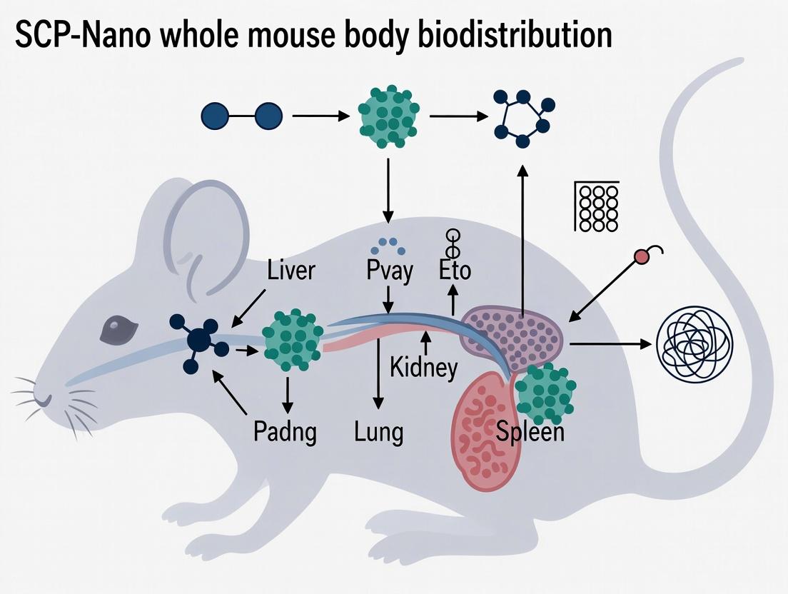
This technical guide details the SCP-Nano (Single-Cell Perfusion Nanotechnology) platform, a transformative methodology for conducting whole mouse body biodistribution research. By integrating high-fidelity vascular perfusion with barcoded nanoparticle tracers and single-cell resolution analysis, SCP-Nano enables the precise mapping of compound delivery to every organ and cell type in a systemic context. This whiteprames the technology as the core experimental pillar for a thesis advancing quantitative systems pharmacology.
Core Technology Principles
SCP-Nano operates on three integrated pillars:
- Controlled Systemic Perfusion: A pressure- and temperature-stabilized perfusion system clears endogenous blood and delivers a uniform bolus of barcoded nanoparticles (NPs) or nanoparticle-conjugated therapeutics throughout the entire murine circulatory system.
- Nano-Barcoding: Each nanoparticle batch is conjugated with a unique oligonucleotide or isotopic barcode, allowing for multiplexed tracking of multiple experimental conditions (e.g., different drug formulations, time points) in a single animal.
- Single-Cell Disaggregation & Analysis: Perfused tissues are dissociated into single-cell suspensions, where the barcode signal is quantified per cell via next-generation sequencing (NGS) or mass cytometry (CyTOF), alongside deep phenotypic profiling.
Key Experimental Protocols
Protocol 3.1: Whole-Body Perfusion with Barcoded Nanoparticles
Objective: To achieve complete vascular replacement with a homogenously distributed barcoded nanoparticle suspension. Materials: See "The Scientist's Toolkit" (Section 7). Procedure:
- Anesthetize mouse (e.g., C57BL/6) and secure in supine position.
- Perform a midline thoracotomy. Cannulate the left ventricle with a 24G catheter. Create an outflow incision in the right atrium.
- Perfuse with 20 mL of pre-warmed (37°C) 1X DPBS + 5 U/mL heparin at a constant pressure of 100 mmHg using a micro-perfusion pump to clear blood.
- Immediately follow with perfusion of 10 mL of the barcoded nanoparticle suspension (e.g., 1x10^11 particles/mL in DPBS) under identical pressure/temperature conditions.
- Harvest target organs (brain, heart, lungs, liver, spleen, kidneys) and rinse in cold PBS. Snap-freeze in liquid N2 or process immediately for disaggregation.
Protocol 3.2: Tissue Disaggregation & Single-Cell Library Preparation
Objective: To generate viable single-cell suspensions from perfused tissues for barcode and transcriptomic analysis. Procedure (for Liver tissue):
- Mechanically dissociate harvested liver using a GentleMACS dissociator with the appropriate enzymatic cocktail (e.g., Liver Dissociation Kit, 37°C for 30 min).
- Pass suspension through a 70-µm cell strainer. Quench enzyme activity with complete medium.
- Perform RBC lysis (if needed) and wash cells twice in PBS + 0.04% BSA.
- Count and assess viability (>85% via trypan blue).
- For sequencing: Use a droplet-based single-cell RNA-seq platform (e.g., 10x Genomics). Generate libraries for both cDNA (cellular transcriptome) and feature barcode (nanoparticle barcode) sequencing concurrently.
- For mass cytometry: Stain cells with a metal-tagged antibody panel for cell phenotyping. The nanoparticle barcode is intrinsically detected via its isotopic label.
Table 1: Representative SCP-Nano Biodistribution Data (Hypothetical Study) Nanoparticle Formulation: PEGylated Liposomal Doxorubicin (PLD) vs. SCP-Nano Barcoded Gold Nanoparticle (AuNP). n=5 mice/group. Perfusion time: 10 minutes. Data presented as mean (SD).
| Target Organ | PLD (ng drug/mg tissue) | SCP-Nano AuNP (Barcode Reads/1000 cells) | Primary Cell Types Targeted (from scRNA-seq) |
|---|---|---|---|
| Liver | 125.4 (18.7) | 15,320 (2,110) | Kupffer cells (85%), Hepatocytes (12%), LSECs (3%) |
| Spleen | 98.2 (22.1) | 8,450 (1,540) | Red pulp macrophages (65%), B cells (30%) |
| Kidney | 12.5 (3.4) | 890 (210) | Proximal tubule epithelial cells (78%) |
| Lung | 25.6 (5.9) | 1,250 (380) | Alveolar macrophages (92%) |
| Heart | 5.1 (1.2) | 105 (45) | Cardiomyocytes (99%) |
| Brain | 0.8 (0.3) | 22 (8) | Microglia (>95%) |
Table 2: Comparison of SCP-Nano with Traditional Biodistribution Methods
| Parameter | Traditional Radioisotope/LC-MS | SCP-Nano Platform |
|---|---|---|
| Resolution | Whole organ homogenate | Single cell |
| Multiplexing Capacity | Low (1-2 labels/study) | High (1000+ barcodes/study) |
| Cell Phenotype Correlation | No, requires separate IHC | Yes, intrinsic to assay |
| Quantitative Throughput | High samples, low parameters | High parameters (full transcriptome + barcode) |
| Key Metric | Concentration per gram tissue | Barcode UMI count per cell type |
Visualized Workflows and Pathways
Title: SCP-Nano Core Experimental Workflow
Title: SCP-Nano Data Analysis Pipeline
Key Signaling Pathways in Nanoparticle-Cell Interaction
Title: NP Delivery & Clearance Pathways
The Scientist's Toolkit: Essential Research Reagents & Materials
| Item Name | Supplier (Example) | Function in SCP-Nano Protocol |
|---|---|---|
| Micro-Perfusion Pump (Pump 11 Elite) | Harvard Apparatus | Provides precise, pulse-free pressure control (80-120 mmHg) for consistent whole-body perfusion. |
| Barcoded Nanoparticle Library (SomaCode) | NanoString / Custom Synthesis | Pre-fabricated nanoparticles with unique DNA barcodes for multiplexed, quantitative tracking. |
| GentleMACS Octo Dissociator | Miltenyi Biotec | Standardized mechanical tissue dissociation to generate high-viability single-cell suspensions. |
| Chromium Next GEM Chip K (10x Genomics) | 10x Genomics | Microfluidic device for partitioning single cells and barcoded nanoparticles into droplets for sequencing. |
| Cell Hashtag Oligonucleotides (TotalSeq) | BioLegend | Allows sample multiplexing by staining cells from different organs/mice with unique barcoded antibodies. |
| CETAC Heparin | Medefil | Anticoagulant in pre-perfusion buffer to prevent clot formation and ensure complete vascular clearance. |
| Collagenase D | Roche | Critical enzyme for tissue-specific digestion (e.g., liver, tumor) during single-cell preparation. |
| Live/Dead Fixable Aqua Stain | Thermo Fisher | Fluorescent viability dye for flow cytometry to exclude dead cells prior to library construction. |
Why Whole-Body Biodistribution is a Critical Pillar in Nanoparticle Drug Development
The advent of nanoparticle-based therapeutics promises a paradigm shift in drug delivery, offering the potential for targeted delivery, reduced systemic toxicity, and enhanced therapeutic efficacy. However, the translation of these promising platforms from benchtop to bedside is intrinsically linked to a comprehensive understanding of their in vivo journey. Whole-body biodistribution research is not merely an auxiliary study; it is a critical, non-negotiable pillar in nanoparticle drug development. This whitepaper, framed within the broader thesis of SCP-Nano biodistribution research, delineates why mastering biodistribution dictates success and provides a technical guide for its rigorous assessment.
The Core Quantitative Challenge: From Injection to Target
The primary goal of a nanoparticle therapeutic is to maximize delivery to the disease site while minimizing accumulation in healthy organs (e.g., liver, spleen). This is quantified through biodistribution studies, which provide essential pharmacokinetic (PK) and pharmacodynamic (PD) parameters.
Table 1: Core Biodistribution & PK Metrics for Nanoparticle Evaluation
| Metric | Definition | Typical Desired Outcome for SCP-Nano | Measurement Method |
|---|---|---|---|
| % Injected Dose per Gram (%ID/g) | Percentage of the administered dose present per gram of tissue at a given time. | High in target tissue; Low in liver/spleen. | Gamma counting (radionuclides), fluorescence quantification, ICP-MS (for inorganic NPs). |
| Target-to-Background Ratio (TBR) | Ratio of %ID/g in target tissue to %ID/g in a key off-target organ (e.g., liver). | >1, with higher values indicating superior specificity. | Calculated from %ID/g data. |
| Area Under the Curve (AUC) | The integral of concentration over time in a specific organ, reflecting total exposure. | High in target; Low in clearance organs. | Non-compartmental analysis of time-course data. |
| Clearance Half-life (t1/2) | Time for systemic concentration to reduce by half. | Optimized for therapeutic window: long enough for efficacy, short enough to avoid chronic toxicity. | PK analysis of blood/plasma data. |
| Maximum Concentration (Cmax) | Peak concentration observed in an organ or plasma. | Below toxicity threshold in off-target organs. | Direct from time-course data. |
Table 2: Common Biodistribution Patterns of Nanoparticles & Implications
| Nanoparticle Type | Typical Primary Accumulation Sites | Primary Clearance Mechanism | Key Development Challenge |
|---|---|---|---|
| PEGylated Liposomes | Liver, Spleen (Mononuclear Phagocyte System - MPS), tumors via EPR effect. | Hepatic clearance, phagocytosis. | Accelerated Blood Clearance (ABC) phenomenon upon repeated dosing. |
| Polymeric NPs (PLGA, etc.) | Liver, Spleen. Degradation-dependent. | Opsonization, MPS uptake, renal (if small fragments). | Controlling degradation kinetics and release profile. |
| Inorganic NPs (Gold, Silica) | Liver, Spleen - often persistent. | Very slow hepatic/biliary clearance; potential long-term retention. | Long-term toxicity from biocumulation. |
| SCP-Nano Platform (Thesis Context) | Data Objective: Demonstrate redirected biodistribution away from MPS towards target tissues via surface engineering. | Goal: Active targeting + stealth properties to modulate clearance. | Balancing stealth coatings with active targeting ligand functionality. |
Experimental Protocols for Whole-Body Biodistribution
Protocol 1: Quantitative Biodistribution Using Radiolabeling (Gold Standard)
Objective: To obtain absolute, quantitative biodistribution data across all major organs.
- Nanoparticle Labeling: Radiolabel SCP-Nano construct (e.g., with ^111In, ^125I, ^89Zr, ^64Cu) via chelation or direct incorporation. Purify to remove free radionuclide.
- Animal Dosing: Administer a known dose (e.g., 100 µL, 1 mg/kg, ~100 µCi) via relevant route (e.g., intravenous tail-vein injection) to healthy or disease-model mice (n=5 per time point).
- Time-Course Sacrifice: Euthanize animals at predetermined time points (e.g., 1, 4, 24, 72 hours post-injection).
- Organ Harvest & Weighing: Systematically harvest blood, heart, lungs, liver, spleen, kidneys, target tissue (e.g., tumor), and a sample of muscle and bone. Weigh each organ/tissue precisely.
- Gamma Counting: Place each sample in a gamma counter. Measure radioactivity, correcting for background, decay, and organ weight.
- Data Analysis: Calculate %ID/g for each organ. Plot biodistribution profiles and calculate PK parameters (AUC, TBR).
Protocol 2: Semi-Quantitative/Imaging-Based Biodistribution
Objective: To visualize spatial distribution and obtain semi-quantitative data in real-time.
- Nanoparticle Labeling: Label SCP-Nano with a near-infrared (NIR) fluorophore (e.g., Cy5.5, DyLight 750) or a CT/MRI contrast agent.
- In Vivo Imaging: At set time points post-injection, anesthetize mice and image using:
- Fluorescence Molecular Tomography (FMT) or Optical Imaging: Provides 3D quantification of fluorescence signal in deep tissues.
- Micro-CT/PET/SPECT: For high-resolution anatomical/functional co-registration.
- Ex Vivo Validation: After terminal imaging, harvest organs and image ex vivo with a high-resolution optical imager to correlate signals with Protocol 1 data.
Visualizing the Biodistribution Workflow & Fate
SCP-Nano In Vivo Journey & Analysis
The Scientist's Toolkit: Key Research Reagent Solutions
Table 3: Essential Reagents & Materials for Biodistribution Studies
| Item | Function & Relevance |
|---|---|
| Radionuclides (^111In, ^89Zr, ^64Cu) | Gold-standard for quantitative, whole-body biodistribution and PK studies via gamma counting/PET imaging. |
| Near-Infrared (NIR) Fluorophores | Enable non-invasive, longitudinal in vivo optical imaging of nanoparticle distribution (semi-quantitative). |
| Desferrioxamine (DFO) & Other Chelators | Critical for stable conjugation of radionuclides (e.g., ^89Zr) to nanoparticle surfaces without leakage. |
| PEGylation Reagents (mPEG-NHS) | Used to create "stealth" coatings on SCP-Nano to reduce opsonization and prolong circulation half-life. |
| Active Targeting Ligands | Antibodies, peptides, or small molecules conjugated to SCP-Nano to direct binding to diseased tissue. |
| ICP-MS Standard Solutions | Essential for calibrating inductively coupled plasma mass spectrometry used to quantify inorganic nanoparticles (e.g., Au, Si). |
| In Vivo Imaging Systems (FMT, PET/CT) | Instruments for non-invasive, real-time visualization and quantification of nanoparticle distribution. |
| Gamma Counter | Foundational instrument for measuring radioactivity in harvested tissues to calculate %ID/g. |
Whole-body biodistribution is the definitive map of a nanoparticle's in vivo behavior. It bridges the gap between elegant in vitro design and tangible therapeutic outcome. For the SCP-Nano platform, rigorous biodistribution research is the critical feedback loop that informs iterative design—guiding surface chemistry, targeting strategy, and dosage regimen. Without this pillar, nanoparticle development proceeds blindly, risking clinical failure due to unforeseen toxicity, insufficient delivery, or rapid clearance. Mastering biodistribution is not an option; it is the cornerstone of rational and successful nanomedicine development.
In the context of SCP-Nano whole mouse body biodistribution research, pharmacokinetic (PK) parameters are critical for quantifying the systemic exposure and disposition of novel nanocarriers and their therapeutic payloads. This whitepaper provides an in-depth technical guide to the four core PK parameters—Area Under the Curve (AUC), Maximum Concentration (Cmax), Time to Maximum Concentration (Tmax), and Clearance (CL)—detailing their calculation, physiological significance, and measurement within advanced biodistribution studies.
Core Pharmacokinetic Parameters: Definitions and Significance
Area Under the Curve (AUC): The integral of the drug concentration-time profile from time zero to infinity (AUC₀–∞) or to the last measurable time point (AUC₀–t). It represents the total systemic exposure to the drug or nanoparticle.
Maximum Concentration (Cmax): The peak observed concentration in plasma or tissue following administration. For SCP-Nano constructs, this indicates initial distribution intensity.
Time to Maximum Concentration (Tmax): The time point at which Cmax is observed. It is a marker of the rate of absorption or distribution.
Clearance (CL): The volume of plasma or blood from which the drug or nanoparticle is completely removed per unit time. It is a measure of the body's efficiency in eliminating the substance.
The following table summarizes typical PK parameter ranges reported in recent literature for polymeric and lipid-based nanoparticles in murine models, providing a benchmark for SCP-Nano research.
Table 1: Representative PK Parameters for Nanocarriers in Mouse Studies (IV Administration)
| Parameter | Typical Range (Polymeric NPs) | Typical Range (Lipid NPs) | Key Influencing Factors |
|---|---|---|---|
| AUC₀–∞ (mg·h/L) | 120 - 350 | 80 - 200 | Surface coating, particle size, targeting ligands |
| Cmax (mg/L) | 25 - 60 | 15 - 40 | Dose, injection rate, opsonization |
| Tmax (h) | 0.08 - 0.25 (immediate for IV) | 0.08 - 0.25 | Administration route, absorption kinetics |
| Clearance (mL/h/kg) | 30 - 100 | 50 - 150 | RES uptake, renal/hepatobiliary elimination |
Table 2: Comparison of Key PK Parameters for Different Administration Routes
| Route | Typical Tmax (h) | Bioavailability (F%)* | Primary Use in Biodistribution |
|---|---|---|---|
| Intravenous (IV) | ~0.08 (2 min) | 100% (by definition) | Full PK profile, clearance studies |
| Subcutaneous (SC) | 2 - 6 | 60 - 90% | Sustained release assessment |
| Intraperitoneal (IP) | 0.25 - 1 | 70 - 100% | Common for rodent therapeutics |
*Bioavailability is highly formulation-dependent.
Experimental Protocols for Parameter Measurement
Integrated PK-Biodistribution Study Workflow for SCP-Nano Constructs
Objective: To simultaneously characterize plasma PK and whole-body biodistribution of radiolabeled or fluorescently tagged SCP-Nano particles.
Materials: See "The Scientist's Toolkit" below.
Protocol:
- Formulation & Labeling: Prepare SCP-Nano construct. Incorporate a radioisotope (e.g., ⁸⁹Zirconium for PET, ¹¹¹Indium for SPECT) or a near-infrared (NIR) dye (e.g., DiR, Cy7) for sensitive detection.
- Animal Dosing: Administer a single bolus dose (e.g., 5 mg/kg nanoparticle) to cohorts of mice (n=5-6 per time point) via the chosen route (typically IV via tail vein).
- Serial Blood Sampling: At pre-defined time points (e.g., 2 min, 15 min, 30 min, 1h, 2h, 4h, 8h, 24h, 48h), collect blood via retro-orbital or submandibular route into heparinized tubes. Centrifuge immediately (5,000xg, 5 min, 4°C) to obtain plasma.
- Terminal Tissue Harvest: Euthanize mice at each corresponding time point. Perfuse with saline via the left ventricle to clear blood from organs. Harvest key organs (liver, spleen, kidneys, heart, lungs, brain, tumor) and weigh them.
- Sample Analysis:
- Radioactive Samples: Count plasma and homogenized tissues in a gamma counter. Express data as % Injected Dose per gram of tissue (%ID/g) or %ID per organ.
- Fluorescent Samples: Image excised organs using an in vivo imaging system (IVIS). Quantify fluorescence intensity and convert to %ID/g using a standard curve.
- Data Processing: Plot mean plasma concentration vs. time. Use non-compartmental analysis (NCA) with software (e.g., Phoenix WinNonlin, PK-Solver) to calculate AUC (trapezoidal rule), Cmax, Tmax (observed directly), and Clearance (CL = Dose / AUC₀–∞).
Title: SCP-Nano PK & Biodistribution Experimental Workflow
The Scientist's Toolkit: Essential Research Reagents and Materials
Table 3: Key Research Reagent Solutions for SCP-Nano PK/Biodistribution Studies
| Item | Function/Brief Explanation |
|---|---|
| SCP-Nano Construct | The core nanoparticle (e.g., polymer, lipid, inorganic) being evaluated for drug delivery. |
| Chelator-Radionuclide (e.g., DFO-⁸⁹Zr) | Enables stable radiolabeling for quantitative, deep-tissue PET imaging and ex vivo counting. |
| Near-Infrared Dye (e.g., Cy7-NHS ester) | Covalent labeling for real-time fluorescence imaging and ex vivo organ quantification. |
| Heparinized Micro-hematocrit Tubes | For consistent collection of blood samples without coagulation. |
| Phosphate-Buffered Saline (PBS) | For perfusion to clear blood, dilutions, and as a formulation vehicle. |
| Tissue Homogenization Buffer | Typically PBS with protease inhibitors, to prepare uniform tissue lysates for counting. |
| Gamma Counter | Instrument to measure radioactivity in plasma and tissue samples with high sensitivity. |
| In Vivo Imaging System (IVIS) | For non-invasive longitudinal fluorescence imaging and ex vivo organ imaging. |
| PK Analysis Software (WinNonlin, PK-Solver) | Essential for applying NCA models to calculate precise PK parameters from raw data. |
Interparameter Relationships and Physiological Pathways
Clearance is the fundamental parameter linking dose to exposure (AUC). The relationship between AUC, Clearance, and Volume of Distribution (Vd) underpins PK analysis. The following diagram illustrates the primary elimination pathways for nanoparticles, which directly determine their clearance rate.
Title: Primary Clearance Pathways for SCP-Nano Constructs
Data Integration in the Broader Thesis
Within a thesis on SCP-Nano whole-body biodistribution, these PK parameters are not endpoints but bridging metrics. They connect the physicochemical properties of the nanoparticle (size, charge, surface ligand) to its in vivo fate. High RES clearance correlates with high liver/spleen AUC (organ exposure). A prolonged terminal half-life, derived from the clearance and volume of distribution, may indicate successful evasion of the RES and potential for enhanced permeability and retention (EPR) in tumors. Thus, meticulous measurement of AUC, Cmax, Tmax, and CL provides the quantitative framework for evaluating the success of SCP-Nano engineering strategies and predicting therapeutic efficacy.
The Role of the EPR Effect and Active Targeting in Mouse Models
This whitepaper details the roles of the Enhanced Permeability and Retention (EPR) effect and active targeting strategies within mouse models, framed within the context of SCP-Nano whole mouse body biodistribution research. The objective is to guide researchers in designing and interpreting preclinical studies for nanomedicine development.
Foundational Concepts
The Enhanced Permeability and Retention (EPR) Effect
The EPR effect is a passive targeting mechanism whereby macromolecules and nanoparticles preferentially accumulate in tumor tissue due to pathological characteristics of tumor vasculature and impaired lymphatic drainage.
Key Physiological Drivers:
- Vasculature Defects: Inefficient angiogenesis leads to wide fenestrations (100-780 nm) in tumor endothelial linings.
- Lack of Lymphatic Drainage: Reduced clearance from the tumor interstitium enhances retention.
- Inflammatory Mediators: Factors like VEGF, bradykinin, and peroxynitrite enhance vascular permeability.
Active Targeting
Active targeting involves the surface functionalization of nanoparticles with ligands (e.g., antibodies, peptides, aptamers) that bind specifically to antigens or receptors overexpressed on target cells (e.g., tumor cells, endothelial cells), enhancing cellular uptake and specificity beyond the EPR effect.
Comparative Analysis in Murine Models
Data synthesized from recent literature (2022-2024) comparing passive (EPR-only) and active targeting strategies in murine tumor models.
Table 1: Biodistribution & Efficacy Comparison of Targeting Modalities
| Parameter | EPR-Based (Passive) Targeting | Active Targeting (e.g., anti-HER2, RGD, Folate) | Notes & Key Conditions |
|---|---|---|---|
| Avg. % Injected Dose/g in Tumor | 0.5 - 3.5 %ID/g | 2.0 - 8.5 %ID/g | Highly dependent on nanoparticle size (typically 30-150 nm optimal for EPR) and tumor model. |
| Tumor-to-Muscle Ratio | 3 - 8 | 10 - 25 | Active targeting significantly improves signal-to-background. |
| Avg. % Injected Dose/g in Liver | 15 - 35 %ID/g | 18 - 40 %ID/g | Active ligands can sometimes increase RES uptake; requires surface engineering (e.g., PEGylation). |
| Cellular Internalization | Low (primarily extracellular) | High (receptor-mediated endocytosis) | Critical for delivering internalizing payloads (e.g., gene therapeutics). |
| In Vivo Therapeutic Efficacy | Moderate tumor growth inhibition | Enhanced tumor growth inhibition & occasional regression | Often tested in xenograft models (e.g., CT26, 4T1, MDA-MB-231). |
| Key Influencing Variables | Tumor type, vascularization, stroma density, nanoparticle size & surface charge. | Receptor density, ligand affinity, binding site accessibility, ligand orientation. | Inter-animal and inter-tumor heterogeneity major challenges for EPR. |
Methodological Protocols for Key Experiments
Protocol: Evaluating EPR Effect with Fluorescent or Radiolabeled Nanoparticles
Objective: Quantify passive accumulation of nanoparticles in tumors and major organs.
Materials:
- Mouse Model: Immunocompromised mice (e.g., nude, NSG) bearing subcutaneous or orthotopic human tumor xenografts (tumor volume ~200-300 mm³).
- Nanoparticles: 100 nm diameter fluorescent liposomes (e.g., DiR-labeled) or PEGylated polystyrene nanoparticles.
- Imaging System: IVIS Spectrum or small animal PET/CT.
Procedure:
- Inject 100 µL of nanoparticle suspension (~5 nmol of dye or 10 µCi radiolabel) via tail vein.
- At predetermined time points (e.g., 1, 4, 24, 48, 72h), anesthetize mice (2% isoflurane).
- Acquire in vivo whole-body fluorescence or PET images.
- Euthanize mice, harvest tumors and organs (liver, spleen, kidneys, heart, lungs, muscle).
- Weigh tissues and quantify fluorescence ex vivo using a plate reader or measure radioactivity with a gamma counter.
- Calculate %ID/g for each tissue.
Protocol: Assessing Active TargetingIn Vivowith Competitive Blocking
Objective: Confirm specific receptor-mediated targeting.
Materials:
- Actively targeted nanoparticles (e.g., conjugated with cRGDfK peptide targeting αvβ3 integrin).
- Free targeting ligand (e.g., excess cRGDfK peptide) for blocking studies.
Procedure:
- Divide tumor-bearing mice into two groups (n=5): Test Group and Blocking Group.
- Blocking Group: Pre-inject with 100 µL of 10 mM free ligand solution 15 minutes prior to nanoparticle administration.
- Both Groups: Inject with the same dose of actively targeted nanoparticles.
- Image and harvest at peak accumulation time (e.g., 24h post-nanoparticle injection) as per Protocol 3.1.
- Significantly reduced tumor uptake in the blocking group confirms receptor-specific active targeting.
Visualization of Mechanisms and Workflows
Diagram 1: EPR vs Active Targeting Mechanisms
Diagram 2: In Vivo Targeting Study Workflow
The Scientist's Toolkit: Research Reagent Solutions
Table 2: Essential Materials for Mouse Model Targeting Studies
| Item | Function & Rationale | Example Product/Type |
|---|---|---|
| Fluorescent Liposomes (100 nm) | Standardized, commercially available nanoparticles for validating EPR effect and baseline biodistribution. | FormuMax F60103; encapsulating DiD, DiR, or ICG dyes. |
| PEGylation Reagents | Conjugate polyethylene glycol (PEG) to nanoparticle surfaces to reduce opsonization, prolong circulation, and enhance EPR. | Methoxy-PEG-SH (mPEG-Thiol), MW: 2000-5000 Da. |
| Biotin-PEG-NHS Ester | A versatile heterobifunctional linker for conjugating streptavidin-labeled targeting ligands to amine-bearing nanoparticles. | Thermo Fisher Scientific 21343. |
| cRGDfK Peptide | A cyclic peptide with high affinity for αvβ3 integrin, commonly used to target tumor angiogenesis or glioblastoma. | Peptide sequences from vendors like PeptideGen. |
| Anti-HER2 scFv or Affibody | Small, high-affinity targeting ligands for HER2-positive breast cancer models (e.g., BT-474). | Recombinant proteins from AcroBiosystems. |
| Near-Infrared (NIR) Dyes | For in vivo fluorescence imaging; minimizes tissue autofluorescence. | Indocyanine Green (ICG), IRDye 800CW. |
| Matrigel | Basement membrane matrix for co-injection with tumor cells to enhance engraftment and promote vascularization. | Corning Matrigel Matrix, Phenol Red-free. |
| IVIS Imaging System | Platform for non-invasive, longitudinal quantification of fluorescent nanoparticle biodistribution. | PerkinElmer IVIS Spectrum. |
| Radioisotope Labeling Kits | For radiolabeling nanoparticles (e.g., with Zr-89, Cu-64) for quantitative PET imaging and dosimetry. | [89Zr]Zr-DFO or [64Cu]Cu-NOTA conjugation kits. |
Essential Mouse Strains and Models for Standardized Biodistribution Studies
Within the burgeoning field of nanomedicine, the development of Systemic Circulating Particulates (SCP-Nano) demands rigorous and standardized preclinical evaluation. A cornerstone of this evaluation is the whole mouse body biodistribution study, which quantifies the temporal and spatial accumulation of a nanoparticle formulation across organs and tissues. The selection of an appropriate mouse strain and model is not a trivial decision; it fundamentally influences pharmacokinetics, immune recognition, and organ uptake profiles. This guide details the essential mouse strains and models, framing their use within the context of a robust SCP-Nano biodistribution research thesis, which posits that standardized model selection is critical for generating reproducible, translatable data in nanocarrier development.
Foundational Mouse Strains for Biodistribution Studies
The choice of immunocompetent versus immunocompromised strains is the primary decision point, dictated by the nature of the SCP-Nano payload (e.g., chemotherapeutic, nucleic acid) and experimental goals (e.g., evaluation of innate immune clearance).
Immunocompetent Strains
These strains possess a fully functional immune system, crucial for studying nanoparticle interactions with plasma proteins (opsonization) and clearance by the mononuclear phagocyte system (MPS).
- C57BL/6 (B6): The most widely used inbred strain. Its well-characterized genetics and robust Th1-biased immune response make it the gold standard for foundational biodistribution studies. It is particularly useful for studying liver and spleen Kupffer cell/sinusoidal endothelial uptake.
- BALB/c: Another common inbred strain, with a Th2-biased immune response. Often preferred in oncology models and for studying nanoparticle accumulation in tumors derived from this background. Comparisons between B6 and BALB/c can reveal strain-specific pharmacokinetic differences.
- CD-1 (ICR): An outbred, genetically heterogeneous stock. Its genetic diversity may better represent population variation, useful for assessing biodistribution consistency across a broader genetic background.
Immunocompromised Strains
Essential for studying human cell-derived xenografts (e.g., patient-derived tumors) without immune rejection, but also valuable for studying nanoparticle fate in the absence of adaptive immunity.
- Nude (Foxn1nu): Lacks a functional thymus, resulting in an absence of T-lymphocytes. Retains functional B-cells and a potent innate immune system (NK cells, macrophages). Useful for studies where T-cell immunity is a major confounder.
- NOD-scid IL2Rγnull (NSG): Severely immunocompromised due to defects in T, B, and NK cells, and impaired cytokine signaling. Minimizes nanoparticle clearance by adaptive immunity, allowing isolation of innate/physical clearance mechanisms. The model of choice for human tumor xenograft studies.
Table 1: Core Mouse Strains for Standardized Biodistribution Studies
| Strain | Immune Status | Key Genetic/Physiological Traits | Primary Application in SCP-Nano Research | Key Considerations |
|---|---|---|---|---|
| C57BL/6 | Immunocompetent | Inbred, Th1 bias, well-defined MPS | Gold standard; baseline PK/BD; liver/spleen uptake studies | High lipopolysaccharide sensitivity; standard for genetic engineering. |
| BALB/c | Immunocompetent | Inbred, Th2 bias, docile temperament | Comparative studies to B6; tumor models (e.g., 4T1); antibody production | Different MPS activity vs. B6 can alter nanoparticle clearance. |
| CD-1 (ICR) | Immunocompetent | Outbred, genetically heterogeneous | Assessing BD variability; general toxicology & safety profiling | Higher litter sizes; less genetic uniformity than inbred strains. |
| Nude | T-cell Deficient | Foxn1 mutation; functional B-cells & NK cells | Human tumor xenografts; studies minimizing T-cell effects | Requires sterile housing; retains robust innate immunity. |
| NSG | Severely Compromised | Prkdcscid + Il2rgnull; lacks T, B, NK cells | Human immune system (HIS) models; human tumor xenografts | Extreme susceptibility to infection; allows engraftment of human cells. |
Specialized Disease Models for Targeted Biodistribution
To evaluate SCP-Nano targeting efficiency, biodistribution must be assessed in the context of pathology.
Oncology Models
- Syngeneic Tumor Models: Tumor cell lines derived from the same mouse strain (e.g., B16-F10 melanoma in C57BL/6, 4T1 mammary carcinoma in BALB/c). They have an intact tumor microenvironment (TME) and immune system, ideal for studying immune-modulatory nanoparticles and the Enhanced Permeability and Retention (EPR) effect.
- Xenograft Models: Human tumor cells implanted in immunocompromised hosts (e.g., NSG). Used to study nanoparticle behavior against human-specific targets. Orthotopic (tumor in native tissue) vs. subcutaneous implants significantly affect biodistribution.
- Genetically Engineered Mouse Models (GEMMs): Spontaneous tumor models (e.g., KrasG12D; Trp53R172H lung cancer) with authentic TME and intact immune systems. Represent the most clinically relevant but complex and variable model for BD studies.
Inflammation & Infection Models
Models like dextran sulfate sodium (DSS)-induced colitis or localized bacterial infection can be used to study nanoparticle extravasation at sites of enhanced vascular permeability.
Organ-Specific Injury Models
Liver fibrosis models (e.g., CCl4 administration) or models of brain barrier disruption (e.g., experimental autoimmune encephalomyelitis) are critical for understanding how disease state alters nanoparticle accumulation in target organs.
Table 2: Key Disease Models for Targeted Biodistribution Assessment
| Model Type | Example Model | Host Strain | Relevance to SCP-Nano BD Studies | Key Pathophysiological Feature |
|---|---|---|---|---|
| Syngeneic Tumor | B16-F10 melanoma | C57BL/6 | Studies of EPR effect, immune-oncology NPs | Intact, immunocompetent TME. |
| Heterotopic Xenograft | MDA-MB-231 breast cancer (s.c.) | NSG | Evaluating targeting to human antigens | Stroma is mouse-derived; limited TME. |
| Orthotopic Xenograft | Patient-derived glioma (brain) | NSG | Assessing brain tumor penetration & BD | More relevant anatomy & microenvironment. |
| GEMM | KrasG12D/p53fl/fl lung | Mixed/C57BL/6 | Gold standard for TME & therapy response | Authentic, heterogeneous, immunocompetent. |
| Inflammatory | DSS-Induced Colitis | C57BL/6, BALB/c | NPs targeting inflamed vasculature | Disrupted epithelial/endothelial barriers. |
Experimental Protocol: Standardized Biodistribution Workflow for SCP-Nano
Objective: Quantify the percentage of injected dose (%ID) of a radiolabeled or fluorescently labeled SCP-Nano formulation per gram of tissue over time.
Materials: See "The Scientist's Toolkit" below. Animals: Selected based on Tables 1 & 2. N ≥ 5 per time point. Acclimate for 1 week.
Procedure:
- SCP-Nano Administration: Via lateral tail vein injection (for systemic distribution) at a standardized volume (e.g., 5 µL/g body weight) and dose (e.g., 5 mg nanoparticles/kg). Record exact injected volume and dose for each animal.
- Temporal Cohorts: Euthanize cohorts at pre-defined time points (e.g., 5 min, 1 h, 4 h, 24 h, 7 d) post-injection via approved method (e.g., CO2 overdose, followed by cervical dislocation).
- Perfusion & Tissue Harvest: Perform transcardial perfusion with >20 mL of cold PBS (pH 7.4) via the left ventricle to clear blood from the vasculature. Systematically harvest organs: blood (via cardiac puncture prior to perfusion), heart, lungs, liver, spleen, kidneys, brain, and any tumor or target tissue. Weigh each organ/tissue precisely.
- Quantification:
- Radiolabel (e.g., 125I, 111In, 89Zr): Place tissues in gamma counter tubes. Measure radioactivity (Counts Per Minute, CPM) for each sample and a dilution series of the injected dose standard. Correct for isotope decay and background. Calculate %ID/g = (CPMtissue / weighttissue) / (Total Injected CPM) * 100.
- Fluorescent Label (e.g., Cy5.5, DiR): Homogenize tissues in a known volume of lysis buffer. Measure fluorescence of clarified lysates using a plate reader. Compare to a standard curve of the nanoparticle in matched tissue homogenate from a control mouse to correct for matrix effects.
- Data Analysis: Express data as mean %ID/g ± SEM. Perform statistical analysis (e.g., ANOVA) between time points, organs, or treatment groups. Key pharmacokinetic parameters: Area Under the Curve (AUC) for each organ, peak concentration (Cmax), and time to peak (Tmax).
Standard SCP Nano Biodistribution Workflow
SCP-Nano Clearance and Signaling Pathways
A key thesis in SCP-Nano research is understanding the signaling that governs MPS recognition and clearance. Opsonization (complement, immunoglobulins) leads to phagocytosis via receptor-mediated endocytosis.
MPS Clearance Pathway for SCP Nano
The Scientist's Toolkit: Essential Research Reagents & Materials
Table 3: Key Reagents for SCP-Nano Biodistribution Studies
| Item | Function in Biodistribution Studies | Example/Notes |
|---|---|---|
| DOTA-NHS Ester | Chelator for radiometal labeling (111In, 64Cu, 89Zr) of amine-containing nanoparticles. | Enables stable in vivo tracking via PET/SPECT. |
| Cy5.5 NHS Ester | Near-infrared fluorescent dye for optical imaging; minimal tissue autofluorescence. | For ex vivo tissue quantification and in vivo imaging. |
| Phosphate Buffered Saline (PBS) | Vehicle for nanoparticle formulation and dilution; used for systemic perfusion. | Must be sterile, endotoxin-free. |
| Heparinized Capillary Tubes | Collection of blood samples for plasma pharmacokinetic analysis. | Prevents clotting for accurate plasma separation. |
| Tissue Homogenization Buffer | Lysis buffer for fluorescent nanoparticle extraction from tissues. | Often contains detergent (e.g., Triton X-100) and protease inhibitors. |
| Gamma Counter | Instrument for measuring gamma radiation from radiolabeled tissues. | Essential for calculating %ID/g with radionuclides. |
| IVIS Spectrum or similar | In vivo imaging system for real-time, non-invasive fluorescence tracking. | Provides longitudinal data in live animals. |
| Isoflurane/Oxygen Mix | Safe and controllable anesthesia for injection and in vivo imaging procedures. | Preferred over injectable anesthetics for short procedures. |
A Step-by-Step Protocol for SCP-Nano Biodistribution Studies in Mice
This guide details the essential first phase of a comprehensive thesis investigating the whole-body biodistribution of Single-Chain Polymer (SCP) nanoparticles in murine models. The precision of this pre-injection phase directly dictates the validity, reliability, and interpretability of subsequent in vivo biodistribution studies. This document provides standardized protocols for formulating SCP-Nano particles, radiolabeling them for sensitive tracking, and performing a rigorous suite of physicochemical and biological characterizations.
Nanoparticle Formulation
SCP nanoparticles are synthesized via controlled reversible addition-fragmentation chain-transfer (RAFT) polymerization, followed by self-assembly in selective solvents.
Protocol: Synthesis and Self-Assembly of SCP-Nano Particles
Polymer Synthesis:
- Purge a reaction vessel with nitrogen for 30 minutes.
- Dissolve chain transfer agent (CTA, e.g., 4-cyano-4-[(dodecylsulfanylthiocarbonyl)sulfanyl]pentanoic acid, 1 eq), monomer (e.g., poly(ethylene glycol) methyl ether acrylate, 200 eq), and initiator (e.g., 2,2'-Azobis(2-methylpropionitrile), 0.2 eq) in anhydrous 1,4-dioxane.
- Seal the vessel and immerse in an oil bath at 70°C for 18 hours under constant stirring (300 rpm).
- Terminate the reaction by rapid cooling and exposure to air. Precipitate the polymer into cold diethyl ether, followed by filtration and vacuum drying.
Nanoparticle Self-Assembly via Nano-precipitation:
- Dissolve 10 mg of the dried SCP polymer in 1 mL of a water-miscible organic solvent (e.g., acetone, DMSO).
- Using a syringe pump set to a rate of 1 mL/hour, slowly inject this solution into 10 mL of vigorously stirred (800 rpm) deionized water or PBS (pH 7.4).
- Allow the suspension to stir uncovered for 12 hours to evaporate the organic solvent.
- Pass the resulting colloidal suspension through a 0.22 µm polyethersulfone (PES) filter to remove any aggregates. Store at 4°C.
Radiolabeling for Biodistribution Tracking
For quantitative whole-body biodistribution, radiolabeling with a gamma-emitting isotope like Zirconium-89 (⁸⁹Zr) is preferred due to its suitable half-life (78.4 hours) for longitudinal studies.
Protocol: Chelator-Based Radiolabeling with ⁸⁹Zr
Chelator Conjugation: Functionalize a portion of the SCP nanoparticles with desferrioxamine (DFO) p-isothiocyanate. Incubate SCP-Nano (5 mg/mL in 0.1 M bicarbonate buffer, pH 8.5) with DFO-p-SCN (10 molar eq) for 2 hours at room temperature. Purify via size-exclusion chromatography (PD-10 column) into 0.25 M HEPES buffer (pH 7.0).
⁸⁹Zr Labeling:
- Adjust the pH of the purified DFO-SCP-Nano solution to 6.8-7.2.
- Add ⁸⁹Zr-oxalate (37-74 MBq) to the nanoparticle solution (1-2 mg/mL). Incubate at 37°C for 1 hour with gentle shaking.
- Quality Control: Determine radiochemical purity (RCP) by instant thin-layer chromatography (iTLC) using a 50 mM diethylenetriaminepentaacetic acid (DTPA) solution as the mobile phase. A successful labeling yields RCP >95%.
Table 1: Critical Quality Control Metrics for Radiolabeled SCP-Nano
| Parameter | Target Specification | Analytical Method | Purpose |
|---|---|---|---|
| Radiochemical Purity (RCP) | ≥ 95% | iTLC / Size-Exclusion HPLC | Ensures radioactivity is nanoparticle-bound, not free isotope. |
| Specific Activity | 10 - 20 MBq/mg | Gamma Counter | Balances detection sensitivity with potential radiotoxicity. |
| Post-Labeling Size | ΔD < 10% from pre-label | DLS | Confirms labeling process does not induce aggregation. |
| Stability in Serum (24h) | RCP > 90% | Incubation in mouse serum @ 37°C, followed by iTLC | Predicts integrity during circulation. |
Pre-Injection Characterization
A multi-faceted characterization profile is mandatory prior to any in vivo application.
Key Experimental Protocols:
Hydrodynamic Diameter & Polydispersity (DLS): Dilute SCP-Nano suspension 1:50 in PBS. Measure using a dynamic light scattering instrument at 25°C with a 173° backscatter angle. Report the Z-average diameter and Polydispersity Index (PdI) from triplicate measurements.
Surface Charge (Zeta Potential): Dilute sample in 1 mM KCl, pH 7.0. Measure electrophoretic mobility using a phase analysis light scattering (M3-PALS) technique. Convert to zeta potential via the Smoluchowski model.
Morphology (Transmission Electron Microscopy - TEM): Apply 5 µL of sample (0.1 mg/mL) onto a carbon-coated copper grid, blot after 60 seconds, and stain with 2% uranyl acetate for 30 seconds. Image at an accelerating voltage of 80 kV.
Sterility & Endotoxin Testing:
- Sterility: Filter sterilize (0.22 µm) the final formulation. Inoculate aliquots into Fluid Thioglycollate Medium and Soybean-Casein Digest Medium. Incubate for 14 days at 32.5°C and 22.5°C, respectively.
- Endotoxin (LAL Test): Use a chromogenic Limulus Amebocyte Lysate assay. Sample endotoxin levels must be <5.0 EU/kg mouse body weight for injectable solutions.
Table 2: Pre-Injection Physicochemical Characterization Specifications
| Characterization Parameter | Target Range for Murine Biodistribution Studies | Method |
|---|---|---|
| Hydrodynamic Diameter | 20 - 150 nm (monomodal) | Dynamic Light Scattering (DLS) |
| Polydispersity Index (PdI) | < 0.2 | DLS |
| Zeta Potential (in PBS, pH 7.4) | -30 mV to +10 mV (context-dependent) | Electrophoretic Light Scattering |
| Morphology | Spherical, uniform (visual confirmation) | Transmission Electron Microscopy (TEM) |
| Endotoxin Level | < 0.5 EU/mL | Limulus Amebocyte Lysate (LAL) Assay |
| Osmolarity | 280 - 320 mOsm/kg | Osmometer |
| pH | 7.0 - 7.4 | pH Meter |
SCP Nano Formulation and QC Workflow
The Scientist's Toolkit: Essential Research Reagents & Materials
Table 3: Key Reagents and Materials for Protocol Phase 1
| Item | Function / Role in Protocol | Example Product / Note |
|---|---|---|
| RAFT Chain Transfer Agent (CTA) | Controls polymer growth, defines end-group for functionalization. | 4-cyano-4-[(dodecylsulfanylthiocarbonyl)sulfanyl]pentanoic acid. |
| DFO-p-SCN | Chelator that tightly binds ⁸⁹Zr for stable radiolabeling. | Desferrioxamine B p-isothiocyanate. Must be stored desiccated at -20°C. |
| Zirconium-89 (⁸⁹Zr) | Positron-emitting radioisotope for quantitative PET imaging and ex vivo biodistribution. | Supplied as ⁸⁹Zr-oxalate in 1 M oxalic acid. Requires neutralization before use. |
| Size-Exclusion Chromatography Columns | Purifies conjugated nanoparticles and removes unreacted small molecules. | PD-10 Desalting Columns (Sephadex G-25). |
| Dynamic Light Scattering (DLS) Cells | Holds sample for hydrodynamic size and polydispersity measurements. | Disposable micro cuvettes (low volume, ~50 µL). |
| LAL Reagent | Detects bacterial endotoxins to ensure injectable safety. | Chromogenic endpoint assay kit for quantitative analysis. |
| Polyethersulfone (PES) Syringe Filters | Sterilizes and removes aggregates from final nanoparticle suspension. | 0.22 µm pore size, 25 mm diameter. |
| Carbon-Coated TEM Grids | Supports nanoparticle sample for high-resolution morphological imaging. | 200-400 mesh copper grids. |
How Pre Injection Traits Impact Biodistribution
This document details the second phase of the experimental protocol for SCP-Nano whole mouse body biodistribution research. The primary objective is to characterize the pharmacokinetic and biodistribution profiles of SCP-Nano constructs following administration via intravenous (IV), intraperitoneal (IP), and oral (PO) routes. Data from this phase are critical for determining the optimal delivery method for downstream therapeutic efficacy studies.
Administration Route Methodologies
Intravenous (IV) Injection
Objective: Achieve immediate and complete systemic circulation for maximal tissue exposure.
- Mouse Preparation: Animals are placed in a restrainer designed for tail vein access. The tail is warmed for 1-2 minutes using a heat lamp or warm water (∼40°C) to induce vasodilation.
- Injection Protocol: Using a 0.3-1.0 mL insulin syringe with a 29-30 gauge needle, the SCP-Nano formulation is injected into one of the two lateral tail veins. A successful injection is indicated by a lack of resistance and no immediate blanching or swelling.
- Volume & Rate: Standard injection volume is 5 mL/kg of mouse body weight. The bolus is administered slowly over 10-15 seconds. For a 25g mouse, this equates to 125 µL.
Intraperitoneal (IP) Injection
Objective: Utilize the peritoneal cavity as a depot for systemic absorption via the mesenteric vasculature.
- Mouse Restraint: The mouse is gently scruffed and held in a head-down position to shift organs cranially.
- Injection Protocol: The needle (25-27 gauge) is inserted at a 30-45 degree angle into the lower left quadrant of the abdomen to avoid vital organs (cecum, spleen, liver). Aspiration is performed before injection to ensure no organ or blood vessel puncture.
- Volume & Rate: A larger volume of up to 10 mL/kg can be administered. The injection is performed steadily over a few seconds, followed by gentle massaging of the site.
Oral Gavage (PO)
Objective: Assess gastrointestinal absorption and first-pass metabolism.
- Mouse Restraint: The mouse is scruffed firmly to immobilize the head, with the body held upright.
- Gavage Protocol: A ball-tipped, curved gavage needle (20-22 gauge, 1-1.5 inches long) is gently passed down the esophagus. Correct placement is confirmed by smooth passage and lack of resistance.
- Volume & Formulation: Maximum recommended volume is 10 mL/kg. The SCP-Nano formulation may require adaptation (e.g., viscosity adjustment, inclusion of mucoadhesive agents) to ensure consistent delivery and absorption.
Dosage Considerations and Rationale
Dosage is calculated based on the target dose of the active pharmaceutical ingredient (API) encapsulated within or conjugated to the SCP-Nano particle. The following table summarizes the key parameters for a standard study using a 25g mouse.
Table 1: Administration Route Parameters for SCP-Nano Biodistribution
| Parameter | Intravenous (IV) | Intraperitoneal (IP) | Oral (PO) |
|---|---|---|---|
| Typical Needle Gauge | 29-30 G | 25-27 G | 20-22 G (ball-tip) |
| Max Injection Volume (for 25g mouse) | 125 µL (5 mL/kg) | 250 µL (10 mL/kg) | 250 µL (10 mL/kg) |
| Injection Site | Lateral Tail Vein | Lower Left Abdominal Quadrant | Esophagus to Stomach |
| Bioavailability (Typical Range for Nano) | 100% (by definition) | 50-85% | 5-30% |
| Peak Plasma Time (Tmax) | Immediate (End of Bolus) | 10-30 minutes | 1-4 hours |
| Key Advantage | Complete, controlled systemic delivery | Easier technique, good absorption | Non-invasive, clinically relevant |
| Primary Limitation | Technically challenging, stress | Risk of organ puncture, variable PK | Low/variable absorption, first-pass effect |
| Formulation Criticality | High (must be sterile, isotonic, small particle size) | Moderate (must be sterile, non-irritant) | Very High (must withstand low pH, enzymes, permeability) |
Dosage Calculation Example: If the target API dose is 5 mg/kg, and the SCP-Nano formulation has an API loading of 10% (w/w):
- Required nanoparticle mass per kg = 5 mg/kg API ÷ 0.10 = 50 mg nanoparticles/kg.
- For a 25g (0.025 kg) mouse: Total nanoparticle mass = 50 mg/kg * 0.025 kg = 1.25 mg.
- If the formulation concentration is 10 mg/mL, the volume to administer IV is 1.25 mg ÷ 10 mg/mL = 0.125 mL (125 µL).
Experimental Protocol for Comparative Biodistribution
This protocol is executed at time points post-administration (e.g., 5 min, 1h, 6h, 24h) to generate kinetic data.
1. Tissue Harvest and Processing:
- Euthanize mouse via CO₂ asphyxiation or approved anesthetic overdose followed by cervical dislocation.
- Perform systemic perfusion with 10-20 mL of ice-cold PBS via cardiac puncture to clear blood from organs.
- Harvest target organs (blood, liver, spleen, kidneys, heart, lungs, brain, intestines). Weigh each organ precisely.
- Homogenize tissues in lysis buffer (e.g., RIPA buffer with protease inhibitors) using a bead homogenizer or mechanical homogenizer. Centrifuge at 12,000 x g for 15 min at 4°C. Collect supernatant.
2. Quantification of SCP-Nano (or API) Concentration:
- Method A: Fluorescence (if nanoparticle is labeled): Measure fluorescence in supernatants using a plate reader. Compare to a standard curve of the SCP-Nano construct in matching tissue homogenates from untreated mice.
- Method B: Mass Spectrometry (for API quantification): Use LC-MS/MS with stable isotope-labeled internal standards. Extract API from tissue homogenates using protein precipitation or solid-phase extraction. Validate method for each tissue matrix.
- Data Normalization: Express concentration as
(Amount of SCP-Nano in organ) / (Mass of organ)(e.g., ng/mg tissue) or as% of Injected Dose per gram of tissue (%ID/g).
Table 2: Example Biodistribution Data (%ID/g) at 1 Hour Post-Dose
| Organ/Tissue | IV Route (Mean ± SD) | IP Route (Mean ± SD) | PO Route (Mean ± SD) |
|---|---|---|---|
| Blood | 25.4 ± 3.1 | 8.2 ± 1.5 | 0.5 ± 0.2 |
| Liver | 35.6 ± 4.8 | 22.7 ± 3.3 | 1.8 ± 0.6 |
| Spleen | 12.3 ± 2.1 | 4.5 ± 1.1 | 0.2 ± 0.1 |
| Kidneys | 10.8 ± 1.7 | 5.3 ± 0.9 | 0.4 ± 0.2 |
| Lungs | 8.9 ± 1.4 | 3.1 ± 0.8 | 0.1 ± 0.05 |
| Brain | 0.5 ± 0.2 | 0.1 ± 0.05 | Below LOQ |
| Heart | 4.2 ± 0.9 | 1.8 ± 0.5 | 0.2 ± 0.1 |
Visualizing Biodistribution Pathways and Workflow
Title: SCP-Nano Administration and Biodistribution Workflow
Title: Key Pathways Governing Nano-Bioavailability and Fate
The Scientist's Toolkit: Research Reagent Solutions
Table 3: Essential Materials for SCP-Nano Administration & Biodistribution Studies
| Item | Function/Brief Explanation |
|---|---|
| Fluorescently-labeled SCP-Nano (e.g., Cy5.5, DiR) | Enables real-time in vivo imaging and ex vivo fluorescence quantification of biodistribution without complex sample prep. |
| LC-MS/MS Grade Solvents (Acetonitrile, Methanol) | Essential for sensitive and specific mass spectrometry-based quantification of the API released from SCP-Nano in tissues. |
| Sterile, Isotonic Formulation Buffer (PBS, 5% Dextrose) | Vehicle for IV injection; must be particle-stable, isotonic, and pyrogen-free to prevent adverse reactions. |
| Mucoadhesive Polymer (e.g., Chitosan, PAA) | Added to oral formulations to increase GI residence time and improve nanoparticle absorption. |
| Protease/Phosphatase Inhibitor Cocktail | Added to tissue lysis buffer to prevent degradation of protein-based SCP-Nano or conjugated targeting ligands during homogenization. |
| PEGylated Lipid or Polymer | Common coating material to reduce opsonization and prolong SCP-Nano circulation time, especially for IV route. |
| Collagenase/DNase I Mix | For gentle dissociation of tissues (e.g., tumor, liver) to analyze cellular-level SCP-Nano uptake via flow cytometry. |
| Stable Isotope-Labeled API (Internal Standard) | Critical for accurate and precise LC-MS/MS quantification, correcting for matrix effects and recovery losses. |
| In Vivo Imaging System (IVIS) | For non-invasive, longitudinal tracking of fluorescently-labeled SCP-Nano biodistribution in live animals. |
| Tail Vein Injector Restrainer & Heat Lamp | Standardized equipment to reduce stress and ensure consistent, successful IV tail vein injections. |
This document details the critical third phase of a comprehensive experimental protocol designed for systemic biodistribution research of engineered nanomaterials (SCP-Nano) in murine models. Within the broader thesis framework, this phase bridges the in vivo administration (Phase 2) and downstream analytical quantification (Phase 4). Its precise execution is paramount for generating temporally resolved, spatially accurate data on SCP-Nano accumulation, clearance, and potential toxicity across all major organ systems. The selection of optimal time-points and the application of consistent, humane euthanasia followed by systematic tissue harvest are foundational to data integrity and ethical compliance.
Rationale for Time-Point Selection
Time-point selection is driven by the pharmacokinetic (PK) and pharmacodynamic (PD) profile of the SCP-Nano construct under investigation. The objective is to capture key phases of the biodistribution lifecycle.
Core Principles:
- Early Phase (Minutes to 1-2 Hours): Captures the distribution phase, highlighting vascular circulation, early endothelial interactions, and initial organ uptake (e.g., liver, spleen, lungs).
- Mid Phase (Hours to 24-48 Hours): Represents the primary equilibrium phase, where maximum tissue accumulation often occurs, and active targeting (if applicable) is most evident.
- Late Phase (Days to 1-2 Weeks): Captures the clearance phase, revealing nanoparticle persistence, degradation, and potential redistribution. Critical for assessing long-term safety profiles.
Recommended Time-Point Schema
Based on current literature and standard practices in nanomedicine PK studies, the following multi-tiered schema is proposed. The exact points must be tailored to the specific SCP-Nano properties (e.g., material, size, surface charge, targeting moiety).
Table 1: Standardized Time-Point Selection Framework for SCP-Nano Biodistribution
| Phase | Post-IV Injection Time-Points | Biological Processes Monitored | Key Tissues of Interest |
|---|---|---|---|
| Distribution | 5 min, 15 min, 30 min, 1 h | Vascular circulation, first-pass clearance, rapid RES uptake. | Blood, Lung, Liver, Spleen, Kidney. |
| Equilibrium | 4 h, 8 h, 24 h, 48 h | Maximized extravasation & target site accumulation, active targeting peak. | Tumor (if applicable), Target Organ, Liver, Spleen, Lymph Nodes. |
| Clearance | 72 h (3d), 168 h (7d), 336 h (14d) | Long-term persistence, biodegradation, renal/hepatobiliary excretion. | Liver, Spleen, Bone Marrow, Excreta (feces/urine if collected). |
Humane Euthanasia: Methodology and Protocols
Euthanasia must be performed in accordance with approved Institutional Animal Care and Use Committee (IACUC) protocols and the AVMA Guidelines for the Euthanasia of Animals (2020). The method must ensure rapid loss of consciousness and death while minimizing stress, pain, and the release of stress-related biochemicals that could alter tissue physiology or nanoparticle distribution.
Primary Recommended Method: Controlled Inhalation Anesthetic Overdose Followed by Exsanguination
This method is preferred for tissue harvest as it minimizes physiological stress and allows for clean tissue collection.
Experimental Protocol:
- Preparation: Place the mouse in a clear induction chamber connected to a calibrated isoflurane vaporizer or a compressed CO₂ delivery system.
- Induction: Administer 5% isoflurane in medical-grade oxygen at a flow rate of 1-2 L/min, or administer 100% CO₂ at a displacement rate of 30-70% of the chamber volume per minute.
- Loss of Consciousness: Confirm the absence of the righting reflex and pedal withdrawal reflex.
- Euthanasia: For isoflurane: Maintain exposure for at least 1 minute after respiratory arrest. For CO₂: Maintain exposure for at least 2 minutes after respiratory arrest.
- Exsanguination/Cardiac Perfusion (Critical Step): Immediately following confirmed death, perform terminal cardiac puncture to draw blood or proceed to transcardial perfusion with phosphate-buffered saline (PBS) to clear the vascular compartment of blood and non-sequestered nanoparticles. This is essential for accurate quantification of tissue-localized SCP-Nano versus blood-pool signal.
- Secondary Physical Method: Apply bilateral thoracotomy or cervical dislocation as a confirmatory method per AVMA guidelines.
Systematic Tissue Harvest and Processing Workflow
A consistent, ordered harvest is crucial to prevent cross-contamination and ensure tissue integrity.
Diagram Title: SCP-Nano Biodistribution Study Tissue Harvest Workflow
Detailed Harvest Protocol
- Blood Collection: Using a 25G needle and syringe coated with an anticoagulant (e.g., EDTA-K2), perform terminal cardiac puncture. Transfer blood to microcentrifuge tubes for plasma/serum separation.
- Vascular Perfusion: Cannulate the left ventricle, incise the right atrium, and perfuse with 20-30 mL of ice-cold PBS at a steady, low pressure until effluent is clear and organs (especially liver) pale.
- Organ Harvest Sequence: Follow the order in the diagram to minimize cross-contamination. Use dedicated, clean instruments for each organ or clean thoroughly between samples.
- Tissue Processing: Weigh each tissue immediately. For biodistribution analysis (e.g., qPCR, radiotracer counting, ICP-MS), snap-freeze tissues in liquid nitrogen and store at -80°C. For histology, place in appropriate fixative (e.g., 10% NBF for light microscopy, glutaraldehyde for TEM).
The Scientist's Toolkit: Essential Research Reagent Solutions
Table 2: Key Materials for Tissue Harvest and Processing in Biodistribution Studies
| Item / Reagent | Function / Purpose | Key Considerations |
|---|---|---|
| Isoflurane or Compressed CO₂ | Humane euthanasia via inhalation overdose. | Preferred for minimizing stress. Must be used with appropriate scavenging systems. |
| Phosphate-Buffered Saline (PBS), Ice-cold | Vascular perfusion to clear blood and non-sequestered nanoparticles from tissues. | Critical for reducing background signal. Must be calcium/magnesium-free for some applications. |
| EDTA-K2 Coated Blood Collection Tubes | Anticoagulation for plasma collection from terminal bleed. | Prevents clotting for subsequent analysis of circulating SCP-Nano. |
| RNAlater Stabilization Solution | Stabilizes RNA in harvested tissues for downstream gene expression analysis (e.g., NanoString). | Use if analyzing nanoparticle-induced transcriptional changes. |
| 10% Neutral Buffered Formalin (NBF) | Fixation for paraffin embedding and histological analysis (H&E, IHC). | Standard for light microscopy. Fixation time varies by tissue size. |
| Glutaraldehyde (2.5% in buffer) | Fixation for transmission electron microscopy (TEM) analysis. | Essential for visualizing subcellular localization of SCP-Nano. |
| Liquid Nitrogen & Cryovials | Snap-freezing and long-term storage of tissues for molecular or elemental analysis. | Preserves labile molecules and prevents nanoparticle degradation/redistribution. |
| High-Purity Nitric Acid | Tissue digestion for elemental analysis via ICP-MS (for metallic nanoparticles). | Requires specialized fume hoods and acid digestion systems. |
Data Normalization and Reporting
Accurate data reporting requires normalization to account for biological variables.
Table 3: Standard Data Normalization Methods for Biodistribution Data
| Normalization Method | Calculation | Application |
|---|---|---|
| % Injected Dose per Gram (%ID/g) | (Signal in tissue / Tissue weight) / Total injected signal * 100 | Most common. Allows direct comparison of uptake between different tissues. |
| % Injected Dose per Organ (%ID/organ) | Signal in whole organ / Total injected signal * 100 | Assesses total organ burden. Requires accurate whole-organ harvest and weighing. |
| Tissue-to-Blood Ratio | Signal in tissue (per g) / Signal in blood (per g) | Indicates specificity of tissue accumulation over simple vascular distribution. |
| Tissue-to-Muscle Ratio | Signal in target tissue (per g) / Signal in skeletal muscle (per g) | Useful for evaluating targeted delivery to tumors or specific organs against a low-background tissue. |
This protocol represents a critical phase in a comprehensive thesis investigating the whole-body biodistribution of Subcutaneous Particle (SCP)-based nanoformulations in murine models. Accurate quantification of nanoparticle accumulation in target and off-target tissues is paramount for evaluating therapeutic efficacy and safety. Phase 4, systematic perfusion and sampling, is engineered to eliminate confounding intravascular nanoparticle signal, thereby ensuring that measured tissue concentrations reflect true extravasation and cellular uptake rather than residual blood pool content. The integrity of data generated in subsequent analytical phases (e.g., ICP-MS, fluorescence imaging) is wholly dependent on the rigor of execution detailed herein.
Pre-Perfusion Preparations and Animal Terminal Anesthesia
Materials Preparation
- Perfusion Apparatus: Peristaltic pump calibrated to 3-5 mL/min for mice, with tubing and a blunt 23G butterfly needle or cannula.
- Perfusion Solutions:
- Solution A (Heparinized Saline, ~50 mL): 0.9% NaCl with 10 U/mL heparin, pre-warmed to 37°C.
- Solution B (Fixative if required, ~50 mL): 4% Paraformaldehyde (PFA) in 0.1M phosphate buffer (pH 7.4), chilled to 4°C. Note: Omit for live tissue collection for molecular assays.
- Surgical Tools: Dissection board, forceps (fine and blunt), scissors, hemostats, silk sutures.
Terminal Anesthesia Protocol
- Induce deep anesthesia using 4% isoflurane in 100% oxygen in an induction chamber.
- Maintain at 1.5-2% isoflurane via nose cone. Confirm depth by absence of pedal reflex (firm toe pinch).
- Secure the mouse in a supine position on the dissection board.
Core Perfusion Technique for Vascular Clearance
This procedure must be performed efficiently to prevent clotting and ensure uniform perfusion.
- Thoracotomy: Make a midline skin incision from the lower abdomen to the mandible. Retract the skin. Use blunt scissors to cut through the rib cage laterally from the xiphoid process towards the axillae, then deflect the rib cage upwards to fully expose the heart and lungs.
- Cannulation of the Left Ventricle: Using fine forceps, gently stabilize the heart. Insert the perfusion needle (attached to Solution A line) into the apex of the left ventricle. Advance carefully into the ascending aorta.
- Creation of Outflow Vent: Immediately make a small incision in the right atrium or the posterior vena cava to allow blood and perfusate to escape.
- Initiate Perfusion: Start the peristaltic pump at 3 mL/min. Observe rapid blanching of the liver and ears. Continue until approximately 50 mL of heparinized saline has been perfused and the effluent from the right atrium runs clear (typically 5-7 minutes).
- Optional Fixative Perfusion: For histology, immediately switch to Solution B (PFA) without interrupting flow. Perfuse with 50 mL of fixative. Observe immediate systemic stiffening.
Sequential Tissue Collection and Blood Sampling Protocol
Terminal Blood Collection (Pre-Perfusion)
- Method: Cardiac puncture from the right ventricle immediately following anesthesia but prior to initiating perfusion.
- Volume: Up to 0.8-1.0 mL from a 25g mouse.
- Processing: Transfer to serum separator or K2EDTA tubes. Process for plasma (centrifuge at 2000 x g, 10 min, 4°C) or serum (allow clot, then centrifuge). Aliquots should be stored at -80°C for SCP-Nano analysis.
Post-Perfusion Tissue Harvest
Harvest tissues in a consistent order to minimize cross-contamination and degradation. Weigh each tissue immediately after collection.
Recommended Harvest Order & Notes:
- Brain: Carefully remove skull cap; extract whole brain.
- Major Organs (Heart, Lungs, Liver, Spleen, Kidneys): Excise, removing connective tissue.
- Target Tissues (e.g., Tumor, Injected Muscle): Excise with clear margins.
- Bone (Femur, Tibia): Dissect muscle away; snap-freeze or flush for marrow.
- Lymph Nodes (Inguinal, Axillary, Mesenteric): Identify under dissection microscope.
Tissue Processing Options:
- Snap-Freezing: For homogenization and nucleic acid/protein extraction, place in cryovial and submerge in liquid nitrogen or dry ice/isopentane slurry. Store at -80°C.
- Fixation: For histology, immerse in 4% PFA (10x tissue volume) for 24-48h at 4°C, then transfer to 70% ethanol.
- Fresh Analysis: Process immediately for flow cytometry or primary cell culture.
The Scientist's Toolkit: Research Reagent Solutions
| Item | Function in Protocol | Key Consideration for SCP-Nano Research |
|---|---|---|
| Heparinized Saline | Anticoagulant perfusion fluid to prevent clot formation and clear blood. | Use at physiological temperature to prevent vasoconstriction, ensuring complete vascular clearance of nano-particles. |
| Paraformaldehyde (PFA) 4% | Cross-linking fixative for tissue preservation for immunohistochemistry. | Fixation may alter nanoparticle surface or mask epitopes; validate detection antibodies post-fixation. |
| Phosphate-Buffered Saline (PBS) | Isotonic washing and dilution buffer. | Must be particle-free (0.22µm filtered) to avoid background contamination in sensitive assays like ICP-MS. |
| K2EDTA Blood Collection Tubes | Prevents blood coagulation by chelating calcium. | Preferred over heparin for plasma metal analysis (ICP-MS) as heparin can cause spectral interference. |
| RNAlater Stabilization Solution | Stabilizes cellular RNA in unfixed tissues. | Critical for downstream transcriptomic analysis of nanoparticle-induced effects in target tissues. |
| Protease/Phosphatase Inhibitor Cocktails | Added to tissue homogenates to preserve protein integrity and phosphorylation states. | Essential for analyzing nanoparticle-triggered signaling pathway alterations in biodistribution studies. |
Data Presentation: Key Quantitative Parameters
Table 1: Standardized Perfusion Parameters for Adult C57BL/6 Mouse (25g)
| Parameter | Specification | Rationale |
|---|---|---|
| Perfusion Flow Rate | 3 - 5 mL/min | Mimics physiological cardiac output; prevents edema or incomplete perfusion. |
| Heparinized Saline Volume | 50 mL (~2x blood volume) | Ensures >95% clearance of intravascular blood content. |
| Perfusion Pressure (Approx.) | 80-100 mmHg | Maintained by pump flow rate; prevents capillary damage. |
| Target Tissue Wet Weight (Range) | Liver: 1.0-1.4g; Kidneys: 0.35-0.45g (pair); Spleen: 0.08-0.1g | Baseline for normalizing nano-particle concentration (e.g., ng/g tissue). |
| Maximum Blood Draw Volume | ≤1.0 mL (≤10% of total blood volume) | Ethical and survival limit for terminal collection; ensures sample quality. |
Table 2: Impact of Perfusion on Measured Tissue Signal of SCP-Nano
| Tissue Type | Signal Reduction Post-Perfusion* (Mean ± SD) | Recommended Analysis Method |
|---|---|---|
| Liver | 85% ± 5% (Fluorescence) | ICP-MS for quantitative metal payload. |
| Spleen | 80% ± 7% (Fluorescence) | Flow cytometry on single-cell suspensions. |
| Kidney | 70% ± 10% (Fluorescence) | Tissue homogenization & ELISA/LC-MS. |
| Tumor (Subcutaneous) | 60% ± 15% (Fluorescence) | Ex vivo fluorescence imaging & IHC. |
| Brain | 95% ± 3% (Fluorescence) | Microscale ICP-MS or autoradiography. |
*Hypothetical data based on typical liposomal nanoparticle studies; SCP-Nano values must be empirically determined.
Experimental Protocol: Validation of Perfusion Efficiency
Objective: To quantitatively confirm the clearance of intravascular SCP-Nano signal post-perfusion. Materials: Fluorescently labeled SCP-Nano (e.g., DiD dye), IVIS imaging system or confocal microscope.
Methodology:
- Administer SCP-Nano via relevant route (e.g., IV) to two groups of mice (n=5/group).
- Group 1 (Non-Perfused): At endpoint, euthanize and collect tissues immediately.
- Group 2 (Perfused): Perform the detailed perfusion protocol (Section 3) prior to tissue collection.
- Image whole organs ex vivo using identical imaging parameters (exposure time, f-stop).
- Quantify total radiant efficiency ([p/s]/[µW/cm²]) within a standardized region of interest (ROI) for each organ.
- Calculate percent reduction:
[1 - (Mean Signal_Perfused / Mean Signal_Non-Perfused)] * 100.
Visualized Workflows and Pathways
Title: Whole Mouse Perfusion and Tissue Harvest Workflow for SCP-Nano Studies
Title: Rationale for Perfusion: Isolating True Tissue Uptake Signal
Within the context of a thesis on SCP-Nanoparticle (NP) whole mouse body biodistribution research, Phase 5 represents the critical data acquisition stage. Following the in vivo administration of labeled SCP-NPs and tissue harvesting, this phase focuses on extracting quantitative data on NP accumulation and potential biological effects. The selection of an appropriate quantification method is dictated by the NP's label or intrinsic properties, desired sensitivity, and the type of data (mass, concentration, biological activity). This guide details four cornerstone techniques: Gamma Counting, Fluorescence, Inductively Coupled Plasma Mass Spectrometry (ICP-MS), and Enzyme-Linked Immunosorbent Assay (ELISA).
Core Quantitative Methodologies
Gamma Counting for Radiolabels
- Principle: Measures gamma-ray emission from radioisotopes (e.g., ¹¹¹In, ⁹⁹mTc, ⁸⁹Zr) directly conjugated to or encapsulated within the SCP-NP.
- Application in Thesis: The gold standard for ex vivo quantitative biodistribution, providing direct, sensitive quantification of the radiolabeled NP's tissue concentration.
Detailed Experimental Protocol:
- Sample Preparation: Precisely weigh all harvested tissues (organs, blood, tumor) and place them in gamma counting tubes.
- Standard Preparation: Prepare a dilution series of the initial injected dose (ID) to create a calibration curve (e.g., 10%, 1%, 0.1% of total ID).
- Counting: Load samples and standards into a shielded, well-type gamma counter (e.g., PerkinElmer Wizard2). Use appropriate energy windows for the specific isotope (e.g., 171 keV and 245 keV for ¹¹¹In).
- Data Analysis: Subtract background counts. Using the standard curve, convert counts per minute (CPM) for each sample to percentage of injected dose per gram of tissue (%ID/g). Calculate total organ uptake by multiplying by organ weight.
Key Quantitative Data Table: Gamma Counting (Representative ¹¹¹In-Labeled SCP-NP Data)
| Tissue / Organ | Mean Counts per Minute (CPM) | Weight (g) | % Injected Dose per Gram (%ID/g) | % Injected Dose per Organ |
|---|---|---|---|---|
| Liver | 45,250 | 1.05 | 12.5 ± 1.8 | 13.1 |
| Spleen | 22,100 | 0.09 | 8.9 ± 0.9 | 0.8 |
| Kidneys | 15,400 | 0.32 | 2.1 ± 0.3 | 0.67 |
| Tumor | 8,850 | 0.25 | 1.4 ± 0.2 | 0.35 |
| Blood | 1,250 | (per 0.1g) | 0.25 ± 0.05 | N/A |
| Muscle | 450 | 0.5 | 0.08 ± 0.02 | 0.04 |
Fluorescence Spectroscopy & Imaging
- Principle: Detects emitted light from fluorophores (e.g., Cy5.5, DiR, quantum dots) attached to SCP-NPs upon excitation.
- Application in Thesis: Used for semi-quantitative tissue homogenate analysis and qualitative/quantitative whole-organ imaging (via IVIS).
Detailed Experimental Protocol (Homogenate Analysis):
- Tissue Processing: Homogenize weighed tissues in PBS or a suitable buffer (1:4 w/v) using a bead homogenizer. Centrifuge to clarify.
- Standard Curve: Serially dilute a known quantity of the fluorescent SCP-NP in control tissue homogenate.
- Measurement: Transfer supernatant and standards to a black-walled 96-well plate. Measure fluorescence using a plate reader with appropriate filters (e.g., Ex/Em 675/720 nm for DiR).
- Analysis: Subtract autofluorescence from control tissues. Use the standard curve to determine NP concentration in samples, expressed as fluorescence units/g or µg/g.
Inductively Coupled Plasma Mass Spectrometry (ICP-MS)
- Principle: Atomizes and ionizes samples in a high-temperature argon plasma, then detects elemental ions by mass/charge ratio. Quantifies elemental tags (e.g., Gold, Lanthanides) in SCP-NPs.
- Application in Thesis: Provides ultra-sensitive, absolute quantification of inorganic NP cores or elemental reporters, independent of biochemical context.
Detailed Experimental Protocol:
- Digestion: Precisely weigh tissues (~50-100 mg) into trace-metal-free tubes. Add 1 mL of concentrated nitric acid (HNO₃) and digest overnight at room temperature, then at 70-80°C for 2-4 hours until clear.
- Dilution: Cool and dilute digestate 10- to 50-fold with ultrapure 2% HNO₃ / 0.5% HCl. Include internal standards (e.g., ¹¹⁵In, ¹⁸⁹Re) to correct for instrument drift and matrix effects.
- Calibration: Prepare external calibration standards from certified elemental stock solutions in the same acid matrix.
- ICP-MS Analysis: Analyze using a collision/reaction cell to mitigate polyatomic interferences. Quantify based on calibration curve. Report as ng of element per g of tissue.
Key Quantitative Data Table: ICP-MS vs. Gamma Counting (Dual-Labeled SCP-NP)
| Tissue | ICP-MS (Gold, ng/g) | Gamma Counting (¹¹¹In, %ID/g) | Correlation (R²) |
|---|---|---|---|
| Liver | 1550 ± 210 | 12.5 ± 1.8 | 0.98 |
| Spleen | 980 ± 95 | 8.9 ± 0.9 | 0.99 |
| Kidneys | 180 ± 30 | 2.1 ± 0.3 | 0.97 |
| Tumor | 95 ± 15 | 1.4 ± 0.2 | 0.96 |
Enzyme-Linked Immunosorbent Assay (ELISA)
- Principle: Uses antibody-antigen binding to detect and quantify specific proteins (e.g., cytokines, biomarkers of toxicity) in biological fluids or tissue lysates.
- Application in Thesis: Not for NP quantification directly, but essential for assessing biological response to SCP-NP administration (e.g., immune activation, inflammation, biomarker release).
Detailed Experimental Protocol (Pro-inflammatory Cytokine Assay):
- Sample Prep: Centrifuge blood samples to collect serum. Dilute serum 1:2 or 1:5 in assay diluent.
- Assay Execution: Follow commercial high-sensitivity mouse ELISA kit protocol (e.g., for IL-6, TNF-α). Steps: coat plate with capture antibody, block, add standards and samples, incubate, add detection antibody, add enzyme (HRP) conjugate, develop with TMB substrate, stop with acid.
- Analysis: Measure absorbance at 450 nm (reference 570 nm). Generate a 4- or 5-parameter logistic standard curve. Interpolate sample concentrations in pg/mL.
Visualization of Method Selection & Workflow
Diagram 1: SCP-NP Quantification Method Decision Tree
Diagram 2: Core Quantification Pathways in SCP-NP Research
The Scientist's Toolkit: Essential Research Reagents & Materials
| Item / Solution | Function in Protocol Phase 5 |
|---|---|
| Certified Radioisotope Standards | Essential for calibrating gamma counters and ensuring accurate quantification of %ID/g. |
| Trace-Metal Grade Nitric Acid (HNO₃) | Required for complete tissue digestion prior to ICP-MS analysis to avoid contamination. |
| Multielement Calibration Standard (e.g., 10 ppm) | Used to create the external calibration curve for ICP-MS quantification of various elements. |
| Internal Standard Mix (e.g., ¹¹⁵In, ¹⁹³Ir) | Added to all ICP-MS samples and calibrators to correct for matrix suppression and instrument drift. |
| High-Sensitivity ELISA Kits (Mouse) | Pre-coated, validated kits for quantifying low-abundance serum cytokines (IL-6, TNF-α, IFN-γ). |
| Fluorophore Conjugation Kits (e.g., Cy5.5 NHS-ester) | For covalently labeling SCP-NPs with bright, stable fluorophores for fluorescence-based tracking. |
| Tissue Homogenization Buffer (PBS + Protease Inhibitors) | Preserves protein integrity during tissue processing for both fluorescence and ELISA analyses. |
| Black-Walled 96-Well Plates | Minimizes optical crosstalk for sensitive fluorescence measurements in plate readers. |
| NIST-Traceable Weight Set | Ensures precise tissue weighing, critical for all per-gram normalization calculations. |
| Matrix-Matched Calibration Standards | Standards prepared in control tissue homogenate or acid digestate to mimic sample matrix for accurate quantification. |
This guide details the critical calculation of % Injected Dose per gram of tissue (%ID/g) and per organ, a cornerstone metric in quantitative biodistribution studies. Within the broader thesis on SCP-Nano whole mouse body biodistribution research, these calculations are paramount for evaluating the targeting efficiency, systemic clearance, and potential off-target accumulation of novel SCP (Site-Specific Conjugatable Platform) nanoparticles. Accurate determination of %ID/g enables thesis conclusions regarding the platform's ability to overcome biological barriers and achieve therapeutic indices requisite for clinical translation in oncology and other targeted therapies.
Foundational Formulas & Definitions
Core Calculations:
% Injected Dose per Gram (%ID/g):
%ID/g = (Radioactivity in tissue sample (Bq or CPM) / Tissue sample mass (g)) / (Total Injected Radioactivity (Bq or CPM)) * 100%% Injected Dose per Organ (%ID/organ):
%ID/organ = (Total Radioactivity in the whole organ (Bq or CPM)) / (Total Injected Radioactivity (Bq or CPM)) * 100%Where total organ radioactivity = (Radioactivity per gram) * (Total organ mass). For paired organs (e.g., kidneys), sum both.Standard Uptake Value (SUV) - Common in Nuclear Medicine:
SUV = (Radioactivity per gram of tissue (Bq/g)) / (Injected Radioactivity (Bq) / Body weight (g))
Experimental Protocols for Biodistribution Studies
Protocol A: Ex Vivo Gamma Counting (Radiolabeled SCP-Nano)
- Radio-labeling: SCP-Nanoparticles are labeled with a γ-emitting radionuclide (e.g., ^125I, ^111In, ^89Zr) via chelation or direct conjugation. Radiochemical purity (>95%) is verified by instant thin-layer chromatography (iTLC).
- Dosing: Mice (n=5-8 per time point) receive a single intravenous bolus injection of a known dose (e.g., 100 µL, 1 MBq) of the radiolabeled SCP-Nano formulation via the tail vein. The exact activity in the syringe pre- and post-injection is measured using a dose calibrator.
- Tissue Harvest: At predetermined time points (e.g., 1, 4, 24, 72 h), mice are euthanized. Blood is collected via cardiac puncture. Key organs (heart, lungs, liver, spleen, kidneys, tumor, muscle, bone, brain) are dissected, rinsed in saline, blotted dry, and weighed.
- Radioactivity Measurement: Tissues are placed in pre-weighed tubes and counted in a calibrated gamma counter (e.g., PerkinElmer Wizard2). Counts are corrected for background, decay, and isotope spillover.
- Data Calculation: Use formulas in Section 2 to calculate %ID/g and %ID/organ for each tissue.
Protocol B: Inductively Coupled Plasma Mass Spectrometry (ICP-MS) for Elemental Tags
- Nanoparticle Synthesis: SCP-Nanoparticles are doped with or conjugated to a rare element tag (e.g., Au, Eu, Tb, Lanthanides).
- Dosing & Harvest: As per Protocol A, steps 2 & 3.
- Tissue Digestion: Weighed tissue samples are digested in concentrated nitric acid (e.g., 70% HNO3) at ~65°C for 24-48 hours until clear. Digests are diluted with ultrapure water.
- Elemental Analysis: Samples are analyzed via ICP-MS. A calibration curve from serially diluted standards of the tag element is used for quantification.
- Data Calculation: The mass of the element tag in each tissue is converted to an equivalent nanoparticle mass based on the known tagging ratio. The injected dose is similarly calculated.
%ID/g = (Nanoparticle mass in tissue (g) / Tissue mass (g)) / Total Injected Nanoparticle mass (g) * 100%.
Data Presentation: Representative SCP-Nano Biodistribution Data
Table 1: Hypothetical Biodistribution of ^89Zr-Labeled SCP-Nano-01 in Tumor-Bearing Mice at 24 Hours Post-Injection (Mean ± SD, n=5).
| Tissue | Radioactivity (kBq/g) | Tissue Weight (g) | %ID/g | %ID/Organ |
|---|---|---|---|---|
| Blood | 12.3 ± 2.1 | 1.8 ± 0.2 | 1.54 ± 0.26 | 2.77 ± 0.47 |
| Liver | 185.5 ± 32.4 | 1.05 ± 0.12 | 23.19 ± 4.05 | 24.35 ± 4.25 |
| Spleen | 210.2 ± 45.6 | 0.08 ± 0.02 | 26.28 ± 5.70 | 2.10 ± 0.46 |
| Kidneys | 95.7 ± 18.3 | 0.35 ± 0.04 | 11.96 ± 2.29 | 8.37 ± 1.60 |
| Tumor | 68.4 ± 15.7 | 0.25 ± 0.10 | 8.55 ± 1.96 | 2.14 ± 0.49 |
| Muscle | 3.2 ± 1.1 | 10.0 ± 1.5 | 0.40 ± 0.14 | 4.00 ± 1.40 |
| Brain | 1.5 ± 0.5 | 0.42 ± 0.03 | 0.19 ± 0.06 | 0.08 ± 0.03 |
| Total Injected Dose: | 8000 kBq | ~50% Recovery* |
Table 2: Comparison of Key Pharmacokinetic Parameters for SCP-Nano Variants (24h Data).
| Formulation | Tumor %ID/g | Liver %ID/g | Tumor-to-Liver Ratio | Blood Half-life (h) |
|---|---|---|---|---|
| SCP-Nano-01 (PEGylated) | 8.55 ± 1.96 | 23.19 ± 4.05 | 0.37 | 18.5 |
| SCP-Nano-02 (Active Target) | 15.20 ± 3.41 | 18.50 ± 3.22 | 0.82 | 16.2 |
| SCP-Nano-03 (Rapid Clearance) | 2.10 ± 0.87 | 5.50 ± 1.34 | 0.38 | 2.1 |
*Note: Total recovery often <100% due to uncollected tissues (e.g., carcass, skin, feces), excretion, and technical limits.
Visualization of Workflows & Relationships
Title: SCP-Nano Biodistribution Experimental Workflow
Title: Data Processing Logic for %ID Calculations
The Scientist's Toolkit: Research Reagent Solutions
Table 3: Essential Materials for SCP-Nano Biodistribution Studies.
| Item | Function/Benefit | Example Vendor/Catalog |
|---|---|---|
| Long-lived Radiolabels | Enable multi-day/week tracking; ^89Zr (t1/2=78.4h) for mAbs & nanoparticles; ^111In (t1/2=67.9h) for peptides. | PerkinElmer (iso-Therix), IBA RadioPharma |
| Metal Chelators | Conjugate radiometals stably to nanoparticles; DOTA, NOTA, DFO are standard for lanthanides/actinides. | Macrocyclics, Chematech |
| Gamma Counter | High-throughput, sensitive quantification of γ-emission in tissue samples with multi-isotope correction. | PerkinElmer Wizard2, Hidex AMG |
| ICP-MS System | Ultra-sensitive detection of non-radioactive elemental tags (ppb-ppt level); allows multiplexing. | Agilent 7900, Thermo Fisher iCAP RQ |
| Ultra-Pure Acids | Essential for complete, contaminant-free tissue digestion prior to ICP-MS analysis. | MilliporeSigma (TraceSELECT) |
| Certified Reference Materials | For ICP-MS calibration and quality control, ensuring accurate quantitative data. | NIST, SPEX CertiPrep |
| Biological Standards | Mimic tissue matrix for validating recovery and accuracy of both radio- and elemental assays. | UTAK Laboratories |
Solving Common SCP-Nano Biodistribution Challenges: From Artifacts to Data Variability
Troubleshooting High Background Signal and Non-Specific Binding
Accurate biodistribution analysis of Single-Chain Polymer (SCP) nanoparticles in whole mouse models is foundational for therapeutic and diagnostic development. A core technical impediment in this field is high background signal and non-specific binding (NSB), which can obfuscate the true tissue localization of nanoparticles, leading to false-positive results and incorrect pharmacokinetic profiles. This guide addresses systematic troubleshooting within the experimental pipeline, from nanoparticle formulation to final imaging and quantification.
Nanoparticle-Tissue Interactions
- Electrostatic Interactions: Positively charged SCP-Nano surfaces can bind non-specifically to anionic proteoglycans on cell membranes.
- Hydrophobic Interactions: Insufficient shielding of hydrophobic polymer blocks can promote adhesion to plasma proteins and lipid bilayers.
- Biological Corona Formation: Rapid adsorption of serum proteins (e.g., albumin, immunoglobulins, apolipoproteins) creates a "corona" that dictates subsequent interactions, often leading to scavenger receptor-mediated clearance by the mononuclear phagocyte system (MPS).
Experimental Artifacts
- Tissue Autofluorescence: Endogenous fluorophores (collagen, elastin, flavins, porphyrins) emit in common detection channels (e.g., GFP, Cy5).
- Incomplete Perfusion: Residual blood containing circulating nanoparticles or free dye dramatically increases organ background, especially in liver, spleen, and kidneys.
- Antibody/Probe Issues: Poorly validated primary or secondary antibodies, incorrect blocking, and excessive probe concentrations.
Experimental Protocols for Diagnosis & Mitigation
Protocol 3.1: Ex Vivo Perfusion for Background Reduction
Objective: Remove circulating nanoparticles and blood components from the vasculature.
- Anesthetize mouse according to approved IACUC protocol.
- Open thoracic cavity and insert a perfusion cannula into the left ventricle.
- Make an incision in the right atrium for outflow.
- Perfuse with 20-30 mL of ice-cold 1X PBS (pH 7.4) containing 5 U/mL heparin at a steady flow rate of 5-7 mL/min using a peristaltic pump.
- Continue until the liver and lungs blanch completely and effluent runs clear (~5-10 minutes).
- Harvest organs for immediate analysis or flash-freeze in liquid N₂.
Protocol 3.2: Tissue Clearing for Deep-Tissue Imaging
Objective: Reduce light scattering and autofluorescence for improved signal-to-noise in whole organs. iDISCO-based Protocol:
- Dehydration: Place fixed, perfused organs in a series of methanol-H₂O gradients (20%, 40%, 60%, 80%, 100%, 100%) for 1 hour each at 4°C.
- Bleaching: Incubate in fresh 5% H₂O₂ in methanol overnight at 4°C to reduce autofluorescence.
- Rehydration: Reverse methanol series (100%, 80%, 60%, 40%, 20%, PBS) for 1 hour each.
- Permeabilization & Blocking: Incubate in PBS++ (0.2% Triton X-100, 0.3M Glycine, 20% DMSO) for 2 days.
- Primary/Secondary Labeling: Incubate with specific antibodies or probes for 3-7 days each.
- Clearing: Dehydrate in methanol series again, then transfer to 100% dichloromethane for 2 hours, followed by incubation in dibenzyl ether (DBE) until optically clear.
Table 1: Impact of Surface Charge on SCP-Nano Liver Uptake (Passive Targeting)
| SCP-Nano Zeta Potential (mV) | Polyethylene Glycol (PEG) Density | % Injected Dose per Gram Liver (%ID/g) at 24h | Relative Signal-to-Background Ratio (Tumor vs Liver) |
|---|---|---|---|
| +15 ± 3 | Low (5% wt) | 45.2 ± 6.7 | 1.5:1 |
| +5 ± 2 | Medium (15% wt) | 25.1 ± 4.1 | 3.2:1 |
| -10 ± 3 | High (30% wt) | 12.8 ± 2.8 | 8.7:1 |
| -20 ± 2 | Medium (15% wt) | 32.5 ± 5.3* | 2.1:1 |
*Increased MPS clearance due to scavenger receptor binding.
Table 2: Efficacy of Blocking Agents in Reducing NSB in IHC/IF
| Blocking Agent & Condition | Target Antigen | Background Score (0-5) | Specific Signal Intensity (AU) |
|---|---|---|---|
| 5% BSA / 1X PBS, 1 hr, RT | CD31 | 3.5 | 12500 ± 1500 |
| 5% Normal Goat Serum / 0.1% Triton, 1 hr, RT | CD31 | 2.0 | 11800 ± 2100 |
| 5% BSA / 10% Normal Goat Serum / 0.3M Glycine, 2 hr, 4°C | CD31 | 1.0 | 13200 ± 1100 |
| M.O.M. (Mouse-on-Mouse) Blocking Kit, per protocol | Mouse IgG | 0.5 | 14500 ± 900 |
Visualization of Pathways and Workflows
Diagram 1: Sources of NSB and Background in SCP-Nano Biodistribution
Diagram 2: Systematic Troubleshooting Workflow for Background Issues
The Scientist's Toolkit: Research Reagent Solutions
Table 3: Essential Reagents for Troubleshooting NSB in Biodistribution Studies
| Reagent/Material | Primary Function | Application Notes |
|---|---|---|
| Heparinized PBS | Prevents blood clotting during perfusion for effective vascular clearance. | Use ice-cold. High purity required to avoid endothelial activation. |
| Normal Serum (Species-Matched) | Blocks non-specific binding sites on tissue proteins and Fc receptors. | Must be from the same species as the secondary antibody. Use 2-10%. |
| Fraction V Bovine Serum Albumin (BSA) | General blocking agent and stabilizer; reduces hydrophobic and ionic interactions. | Common at 1-5% in PBS or TBS. May not be sufficient for high-affinity NSB. |
| Triton X-100 or Tween-20 | Non-ionic detergents for permeabilizing membranes and reducing NSB in wash buffers. | Typical use: 0.1-0.5%. Critical for intracellular target accessibility. |
| Glycine | Quenches free aldehydes from tissue fixation (paraformaldehyde), reducing background. | Use 0.1-0.3M in blocking or wash buffers post-fixation. |
| Commercial Mouse-on-Mouse (M.O.M.) Blocking Kit | Blocks endogenous mouse IgG when using mouse primary antibodies on mouse tissue. | Essential for immunohistochemistry in murine biodistribution studies. |
| TrueBlack / Sudan Black B | Lipophilic dyes that quench tissue autofluorescence, particularly in liver and kidney. | Apply to tissue sections after immunolabeling and before mounting. |
| Dibenzyl Ether (DBE) | Final refractive index matching medium for iDISCO-based tissue clearing. | Results in high transparency. Handle with appropriate chemical safety protocols. |
| Spectrally Resolved Control Tissue | Unlabeled or isotype-control injected tissue for signal unmixing. | Critical for defining and digitally subtracting background signatures in imaging. |
Optimizing Perfusion Efficiency to Remove Circulating Nanoparticles
This technical guide details advanced perfusion techniques for the effective removal of circulating nanoparticles in murine models, a critical preparatory step for accurate whole-body biodistribution analysis in SCP-Nano (Single-Cell Perfusion-Nanoparticle) research. Optimized perfusion is essential to distinguish truly tissue-internalized nanoparticles from those merely resident within the vasculature, thereby refining biodistribution data for drug development.
In the context of SCP-Nano whole mouse body biodistribution research, the systemic circulation represents a significant confounding compartment. Without effective clearance, nanoparticles (NPs) within the blood pool can be misattributed to organ uptake during tissue harvesting and analysis, leading to overestimation of targeting efficiency. This guide outlines a physiologically-grounded, protocol-driven approach to maximize perfusion efficiency, ensuring data reflects genuine extravasation and cellular association.
Physiological & Technical Foundations of Perfusion
Hemodynamic Considerations
Effective perfusion requires understanding murine cardiovascular physiology. Key parameters include:
- Mean Arterial Pressure (MAP): ~70-90 mmHg in anesthetized mice. Perfusion pressure should not chronically exceed this to avoid capillary damage and edema.
- Total Blood Volume: ~72-80 mL/kg, approximately 1.5-2.0 mL for a 25g mouse.
- Colloid Osmotic Pressure: Primarily maintained by serum albumin (~25 mmHg). Perfusates must include an oncotic agent to prevent tissue flooding.
Perfusate Composition Criteria
The ideal perfusate clears blood components without inducing tissue artifacts.
Table 1: Comparison of Perfusate Formulations
| Component | Phosphate-Buffered Saline (PBS) | Modified Krebs-Henseleit Buffer | Heparinized Saline (Initial Flush) |
|---|---|---|---|
| Primary Function | Ionic balance, osmolarity | Physiological ion balance, metabolism support | Anticoagulation, initial blood displacement |
| Oncotic Agent | None (risk of edema) | 1% Bovine Serum Albumin (BSA) or 5% Dextran | None |
| Anticoagulant | None | 10 U/mL Heparin | 10-20 U/mL Heparin |
| Optimal Use Case | Brief, low-pressure flush prior to fixation | Primary perfusion for NP clearance (recommended) | Cannulation and initial vascular washout |
| Perfusion Volume | 5-10 mL | 20-30 mL (until effluent is clear) | 2-5 mL |
The Perfusion Efficiency Metric
Perfusion Efficiency (PE) can be quantified as: PE (%) = [1 - (CNPpost / CNPpre)] × 100 Where CNPpost* and *C*NPpre* are nanoparticle concentrations in a representative tissue (e.g., liver or spleen) before and after perfusion, as measured by techniques like inductively coupled plasma mass spectrometry (ICP-MS) for inorganic NPs or fluorescence for labeled NPs.
Core Experimental Protocol: Transcardial Perfusion for NP Clearance
Materials & Animal Preparation
- Anesthesia: Ketamine (100 mg/kg) / Xylazine (10 mg/kg) IP injection.
- Surgical Tools: Dissection scissors, fine forceps, hemostats, 22G-24G intravenous catheter.
- Perfusion Apparatus: Peristaltic pump with flow rate control (or gravity-fed system with pressure monitor), tubing, 3-way stopcock.
- Solutions: (See Table 1): Pre-warmed (37°C) Heparinized Saline (20 U/mL), followed by ice-cold (4°C) Modified Krebs Buffer with 1% BSA and 10 U/mL Heparin.
Step-by-Step Procedure
- Anesthesia & Heparinization: Deeply anesthetize mouse. Administer 100 U heparin IP 5-10 minutes prior to surgery to prevent coagulation.
- Cannulation: Secure mouse dorsally. Perform a thoracotomy to expose the heart. Insert the cannula into the left ventricle, advancing into the ascending aorta. Clamp in place. Immediately make an incision in the right atrium to serve as an outflow.
- Initial Washout: Initiate flow of warm heparinized saline at a low rate (3 mL/min) for 2-3 minutes until outflow from the atrium is mostly clear of blood.
- Primary Perfusion for NP Clearance: Switch to the ice-cold Modified Krebs-BSA perfusate. Increase flow rate gradually to 5-7 mL/min. Perfuse with a total volume of 20-30 mL (or ~1-1.2x total blood volume per minute for 10 minutes). Completion is indicated by blanching of the liver and limbs, and clear effluent.
- Tissue Harvest: Proceed immediately with organ dissection for downstream biodistribution analysis (e.g., gamma counting, ICP-MS, fluorescence imaging).
Validation & Quality Control Methods
- Visual Inspection: Uniform blanching of liver, ears, and paws.
- Hemoglobin Assay: Measure hemoglobin content in perfusate effluent spectrophotometrically at 415 nm; >95% reduction indicates efficient blood removal.
- Circulating NP Quantification: Collect terminal blood sample pre-perfusion and analyze perfusate effluent for NP content to calculate clearance percentage.
- Tissue Histology: H&E staining of perfused tissues should show vasculature free of erythrocytes and no significant edema.
The Scientist's Toolkit: Essential Research Reagents & Materials
Table 2: Key Research Reagent Solutions for Perfusion
| Item | Function & Rationale |
|---|---|
| Heparin (Sodium Salt) | Anticoagulant. Prevents clot formation during and prior to perfusion, ensuring patent vasculature and uniform flow. |
| Bovine Serum Albumin (BSA), Fraction V | Oncotic agent. Maintains colloid osmotic pressure in perfusate, preventing fluid leakage and tissue edema that could trap NPs. |
| Modified Krebs-Henseleit Buffer | Physiological saline. Provides ions (Na+, K+, Ca2+, Mg2+) in concentrations mimicking plasma, maintaining vascular tone and cell integrity during perfusion. |
| Paraformaldehyde (PFA, 4%) | Fixative. Used in a separate, follow-up perfusion protocol if tissue fixation is required for histology after NP clearance. |
| Fluorescently-Labeled Dextran (70 kDa) | Vascular integrity tracer. Can be added to perfusate to confirm uniform vascular filling and detect leaks. |
| Peristaltic Pump with Flow Sensor | Provides consistent, controllable, and pulsatile flow that mimics physiological cardiac output, superior to simple gravity feed. |
Data Interpretation and Integration into SCP-Nano Workflow
Post-perfusion biodistribution data must be interpreted with the understanding that perfusion removes the intravascular fraction. Remaining NP signals represent:
- Extravasated NPs: In tissue interstitium.
- Cell-Internalized NPs: Within tissue parenchymal or immune cells.
- Non-Perfusible NPs: Sequestered in sinusoids or firmly adhered to vascular endothelium (a potential artifact).
Diagram 1: SCP-Nano Workflow with Perfusion
Diagram 2: NP Compartments Pre & Post Perfusion
Advanced Optimization Strategies
- Pressure-Controlled vs. Flow-Controlled Perfusion: Pressure-controlled systems (maintaining ~70-90 mmHg) may better protect delicate capillary beds.
- Temperature Modulation: Starting with warm (37°C) solutions promotes vasodilation and efficient clearance, followed by cold (4°C) solutions to reduce metabolic activity and NP uptake post-perfusion.
- Vasodilators: Including mild vasodilators (e.g., sodium nitroprusside) in the initial perfusate can enhance capillary bed access.
- Perfusate pH and Oxygenation: Maintaining physiological pH (7.4) and oxygenating the perfusate for extended procedures preserves endothelial integrity.
Optimized perfusion is a non-negotiable, foundational step in generating high-fidelity data for SCP-Nano biodistribution studies. By employing a physiologically-informed perfusate and a rigorous, validated protocol, researchers can effectively isolate the tissue-specific fate of nanoparticles from their circulating pool. This precision directly translates to more accurate assessments of targeting efficacy, safety profiles, and overall therapeutic potential in nanomedicine development.
Addressing Issues with Nanoparticle Aggregation and Opsonization
This technical guide is framed within the broader thesis of SCP-Nano (Surface-Controlled Polymeric Nanoparticles) for whole mouse body biodistribution research. A primary obstacle in achieving uniform, predictable, and long-circulating biodistribution is the dual challenge of nanoparticle (NP) aggregation and opsonization. Aggregation alters size distribution, affecting passive targeting and clearance pathways. Opsonization, the adsorption of plasma proteins (e.g., immunoglobulins, complement proteins), marks NPs for rapid sequestration by the mononuclear phagocyte system (MPS), primarily in the liver and spleen. This guide details contemporary strategies and experimental protocols to characterize and mitigate these issues to enhance systemic delivery for biodistribution studies.
Table 1: Common Surface Modifications and Their Impact on Aggregation & Opsonization
| Surface Coating/Polymer | Hydrodynamic Size (nm) Change in Serum* | Zeta Potential (mV) in PBS | Key Opsonins Reduced | Circulation Half-life (Mouse Model)* |
|---|---|---|---|---|
| PEG (Dense Brush) | +2 to +5 | -10 to -20 | IgG, C3, Fibrinogen | 12-24 h |
| Poly(sarcosine) | +3 to +7 | -5 to -15 | IgG, C3 | 10-18 h |
| Zwitterionic PCBMA | +1 to +4 | ≈ 0 | Albumin, C3 | 15-30 h |
| Dextran | +5 to +15 (risk of aggregation) | -15 to -30 | C3, Apolipoproteins | 4-8 h |
| "Stealth" Chitosan | +10 to +30 | +20 to +40 | Variable | 2-6 h |
| Uncoated PLGA | +50 to >1000 (aggregation) | -25 to -40 | All major opsonins | 0.5-2 h |
*Data compiled from recent in vitro serum incubation studies (2022-2024) and representative murine in vivo pharmacokinetic profiles. Actual values depend on core NP properties and coating density.
Table 2: Techniques for Quantifying Aggregation and Opsonization
| Technique | Measured Parameter | Sample Requirement | Throughput | Key Limitation |
|---|---|---|---|---|
| DLS (Dynamic Light Scattering) | Hydrodynamic Diameter, PDI (Polydispersity Index) | Low (µg- mg) | High | Less accurate for polydisperse samples |
| NTA (Nanoparticle Tracking Analysis) | Particle Concentration, Size Distribution | Low | Medium | Lower size limit ~30 nm |
| SEC (Size Exclusion Chromatography) | Aggregated vs. Monomeric Fraction | Medium | Low | Recovery may be incomplete |
| SP-IRIS (Single Particle Interferometric Reflectance Imaging) | Single-particle adsorption count (proteins) | Very Low | High | Requires specific chip functionalization |
| Microscale Thermophoresis | Binding constants (Kd) for opsonin-NP interaction | Low | Medium | Signal interference from serum matrix |
| Quantitative Proteomics (LC-MS/MS) | Identification and quantification of corona proteins | Medium-High | Low | Complex sample preparation, costly |
Experimental Protocols
Protocol 3.1: In Vitro Serum Stability and Aggregation Assay
Objective: To assess NP size stability and aggregation propensity in biological fluid.
- Incubation: Dilute purified SCP-Nano stock in pre-warmed (37°C) complete mouse serum (or PBS control) to a final particle concentration of 1 mg/mL.
- Time Points: Aliquot samples into low-protein-binding Eppendorf tubes. Incubate at 37°C with gentle agitation. Remove samples at t = 0, 0.5, 1, 2, 4, 8, and 24 hours.
- Termination: At each time point, dilute the sample 1:50 in filtered (0.1 µm) PBS or the specific buffer used for DLS analysis to halt protein interactions.
- Analysis: Immediately measure hydrodynamic diameter and PDI via DLS (perform minimum of 3 measurements per sample). A >20% increase in mean diameter or PDI >0.3 indicates significant aggregation.
- Validation: Confirm DLS data for key time points using NTA to visualize population distribution.
Protocol 3.2: Protein Corona Isolation and Analysis for Opsonization
Objective: To isolate and identify proteins adsorbed onto SCP-Nano from plasma.
- Corona Formation: Incubate 1 mL of SCP-Nano suspension (5 mg/mL in PBS) with 4 mL of mouse plasma at 37°C for 1 hour.
- Isolation: Ultracentrifuge the mixture at 100,000 x g for 45 minutes at 4°C using a sucrose cushion (40% w/v) to pellet the NP-corona complex. Carefully discard the supernatant.
- Washing: Gently resuspend the pellet in 5 mL of cold PBS. Repeat ultracentrifugation twice to remove loosely bound proteins.
- Protein Elution: Resuspend the final pellet in 200 µL of 2X Laemmli buffer (with β-mercaptoethanol). Heat at 95°C for 10 minutes to denature and elute proteins from the NPs.
- Analysis:
- Gel Electrophoresis: Run the eluate on a 4-20% gradient SDS-PAGE gel, stain with Coomassie Blue or silver stain.
- Mass Spectrometry: For identification, digest the gel bands or the entire eluate with trypsin and analyze via LC-MS/MS. Use software (e.g., MaxQuant) to quantify relative protein abundance against a plasma proteome database.
Key Signaling Pathways in Opsonization and Clearance
Diagram Title: NP Opsonization and Stealth Coating Disruption Pathway
Experimental Workflow for SCP-Nano Evaluation
Diagram Title: SCP-Nano Biodistribution Study Pipeline
The Scientist's Toolkit: Research Reagent Solutions
| Item (Supplier Examples) | Function in Addressing Aggregation/Opsonization |
|---|---|
| Methoxy-PEG-Thiol (Sigma, Iris Biotech) | Conjugates to gold or other metal NPs via thiol group, forming a dense hydrophilic brush to sterically hinder protein adsorption. |
| DSPE-PEG(2000)-Amine (Avanti Polar Lipids) | A phospholipid-PEG conjugate for embedding into liposomal or polymeric NP membranes, providing a stealth coating and a reactive amine for further functionalization. |
| Zwitterionic SulfoBetaine Silane (Gelest) | A silane coupling agent to create a super-hydrophilic, charge-neutral zwitterionic monolayer on silica or metal oxide NPs, resisting nonspecific protein binding. |
| Size Exclusion Chromatography Columns (e.g., Superose 6 Increase, Cytiva) | For high-resolution purification of NPs from aggregates or unreacted coating materials, crucial pre-injection. |
| Mouse Serum, Strain-Specific (e.g., C57BL/6, BioreclamationIVT) | For in vitro stability and corona studies relevant to the intended mouse biodistribution model. Avoid interspecies variability. |
| Preformed Protein Corona Kits (NanoComposix) | Contains standardized reagents to form a defined human or mouse protein corona on NPs for controlled opsonization studies. |
| Phospholipid Detection Kit (Sigma, for HPLC/MS) | Quantifies lipid stripping from liposomal NPs by serum proteins, a key aggregation/opsonization mechanism. |
| Micro BCA Protein Assay Kit (Thermo Fisher) | Colorimetric quantification of total protein adsorbed onto NPs after corona isolation. |
The pursuit of quantitative, reproducible data in preclinical research is paramount, especially in complex, high-stakes fields like nanoparticle biodistribution and pharmacokinetics. This guide is framed within the broader thesis of SCP-Nano whole mouse body biodistribution research, which aims to define the systemic fate of novel Surface-Charged Polymeric Nanoparticles (SCP-Nano). A primary confounder in such studies is inter-animal variability, which can obscure true treatment effects and compromise data integrity. This whitepaper provides an in-depth technical guide to standardizing surgical and handling procedures, a critical component for minimizing this variability and ensuring that observed differences in biodistribution (e.g., organ accumulation, blood clearance) are attributable to nanoparticle design rather than procedural artifacts.
Inter-animal variability arises from both intrinsic (biological) and extrinsic (procedural) factors. Standardization focuses on mitigating extrinsic factors.
- Intrinsic Factors: Age, sex, genetic background, microbiome, immune status. These are controlled by using inbred strains, specific age ranges, and single-sex studies.
- Extrinsic Factors: These are the target of this guide:
- Animal Handling: Stress from transport, housing changes, and rough handling alters hormone levels (corticosterone) and vascular permeability.
- Anesthesia: Inconsistent depth or duration affects cardiac output, organ perfusion, and drug metabolism.
- Surgical Technique (for terminal procedures): Inconsistent site of injection (e.g., tail vein), injection speed/volume, technique for blood collection, and organ excision/processing.
- Post-Injection Interval Timing: Inaccurate timing between dose administration and sample collection.
- Sample Processing: Inconsistent organ weighing, homogenization, and storage.
Standardized Pre-Experimental Housing and Handling
Acclimatization Protocol: House animals for a minimum of 5-7 days post-delivery in the experimental room under standard conditions (12h light/dark cycle, ad libitum food/water). Handling Desensitization: For 3-5 consecutive days prior to experiment, gently handle each animal for 2-3 minutes daily. For intravenous procedures, habituate mice to tail restraint or a warming chamber (not exceeding 37°C for 5-10 mins) without injection.
Standardized Anesthesia for Surgical Procedures
Unstable anesthesia is a major source of hemodynamic variability. A single, validated protocol must be used for all animals.
Recommended Protocol (Inhalational Isoflurane):
- Agent: Isoflurane (preferred over injectables like ketamine/xylazine due to rapid induction/recovery and tunable depth).
- Induction: Place mouse in an induction chamber with 3-4% isoflurane in 100% oxygen (flow rate: 1 L/min).
- Maintenance: Transfer to a nose cone on a heated surgical pad, maintaining 1.5-2% isoflurane.
- Depth Monitoring: Continuously monitor respiratory rate and response to toe pinch. The surgical plane is characterized by slow, regular breathing and absence of reflex.
- Support: Apply ophthalmic ointment; maintain body temperature at 37±0.5°C using a feedback-controlled heating pad.
Standardized Surgical Protocol for Intravenous Administration & Terminal Blood/Organ Collection
This detailed protocol is critical for SCP-Nano biodistribution studies.
Materials: Pre-warmed restraint device, alcohol swabs, 30G insulin syringe with 0.9% saline-heparin flush, timer, surgical tools (forceps, scissors), pre-weighed collection tubes.
Workflow:
- Animal Preparation: Anesthetize mouse per Section 4. Place in sternal recumbency on heating pad.
- Tail Vein Injection:
- Dilate tail vein using warm gauze.
- Stabilize tail. Insert needle bevel-up at a shallow angle (~10°) into a lateral tail vein.
- Administer SCP-Nano formulation at a fixed volume (e.g., 5 µL/g body weight) at a fixed rate (e.g., 10 µL/sec). Start timer upon completion of injection.
- Apply gentle pressure with sterile gauze for 30 seconds post-injection.
- Maintenance: Maintain anesthesia on heating pad for the prescribed terminal timepoint (e.g., 1h, 4h, 24h).
- Terminal Blood Collection (Cardiac Puncture):
- At t = exact timepoint, ensure surgical plane anesthesia.
- Position mouse supine. Make a transverse incision at the xiphoid process.
- Retract the sternum cranially. Insert a 25G needle attached to a heparinized syringe into the left ventricle.
- Aspirate blood slowly to avoid collapse (target: ~500-800 µL for an adult mouse). Transfer to a pre-labeled K2EDTA tube. Place on ice.
- Perfusion and Organ Harvest:
- Immediately following blood draw, perfuse with 10 mL of ice-cold 1X PBS via the right ventricle to clear blood from organs.
- Harvest organs in a fixed order (e.g., brain, heart, lungs, liver, spleen, kidneys). Use dedicated, clean instruments for each organ.
- Place each organ in a pre-weighed tube, record weight, snap-freeze in liquid nitrogen, and store at -80°C.
Table 1: Key Procedural Variables and Standardization Targets
| Variable | Source of Variability | Standardization Target |
|---|---|---|
| Injection Volume | Dosing error | Fixed volume/body weight (e.g., 5 µL/g) |
| Injection Rate | Vascular shear stress, bolus distribution | Fixed rate via pump or trained operator (e.g., 10 µL/sec) |
| Anesthesia Depth | Cardiac output, organ perfusion | Fixed isoflurane % (1.5-2%), monitored by respiration |
| Time to Euthanasia | PK/biodistribution kinetics | Exact interval (± 1 min) from injection start |
| Blood Collection Volume | Hemodilution for plasma assays | Maximum consistent volume (e.g., ~600 µL) |
| Organ Processing Delay | Metabolic/degradation changes | Immediate weighing & freezing (< 2 mins post-excision) |
| Perfusion Volume/Pressure | Residual blood in organs | Fixed volume (10 mL) at consistent, gentle manual pressure |
Standardized Biodistribution Workflow
The Scientist's Toolkit: Research Reagent Solutions & Essential Materials
Table 2: Essential Materials for Standardized SCP-Nano Biodistribution Studies
| Item / Reagent | Function / Rationale | Key Consideration |
|---|---|---|
| Inbred Mouse Strain (e.g., C57BL/6J) | Minimizes genetic variability in immune response and physiology. | Use age-matched (e.g., 8-12 weeks) and sex-matched cohorts. |
| Isoflurane Vaporizer & Induction Chamber | Provides consistent, tunable anesthesia depth with rapid recovery. | Regular calibration required. Use scavenging system. |
| Feedback-Controlled Heating Pad | Maintains normothermia (37°C), preventing hypothermia-induced changes in metabolism and circulation. | Critical for prolonged anesthesia. |
| Micro-volume Syringe Pump | Ensures absolutely consistent intravenous injection rate and volume. | Eliminates operator-dependent variability in manual injection. |
| Heparinized Saline (10 U/mL) | Pre-flush for injection line; anticoagulant for blood collection tubes. | Prevents clotting in catheter/needle and blood samples. |
| Sterile, Ice-cold 1X PBS | Perfusion buffer to clear residual blood from vasculature of harvested organs. | Reduces background signal from nanoparticles in blood pool. |
| Pre-weighed, DNase/RNase-free Microtubes | For organ collection. Pre-weighing allows immediate net organ weight calculation. | Minimizes sample degradation and cross-contamination. |
| Liquid Nitrogen Dewar | For immediate snap-freezing of tissue to halt enzymatic degradation. | Preserves SCP-Nano integrity and prevents analyte degradation. |
| Bar-coded Sample Tubes & Scanner | Links sample ID to metadata (mouse ID, timepoint, weight) digitally, preventing transcription errors. | Essential for traceability in large studies. |
| Standard Operating Procedure (SOP) Document | Detailed, step-by-step written protocol for all personnel to follow. | The cornerstone of procedural standardization; must be version-controlled. |
Validation of Standardization: Quality Control Metrics
Implement internal QC checks:
- Injection Success Rate: Record every attempt; target >95% first-pass success in tail vein.
- Anesthesia Records: Log induction time, isoflurane percentage, and respiratory rate for each animal.
- Sample Integrity: Record time from euthanasia to freezing for each organ. Monitor organ weights for outliers (e.g., >2 SD from group mean may indicate improper perfusion or disease).
- Data: Calculate coefficient of variation (CV = SD/mean * 100%) for organ weights and key biodistribution parameters (e.g., % injected dose/g) within control groups. Effective standardization reduces CVs.
Table 3: Expected Impact of Standardization on Data Variability
| Metric | High-Variability Scenario (CV%) | Standardized Scenario (Target CV%) | Improvement Driver |
|---|---|---|---|
| Liver %ID/g (1h post-inj.) | 25-40% | <15% | Consistent injection, anesthesia, perfusion |
| Plasma Nano Concentration | 30-50% | <20% | Exact timing, consistent blood draw volume |
| Organ Weights (e.g., Spleen) | 10-15% | <8% | Fixed harvest & processing delay |
| Inter-experiment Reproducibility | Low (p-value drift) | High | Comprehensive SOPs and staff training |
Impact of Standardization on Data Clarity
In SCP-Nano whole-body biodistribution research, where subtle differences in surface charge and polymer chemistry are engineered to alter pharmacokinetic profiles, minimizing inter-animal variability is not merely a best practice—it is a scientific necessity. The rigorous standardization of surgical and handling procedures, as outlined in this technical guide, reduces extrinsic noise to a level where the true biological signal of nanoparticle fate can be detected with confidence. This translates to more reproducible experiments, more reliable data, and ultimately, a stronger thesis capable of informing the rational design of next-generation nanotherapeutics.
Strategies for Improving Tumor Penetration and Reducing Reticuloendothelial System (RES) Uptake
Within the broader thesis on SCP-Nano (Spatially-Controlled Pharmacokinetics Nanocarriers) whole mouse body biodistribution research, a central challenge is the optimization of nanocarrier design to overcome biological barriers. The twin pillars of this challenge are enhancing tumor penetration—ensuring delivery beyond the vascularized periphery into the hypoxic core—and minimizing off-target sequestration by the reticuloendothelial system (RES), primarily in the liver and spleen. This technical guide details current, evidence-based strategies to address these interlinked issues, focusing on actionable experimental parameters validated in preclinical models.
Engineering Strategies for RES Avoidance
The RES, a network of phagocytic cells, rapidly clears conventional nanoparticles from circulation. The following strategies modulate the "protein corona" and subsequent cellular recognition.
- PEGylation (Stealth Effect): Polyethylene glycol (PEG) conjugation creates a hydrophilic, steric barrier that reduces opsonin adsorption and delays macrophage recognition.
- Critical Parameter: PEG density and chain length (MW). Optimal balance is typically achieved with 2-5 kDa PEG at a density of 10-20 chains per 100 nm².
- Surface Charge Neutralization: Highly charged surfaces (positive or negative) promote protein adsorption and Kupffer cell uptake. A near-neutral, slightly negative zeta potential (-10 to 0 mV) in physiological pH is ideal.
- Biomimetic Camouflage: Coating nanoparticles with endogenous cell membranes (e.g., erythrocyte, leukocyte, platelet) or ligands like CD47 ("don't eat me" signal) exploits natural self-markers.
- Size Optimization: While sub-100 nm particles avoid splenic filtration, sizes between 20-50 nm show prolonged circulation times by balancing reduced opsonization with avoidance of hepatic sinusoid fenestrae.
Table 1: Impact of Physicochemical Properties on RES Uptake (Mouse Model Data)
| Parameter | Typical Range Tested | Optimal for Low RES Uptake (Liver+Spleen %ID/g)* | Key Mechanism |
|---|---|---|---|
| Hydrodynamic Size | 20-200 nm | 30-50 nm | Avoids mechanical filtration & reduces opsonization |
| Surface Charge (Zeta) | -40 mV to +30 mV | -10 to 0 mV | Minimizes electrostatic opsonic protein binding |
| PEG Chain Length | 1 kDa - 10 kDa | 2-5 kDa | Optimal steric barrier & solubility |
| PEG Density | 1-30 chains/100nm² | 10-20 chains/100nm² | Maximizes surface coverage without causing aggregation |
*%ID/g: Percentage of Injected Dose per gram of tissue, typically measured at 24h post-injection. Optimal values target <30% ID/g combined liver+spleen.
Strategies for Enhanced Tumor Penetration
Once extravasated via the Enhanced Permeability and Retention (EPR) effect, nanoparticles face high interstitial fluid pressure, dense extracellular matrix (ECM), and heterogeneous blood flow, limiting penetration.
- Size-Shifting Strategies:
- Stimuli-Responsive Size Reduction: Use tumor microenvironment (TME) triggers (e.g., pH 6.5-6.8, matrix metalloproteinases MMP-2/9, redox potential) to dissemble larger (~100 nm) circulating aggregates into smaller (<20 nm) particles for deeper diffusion.
- Charge-Reversal Systems: Surface charge shifts from negative/neutral in circulation to positive upon entering the acidic TME, enhancing interaction with negatively charged cell membranes.
- ECM Remodeling:
- Co-delivery of ECM-Degrading Enzymes: Nanoparticles co-loaded with hyaluronidase, collagenase, or MMP-degrading peptides transiently degrade physical barriers.
- Targeting Cancer-Associated Fibroblasts (CAFs): Modulating CAF activity reduces ECM production and desmoplasia.
- Transcellular Pathways: Functionalization with ligands (e.g., iRGD, TAT peptide) that promote tumor cell binding and subsequent active transport through cells.
Experimental Protocols for SCP-Nano Evaluation
Protocol 1: In Vivo Whole-Body Biodistribution of SCP-Nano
Objective: Quantify nanocarrier accumulation in RES organs and tumors over time.
- Nanocarrier Labeling: Label SCP-Nano with a near-infrared dye (e.g., Cy5.5, DIR; 1-2% w/w) or a radiotracer (⁶⁴Cu, ⁸⁹Zr) for multimodal detection.
- Animal Model: Implant tumor xenografts (e.g., 4T1, U87MG, or patient-derived xenografts) subcutaneously in BALB/c nude or C57BL/6 mice. Proceed when tumors reach 200-300 mm³.
- Administration: Inject 100 µL of labeled SCP-Nano (particle concentration ~10¹¹ particles/mL) via tail vein.
- Time-Point Analysis: At defined intervals (1, 4, 24, 48, 72h), euthanize animals (n=5 per group).
- Ex Vivo Imaging & Quantification: Excise major organs (liver, spleen, kidney, heart, lung, tumor) and image using an in vivo imaging system (IVIS) or gamma counter. Calculate %ID/g for each tissue.
- Histological Validation: Fix tissues, section, and stain with DAPI/H&E. Visualize nanoparticle localization via fluorescence or autoradiography microscopy.
Protocol 2: Tumor Penetration Depth Assay
Objective: Quantify spatial distribution of SCP-Nano within tumor tissue.
- Following Protocol 1, at the 24h time point, harvest tumors.
- Embed tumors in OCT compound, freeze, and prepare 10-20 µm thick cryosections.
- Stain for blood vessels (CD31 antibody) and nuclei (DAPI).
- Acquire high-resolution confocal microscopy images from the tumor periphery to the core.
- Image Analysis: Use software (e.g., ImageJ, Imaris) to calculate the distance of each fluorescent nanoparticle signal from the nearest perfused vessel. Generate a penetration profile histogram.
Visualizing Core Principles & Workflows
Title: SCP-Nano Property Impact on RES Uptake & Tumor Penetration
Title: Whole-Body Biodistribution & Tumor Penetration Assay Workflow
The Scientist's Toolkit: Research Reagent Solutions
Table 2: Essential Materials for SCP-Nano Biodistribution Studies
| Item | Function in Research | Example Product/Catalog |
|---|---|---|
| Heterobifunctional PEG Linkers | Enables controlled PEGylation & conjugation of targeting ligands. | mPEG-NHS (MW 2000-5000), Mal-PEG-NHS (Nanocs) |
| Near-Infrared Lipophilic Dyes | Stable, high-sensitivity labeling of nanocarriers for in vivo & ex vivo optical imaging. | DIR, DiD, DiR (Thermo Fisher); Cy5.5-NHS |
| Methoxy PEG-PLGA Copolymer | Core material for forming biodegradable, PEGylated nanoparticles via nanoprecipitation. | mPEG(5k)-PLGA(15k) (Lactel Absorbable Polymers) |
| Matrigel (Basement Membrane) | For establishing orthotopic or high-density tumor models with relevant ECM. | Corning Matrigel Matrix, Phenol Red-free |
| CD31 (PECAM-1) Antibody | Endothelial cell marker for staining tumor vasculature in penetration assays. | Anti-CD31, clone MEC 13.3 (BioLegend) |
| IVIS Imaging System | Quantitative 2D/3D optical imaging of fluorescent/ bioluminescent signals in whole animals and organs. | PerkinElmer IVIS Spectrum |
| Tumor Dissociation Kit | Enzymatic digestion of tumor tissue into single-cell suspensions for flow cytometry analysis of uptake. | Miltenyi Biotec Tumor Dissociation Kit |
| ICP-MS Standards | For quantitative elemental analysis of inorganic nanoparticle (e.g., Au, Si) biodistribution. | Gold Standard for ICP (TraceCERT, Sigma) |
Data Normalization and Statistical Approaches for Noisy Datasets
The quantification of nanocarrier (SCP-Nano) distribution across murine organ systems generates complex, high-dimensional, and inherently noisy datasets. Sources of noise include biological variability, instrumentation limits of LC-MS/MS and gamma scintigraphy, tissue autofluorescence in imaging, and non-specific background signal. Reliable extraction of pharmacokinetic and biodistribution parameters hinges on robust pre-processing through data normalization and the application of statistical models designed for noise resilience. This guide details protocols and methodologies integral to thesis research on SCP-Nino whole-body biodistribution.
Data Normalization Techniques for Biodistribution Data
Normalization mitigates systematic technical variance, allowing accurate cross-sample and cross-organ comparison.
Standard Curve-Based Normalization
- Protocol: Generate a standard curve for each analyte (e.g., payload drug, radiolabel) in matched biological matrices (e.g., homogenized liver, plasma). Fit a regression model (weighted linear or quadratic) to the concentration-response data. Use the model to convert instrument response (peak area, counts) in unknown samples to absolute concentration. Apply dilution factors from sample preparation.
- Application: Essential for MS and radioactivity data to account for matrix effects and instrument drift.
Biological Normalization Factors
- Protocol: Normalize the measured analyte amount in each tissue sample to a relevant biological parameter.
- Per Gram Tissue: Divide total analyte mass in tissue sample by the exact weight (g) of the harvested tissue piece. Standard for most organs.
- Per Total Organ: Multiply concentration per gram by the average total organ weight for the mouse strain/age. Used for total organ burden estimates.
- To Dose Administered (%ID/g, %ID/organ): Divide by the total injected dose (ID). Enables cross-study comparison.
- To Protein Content (Bradford/Lowry Assay): Useful for cultured cell uptake studies from ex vivo tissues.
Background Subtraction and Signal Thresholding
- Protocol:
- Measure mean signal intensity in control (untreated) tissue regions from ≥5 animals.
- Calculate mean + 2Standard Deviation (SD) of this background.
- Subtract the mean background signal from all experimental sample readings.
- Apply a threshold: signals below (mean + 3SD) are considered below the limit of reliable quantification.
Internal Reference Normalization (for qPCR/RNA-Seq in Mechanistic Studies)
- Protocol: When analyzing tissue cytokine or pathway response to SCP-Nano, use housekeeping genes (e.g., Gapdh, Actb) validated for stability across treatment groups. Calculate ΔCt = Ct(target gene) - Ct(reference gene). Use ΔΔCt method for fold-change analysis.
Table 1: Comparison of Normalization Methods for Biodistribution Data
| Method | Primary Use Case | Advantage | Limitation |
|---|---|---|---|
| Standard Curve | LC-MS/MS, Gamma Counting | Accounts for matrix effects, yields absolute concentration. | Requires analyte-specific standard in matrix. |
| Per Gram Tissue | Organ distribution (Liver, Spleen, Tumor) | Standardizes for sample size variability. | Does not reflect total organ burden. |
| % Injected Dose (%ID/g) | Pharmacokinetics & Cross-study comparison | Normalizes for dosing errors, enables comparison. | Requires precise measurement of injected dose. |
| Background Subtraction | Imaging (IVIS, SPECT), Fluorescence Assays | Reduces non-specific background noise. | Risk of over-subtraction if controls are not representative. |
| Internal Reference | Gene/Protein Expression Analysis | Controls for input material and technical variation. | Requires stable reference under experimental conditions. |
Statistical Approaches for Noisy Datasets
Robust statistical methods distinguish true biodistribution signals from noise.
Outlier Detection and Management
- Protocol (Modified Z-score):
- For each treatment group/organ, calculate the Median Absolute Deviation (MAD).
- Compute modified Z-score: M~i~ = 0.6745 * (x~i~ - median(x)) / MAD.
- Flag data points where |M~i~| > 3.5 as potential outliers.
- Investigate flagged points for technical error; if none, consider robust statistical models instead of removal.
Non-Parametric Tests
- Protocol (Mann-Whitney U / Kruskal-Wallis): Use when data residuals are non-normal (Shapiro-Wilk test p < 0.05) or variance is heterogeneous, common in noisy biological data.
- Rank all data points from lowest to highest, ignoring group.
- Compute test statistic based on sum of ranks for each group.
- Correct for ties. Use Dunn's post-hoc test with Bonferroni adjustment for multiple comparisons.
Mixed-Effects Models
- Protocol: Essential for longitudinal biodistribution studies or nested designs (e.g., multiple samples per organ).
- Define fixed effects: Time point, Dose, Organ.
- Define random effects: Mouse ID (to account for repeated measures), Litter origin.
- Fit model using restricted maximum likelihood (REML) in software (R
lme4, Pythonstatsmodels). - Perform ANOVA on the model to assess significance of fixed effects.
Bootstrap Confidence Intervals
- Protocol: For derived parameters like Target-to-Background Ratio (TBR) or AUC, where traditional error propagation is complex.
- Resample the raw data with replacement (e.g., 10,000 iterations).
- Recalculate the parameter (e.g., TBR) for each resampled dataset.
- Determine the 95% confidence interval from the 2.5th and 97.5th percentiles of the bootstrapped distribution.
Table 2: Statistical Tests for Common Biodistribution Comparisons
| Experimental Comparison | Recommended Primary Test | Key Assumption Checks |
|---|---|---|
| SCP-Nano vs. Free Drug in one organ | Mann-Whitney U test | Normality, Homoscedasticity. Use if violated. |
| Dose-response across >2 groups | Kruskal-Wallis with Dunn's post-hoc | As above. |
| Time-course in same animals | Linear Mixed-Effects Model | Sphericity, random effects structure. |
| Correlation: Organ Exposure vs Efficacy | Spearman's Rank Correlation | Monotonic relationship. Robust to outliers. |
| Compare AUC of two formulations | Bootstrap for CI & Welch's t-test | Compare bootstrapped CIs for difference. |
Experimental Protocols for Key Biodistribution Experiments
Protocol: Ex Vivo Gamma Scintigraphy Quantification
Objective: Quantify radiolabeled (e.g., ^99m^Tc, ^111^In) SCP-Nano distribution. Materials: See Scientist's Toolkit. Method:
- Dosing & Sacrifice: Inject mice intravenously with radiolabeled SCP-Nano. Euthanize at predetermined times (e.g., 1, 4, 24 h). Collect blood via cardiac puncture.
- Organ Harvest: Dissect and weigh all organs of interest (liver, spleen, kidneys, heart, lungs, tumor). Place each in pre-weighed gamma counter tubes.
- Standards Preparation: Dilute a known aliquot of the injectate 1:1000 in matching tubes to create a dose reference standard.
- Counting: Count all samples and standards in a calibrated gamma counter (e.g., PerkinElmer Wizard2). Correct for decay if necessary. Count time should achieve >10,000 counts for key organs.
- Calculation: Calculate %ID/g = (sample counts per minute (CPM) / sample weight (g)) / (standard CPM * dilution factor / total injected volume) * 100.
Protocol: LC-MS/MS Quantification of Payload from Tissue Homogenates
Objective: Measure encapsulated drug payload concentration in tissues. Method:
- Homogenization: Homogenize weighed tissue in 3-4 volumes of ice-cold PBS or acetonitrile:water (1:1) using a bead homogenizer. Centrifuge at 14,000g, 4°C for 15 min.
- Protein Precipitation: Mix supernatant with 2 volumes of ice-cold acetonitrile containing internal standard (IS, e.g., stable-isotope labeled analog). Vortex, incubate (-20°C, 1h), centrifuge.
- Sample Cleanup: Evaporate supernatant under N~2~, reconstitute in mobile phase. Pass through a 0.22 µm PVDF filter or solid-phase extraction (SPE) plate.
- LC-MS/MS Analysis: Inject onto UHPLC system coupled to tandem mass spectrometer. Use a reverse-phase C18 column. Multiple Reaction Monitoring (MRM) mode for analyte and IS.
- Quantification: Using the standard curve prepared in blank tissue matrix, calculate ng of drug per g of tissue. Normalize to IS response to correct for ionization variability.
Visualization: Workflows and Pathways
Data Normalization Workflow for SCP-Nano Datasets
Statistical Test Selection Decision Tree
The Scientist's Toolkit: Research Reagent Solutions
Table 3: Essential Materials for SCP-Nano Biodistribution Studies
| Item | Function in Experiment | Example Product/Note |
|---|---|---|
| Radioisotope Label (^111^In-oxine, ^99m^Tc-HMPAO) | Enables in vivo tracking and ex vivo gamma counting via chelation to nano-carrier. | PerkinElmer NEV111A (^111^In), Curium Technescan HMPAO. |
| LC-MS/MS Internal Standard | Stable isotope-labeled analog of the payload drug. Corrects for matrix effects and ionization efficiency variance during MS quantification. | d~3~- or ^13^C~6~- labeled compound, from Sigma-Aldrich or Cambridge Isotopes. |
| Tissue Homogenization Kit | For efficient and reproducible lysis of diverse tissues (tumor, liver, muscle) to extract analyte. | Precellys Evolution with CK14 tubes (Bertin) or Bead Mill 24 (Fisher). |
| SPE (Solid-Phase Extraction) Plates | Clean-up tissue homogenates prior to LC-MS/MS to remove phospholipids and salts that cause ion suppression. | Waters Oasis PRiME HLB 96-well µElution Plate. |
| Bradford or BCA Protein Assay Kit | Quantify total protein in tissue lysates for normalization in cell uptake or proteomic sub-studies. | Pierce BCA Protein Assay Kit (Thermo Fisher). |
| Validated Housekeeping Gene Assays | For qPCR normalization when analyzing inflammatory or pathway responses in tissues post-SCP-Nano dosing. | TaqMan assays for Gapdh, Actb (Thermo Fisher). Validation via geNorm/NormFinder is critical. |
| Phantom/Calibration Sources (Gamma Counter) | For daily calibration and quality control of gamma counters, ensuring accurate CPM measurement. | ^137^Cs or ^57^Co source with known activity. |
| Blank Matrix (Control Plasma/Tissue) | From untreated animals. Used to prepare standard curves for LC-MS/MS, matching the sample matrix. | Pooled from >10 animals of same strain/age. |
Benchmarking SCP-Nano: Validation Against Imaging and Traditional Biodistribution Methods
This whitepaper provides a technical analysis of the Single-Cell Profiling Nanoplatform (SCP-Nano) against established in vivo optical and tomographic imaging modalities, specifically In Vivo Imaging Systems (IVIS), Positron Emission Tomography/Computed Tomography (PET/CT), and Magnetic Resonance Imaging (MRI). The analysis is framed within a thesis on whole-mouse body biodistribution research, a critical component of preclinical drug and therapeutic agent development. The objective is to compare the fundamental principles, capabilities, and limitations of each technology in providing quantitative, spatially resolved biodistribution data.
SCP-Nano (Single-Cell Profiling Nanoplatform)
SCP-Nano is an emerging ex vivo analytical platform designed for ultra-sensitive, high-resolution biodistribution mapping. It typically involves injecting nanoparticles (NPs) tagged with rare-earth elemental reporters (e.g., lanthanides) or mass cytometry (CyTOF) tags. Post-euthanasia, the entire mouse is processed—often via cryo-sectioning or complete tissue dissociation—and analyzed using inductively coupled plasma mass spectrometry (ICP-MS) or time-of-flight (TOF) detection. This method provides absolute quantification of the NP label at the single-cell or near-single-cell level across all tissues.
Live Imaging Modalities
- IVIS (Bioluminescence/Fluorescence Imaging): Utilizes sensitive CCD cameras to detect visible and near-infrared light emitted from luciferase-based reporters or fluorescent probes within the living animal. Provides rapid, 2D planar or 3D reconstructed data on signal location and intensity.
- PET/CT: Combines the high sensitivity of PET—which detects gamma rays from systemically administered radiotracers (e.g., ¹⁸F, ⁸⁹Zr)—with the anatomical context provided by X-ray CT. Delivers quantitative, 3D tomographic data on radiotracer concentration over time.
- MRI: Uses strong magnetic fields and radio waves to generate high-resolution anatomical images. For biodistribution, it often relies on contrast agents (e.g., gadolinium-based, iron oxide nanoparticles) that alter the magnetic relaxation properties (T1/T2) of water protons in tissues.
Comparative Data Analysis
Table 1: Core Technical Specifications and Performance Metrics
| Parameter | SCP-Nano | IVIS | PET/CT | MRI |
|---|---|---|---|---|
| Primary Readout | Elemental mass / Cell count | Photon flux (p/s/cm²/sr) | Radioactivity (Bq/cc, SUV) | Signal intensity (T1/T2 relaxation) |
| Spatial Resolution | Single-cell (5-20 µm) ex vivo | 1-3 mm (surface-weighted) | 1-2 mm (PET), 50-200 µm (CT) | 50-100 µm (anatomical) |
| Depth Resolution | Full depth (via sectioning) | Poor (surface-weighted) | Excellent (tomographic) | Excellent (tomographic) |
| Sensitivity (Limit of Detection) | Attogram-femtogram (per cell) | 10²-10³ cells (luciferase) | Picomolar (10⁻¹² M) | Micromolar-Millimolar (10⁻⁶-10⁻³ M) |
| Quantification | Absolute, mass-based | Semi-quantitative (relative) | Fully quantitative (absolute) | Semi-quantitative (relative) |
| Temporal Data | Terminal endpoint (single time point) | Real-time, longitudinal (minutes) | Real-time, longitudinal (minutes-hours) | Real-time, longitudinal (minutes-hours) |
| Multiplexing Capacity | High (40+ metal tags) | Low-Medium (4-5 colors) | Low (1-2 isotopes) | Low (1-2 contrast mechanisms) |
| Throughput | Low (days per sample) | High (minutes per scan) | Medium (10-30 min per scan) | Low (20-60 min per scan) |
| Primary Advantage | Ultra-sensitive, single-cell, whole-body map | Fast, inexpensive, functional readouts | Quantitative, deep-tissue, clinical translation | High-resolution anatomy, no ionizing radiation |
| Key Limitation | Terminal, no longitudinal data, complex prep | Poor depth penetration, scattering, autofluorescence | Ionizing radiation, cost, limited probe chemistry | Low sensitivity for contrast agents, cost |
Table 2: Suitability for Biodistribution Study Phases
| Study Phase | SCP-Nano | IVIS | PET/CT | MRI |
|---|---|---|---|---|
| Early Screening (High-Throughput) | Low | High | Medium | Low |
| Pharmacokinetics (Longitudinal) | Low (requires cohort sacrifice) | High | High | Medium |
| Tissue-Specific Uptake | High (definitive enumeration) | Low | Medium-High | Medium |
| Cellular Targeting & Heterogeneity | High (single-cell resolution) | Low | Low | Very Low |
| Clinical Translation Pathway | Low (preclinical tool) | Medium (optical) | High (direct translation) | High (direct translation) |
Experimental Protocols
Protocol 1: SCP-Nano for Whole-Body Biodistribution (Metal-Tagged Nanoparticles)
Objective: To obtain a quantitative, single-cell resolution distribution map of a target nanoparticle across all major organs.
- Probe Preparation: Conjugate nanoparticles with a stable, chelated lanthanide metal tag (e.g., ¹⁶⁵Ho, ¹⁷⁵Lu) using standard bioconjugation chemistry (e.g., NHS-ester, maleimide).
- Animal Dosing: Administer a single dose of tagged NPs (e.g., 5 mg/kg, 100 µL) to C57BL/6 mice (n=5) via intravenous tail-vein injection.
- Terminal Time Point: At the predetermined endpoint (e.g., 24h post-injection), euthanize the mouse via CO₂ asphyxiation followed by cervical dislocation. Perform systemic vascular perfusion with 20 mL of chilled PBS to clear blood-pool signal.
- Whole-Body Processing: Immediately embed the entire mouse in optimal cutting temperature (OCT) compound and rapidly freeze in a dry ice/isopentane slurry. Serially section the entire body at 20 µm thickness using a cryostat-microtome. Alternatively, dissect all organs, create a single-cell suspension via enzymatic digestion (Collagenase IV/DNase I), and filter through a 70-µm cell strainer.
- ICP-MS/CyTOF Analysis: For tissue sections, use laser ablation (LA) to ablate material directly into the ICP-MS. For cell suspensions, stain with additional metal-tagged antibodies for cell phenotyping and analyze via CyTOF.
- Data Analysis: Normalize signals to internal standards. For LA-ICP-MS, reconstruct elemental maps. For CyTOF, use clustering algorithms (e.g., viSNE, PhenoGraph) to identify cell types associated with NP uptake.
Protocol 2: Longitudinal Biodistribution via PET/CT Imaging
Objective: To non-invasively quantify the spatiotemporal distribution of a radiolabeled therapeutic agent.
- Radiolabeling: Label the target therapeutic (e.g., antibody, nanoparticle) with a positron-emitting isotope (e.g., ⁸⁹Zr, t₁/₂=78.4h) using a suitable bifunctional chelator (e.g., deferoxamine).
- Quality Control: Purify the product via size-exclusion chromatography. Confirm radiochemical purity (>95%) and measure specific activity (MBq/µg).
- Animal Preparation: Anesthetize a mouse (isoflurane 2-3% in O₂) and place it in the prone position on a heated bed in the PET/CT scanner.
- Image Acquisition: Administer ~5-10 MBq of the tracer via tail-vein catheter. Initiate a dynamic PET scan (e.g., 0-60 min) followed by static scans at 4h, 24h, 48h, and 72h. Acquire a low-dose CT scan immediately after each PET for anatomical co-registration and attenuation correction.
- Image Reconstruction & Analysis: Reconstruct PET data using an ordered-subset expectation maximization (OSEM) algorithm. Co-register PET and CT images. Define 3D volumes of interest (VOIs) over major organs using CT anatomy. Export mean and maximum standardized uptake values (SUVmean, SUVmax) for each VOI and time point.
Visualizing the Comparative Workflow
Diagram 1: Workflow comparison: Live imaging vs. SCP-Nano.
The Scientist's Toolkit: Key Research Reagent Solutions
| Item | Function | Example/Typical Use |
|---|---|---|
| Polymer-Encapsulated Lanthanide Nanoparticles | Serve as the inert, multichannel carrier for elemental tags in SCP-Nano; allow for surface functionalization with targeting ligands. | Used as the core probe for SCP-Nano biodistribution; tagged with ¹⁶³Dy, ¹⁷⁵Lu. |
| Dual-Modality PET/MRI Contrast Agent | Enables direct correlation of high-sensitivity PET signal with high-resolution MRI anatomy in a single scan session. | Gd³⁺- or Mn²⁺-based nanoparticles co-labeled with ⁶⁴Cu or ⁸⁹Zr. |
| Near-Infrared-II (NIR-II) Fluorescent Dye | Improves IVIS imaging depth and resolution by emitting light in the 1000-1700 nm range, which reduces tissue scattering and autofluorescence. | IRDye 800CW, CH-4T for deep-tissue vascular and tumor imaging in mice. |
| Cryo-Embedding Matrix (OCT Compound) | Preserves the spatial architecture of tissues and whole mice for cryo-sectioning prior to SCP-Nano analysis (e.g., LA-ICP-MS). | Tissue-Tek O.C.T. Compound, used to embed perfused mouse for whole-body sectioning. |
| Bifunctional Chelator for Radiometals | Chemically links positron-emitting isotopes (⁶⁴Cu, ⁸⁹Zr) to biomolecules (antibodies, peptides) for PET tracer synthesis. | p-SCN-Bn-Deferoxamine (DFO) for ⁸⁹Zr labeling of monoclonal antibodies. |
| Cell Staining Buffer for Mass Cytometry | Contains heavy metal intercalator (e.g., ¹⁹³Ir) for DNA labeling and a palladium-based viability stain to identify dead cells in SCP-Nano/CyTOF samples. | Maxpar Cell Staining Buffer & Cell-ID Intercalator-Ir. |
| Multispectral Calibration Beads | Essential for correcting signal overlap and detector sensitivity in IVIS fluorescence imaging, improving quantification accuracy. | FMT-Spectra Calibration Beads for the IVIS Spectrum system. |
The choice between SCP-Nano and live imaging modalities is not one of superiority but of strategic application aligned with study objectives. IVIS, PET/CT, and MRI are indispensable for in vivo, longitudinal pharmacokinetic studies and provide critical data for clinical translation. Their limitations in resolution and cellular detail are, however, significant. SCP-Nano addresses these gaps by offering an unparalleled, definitive map of biodistribution at the single-cell level, making it the ultimate validation tool and a discovery platform for understanding cellular heterogeneity in uptake. A synergistic approach is recommended: using live imaging for longitudinal screening and kinetics, followed by terminal SCP-Nano analysis at key time points to unlock the full cellular and quantitative distribution profile of novel therapeutics. This integrated framework strengthens the validity and depth of preclinical biodistribution research.
Correlating Ex-Vivo %ID/g with In-Vivo Imaging Signal Intensity
Within the broader thesis on SCP-Nano whole mouse body biodistribution research, establishing a robust, quantitative correlation between in-vivo imaging signals (e.g., fluorescence, bioluminescence, radiance) and ex-vivo biodistribution data (%ID/g) is paramount. This technical guide details the methodologies, challenges, and analytical frameworks for validating in-vivo imaging as a predictive tool for pharmacokinetic and biodistribution studies of novel nanoparticle constructs like SCP-Nano.
Non-invasive in-vivo imaging provides longitudinal data on probe location and intensity but is semi-quantitative, influenced by tissue attenuation, scattering, and depth. Ex-vivo gamma counting or fluorescence measurement of harvested organs provides the gold-standard quantitative metric: percentage of injected dose per gram of tissue (%ID/g). The core objective is to derive a reliable conversion function that translates in-vivo pixel intensity or radiance (p/s/cm²/sr) into predicted %ID/g, thereby reducing animal use and enabling accurate longitudinal predictions from a single subject.
Foundational Principles and Key Variables
- Fluorescence Imaging (FLI): Measures radiant efficiency ([p/s/cm²/sr] / [µW/cm²]). Subject to absorption and scattering, especially > 1 mm depth.
- Bioluminescence Imaging (BLI): Measures radiance (p/s/cm²/sr). Not excitation-dependent, but light transmission is highly tissue-dependent.
- Radioisotopic Imaging (e.g., SPECT): Directly measures radionuclide concentration, allowing more linear correlation with %ID/g.
Ex-Vivo Biodistribution Quantification
- %ID/g Calculation:
(Radioactivity or fluorescence in tissue sample / Mass of tissue) / (Total injected radioactivity or fluorescence) * 100% - Provides absolute quantification but is terminal and single-time-point.
Confounding Factors for Correlation
- Tissue Optical Properties: Absorption (hemoglobin, melanin) and scattering vary per organ.
- Depth and Geometry: Signal attenuation is non-linear with depth.
- Probe Pharmacokinetics: Changing clearance rates over time.
- Instrumentation: Camera sensitivity, field of view, and binning settings.
Experimental Protocol for Correlation Studies
Animal Model and Probe Administration
- Animals: Nude or C57BL/6 mice (n=5-8 per time point).
- Probe: SCP-Nano particles labeled with a dual modality tag (e.g., ²⁹⁹Tc or ¹¹¹In for radioactivity/SPECT and Cy5.5 or IRDye800CW for fluorescence).
- Administration: Intravenous injection via tail vein. Standardize dose concentration and volume.
Longitudinal In-Vivo Imaging
- Anesthetize mice using isoflurane.
- Acquire pre-injection background images.
- Image at serial time points (e.g., 1, 4, 24, 48, 72h post-injection).
- For FLI: Use consistent exposure time, f/stop, and binning. Acquire spectral unmixing data if using multiple fluorophores.
- For BLI/Radioluminescence: Acquire images with integration times ensuring no pixel saturation.
- Region of Interest (ROI) Analysis: Draw standardized ROIs over major organs (liver, spleen, kidneys, lungs, tumor) and a background region. Record total flux (p/s) or average radiance for each ROI.
Terminal Ex-Vivo Biodistribution
- Euthanize mice at corresponding imaging time points.
- Perfuse with saline via cardiac puncture to clear blood pool.
- Harvest organs of interest, weigh, and process.
- For Radioactive Probes: Count tissue in a gamma counter. Correct for decay and isotope energy window.
- For Fluorescent Probes: Homogenize tissue, extract fluorophore in a standardized buffer, and measure fluorescence with a plate reader against a standard curve of the injected probe.
- Calculate %ID/g for each organ sample.
Data Analysis and Correlation Modeling
Data Tabulation
Table 1: Representative Correlation Data Set (24h Post-Injection of SCP-Nano)
| Organ/Tissue | In-Vivo Avg. Radiance (p/s/cm²/sr) *10⁸ | Ex-Vivo Fluorescence Intensity (a.u.) | Tissue Weight (g) | Calculated %ID/g | Predicted %ID/g (from Model) |
|---|---|---|---|---|---|
| Liver | 5.82 | 1450 | 1.12 | 12.5 | 12.1 |
| Spleen | 1.95 | 620 | 0.09 | 25.3 | 24.8 |
| Kidneys | 1.21 | 310 | 0.33 | 4.1 | 4.3 |
| Lungs | 0.48 | 95 | 0.16 | 2.1 | 2.3 |
| Heart | 0.12 | 25 | 0.11 | 0.8 | 0.9 |
| Tumor | 3.34 | 880 | 0.45 | 8.7 | 8.5 |
| Muscle | 0.03 | 8 | 0.50 | 0.1 | 0.1 |
Table 2: Key Optical Properties of Mouse Tissues Affecting Signal Correlation
| Tissue | Approximate Reduced Scattering Coefficient (µs') @ 700nm (cm⁻¹) | Approximate Absorption Coefficient (µa) @ 700nm (cm⁻¹) | Estimated Effective Attenuation Coefficient (µeff) (cm⁻¹) |
|---|---|---|---|
| Liver | 12.0 | 0.35 | 2.2 |
| Spleen | 10.5 | 0.30 | 2.0 |
| Kidney | 9.0 | 0.25 | 1.8 |
| Lung | 15.0 | 0.40 | 2.5 |
| Skin | 13.0 | 0.20 | 2.1 |
| Tumor | 8.0 | 0.20 | 1.6 |
| Muscle | 6.0 | 0.15 | 1.3 |
Mathematical Correlation Models
- Simple Linear Regression:
%ID/g = m * (In-Vivo Signal) + c. Often sufficient for superficial structures or single organs. - Depth-Attenuation Corrected Model:
%ID/g = m * (In-Vivo Signal) * exp(µeff * d) + c, wheredis estimated depth andµeffis tissue-specific effective attenuation coefficient. - Multivariate Linear Regression: Incorporates organ-specific correction factors (CF_organ) based on ex-vivo calibration phantoms.
- Validation: Use leave-one-out cross-validation. Report correlation coefficient (R²), slope, intercept, and mean absolute percentage error (MAPE).
The Scientist's Toolkit: Research Reagent Solutions
Table 3: Essential Materials for Correlation Experiments
| Item | Function & Rationale |
|---|---|
| SCP-Nano Conjugate (Dual-Labeled) | Core research nanoparticle; labeling with both a radionuclide (for absolute ex-vivo quantification) and a NIR fluorophore enables direct signal comparison. |
| IVIS Spectrum or Equivalent In-Vivo Imager | Provides quantitative 2D optical imaging capabilities for both fluorescence and bioluminescence, with spectral unmixing to remove autofluorescence. |
| Gamma Counter (e.g., PerkinElmer Wizard²) | Gold-standard instrument for measuring radioactivity in excised tissues to calculate %ID/g for radiolabeled probes. |
| Microplate Fluorescence Reader | Quantifies fluorescence intensity in tissue homogenates when using fluorescent probes, referenced to a standard curve. |
| Calibration Phantom Kit (e.g., Fluorescent Beads in Tissue Mimicking Gel) | Creates standard curves for signal depth attenuation and validates imaging system linearity across expected signal range. |
| Isoflurane Anesthesia System | Provides safe, consistent, and reversible anesthesia for prolonged imaging sessions. |
| Saline & Perfusion Pump | For vascular perfusion post-euthanasia to remove blood-pool signal, ensuring ex-vivo measurements reflect tissue-specific uptake. |
| GraphPad Prism or R/Python with SciPy/StatsModels | Software for performing linear and non-linear regression analysis, generating correlation plots, and calculating statistical significance. |
Signaling Pathways and Experimental Workflow
Diagram Title: Workflow for Correlating In-Vivo and Ex-Vivo Biodistribution Data
Diagram Title: Logical Flow for Predictive Signal Correction Model
Establishing a validated mathematical correlation between in-vivo signal intensity and ex-vivo %ID/g transforms optical imaging from a qualitative localization tool into a quantitative biodistribution predictor. For the SCP-Nano thesis, this calibrated approach enables the non-invasive, longitudinal assessment of particle pharmacokinetics, target organ accumulation, and clearance profiles across multiple mouse cohorts. This rigorous methodology strengthens the validity of conclusions regarding nanoparticle design optimizations and their in-vivo performance.
Within the broader thesis on SCP-Nano (Spatially Confined Photothermal-Nano) whole mouse body biodistribution research, validating novel analytical techniques against established methods is paramount. Traditional tissue homogenization remains the gold standard for quantifying nanocarrier and drug distribution in preclinical models. This technical guide examines the core principles, strengths, and inherent limitations of these foundational methods, providing context for their role in corroborating advanced imaging and spatially-resolved data from SCP-Nano studies.
Traditional Homogenization: Core Methodology
The fundamental protocol involves mechanically disrupting tissue architecture to liberate analytes of interest (e.g., nanoparticles, encapsulated drugs, metabolites) into a homogeneous liquid mixture suitable for quantitative assays (e.g., HPLC-MS, fluorescence spectroscopy, ICP-MS for metals).
Detailed Experimental Protocol for Whole-Body Biodistribution (Mouse)
1. Perfusion and Organ Harvest:
- Anesthetize mouse per IACUC protocol.
- Perform systemic transcardial perfusion with 20-30 mL of chilled phosphate-buffered saline (PBS) to clear blood from the vasculature.
- Dissect and harvest target organs (liver, spleen, kidneys, heart, lungs, brain, tumor) and a blood sample.
- Weigh each tissue sample immediately and record weights.
2. Tissue Homogenization:
- Place tissue in a suitable buffer (e.g., PBS, RIPA for proteins) at a typical mass/volume ratio of 1:4 (w/v).
- Homogenize using a mechanical homogenizer (rotor-stator or bead mill) on ice to prevent degradation. For tough tissues (e.g., tumor, muscle), a pre-digestion with proteinase K may be employed.
- Centrifuge the homogenate (e.g., 10,000 x g, 15 min, 4°C) to pellet cellular debris. The supernatant contains the solubilized analyte.
3. Analytical Quantification:
- For fluorescently-labeled SCP-Nanoparticles: Measure fluorescence in supernatant using a plate reader. Construct a standard curve from spiked control tissue homogenates.
- For drug payloads: Extract drug from supernatant using organic solvents (e.g., acetonitrile) and analyze via HPLC-MS/MS.
- Normalize all data to the weight of the original tissue sample (e.g., % Injected Dose per gram of tissue, %ID/g).
Strengths and Limitations: A Comparative Analysis
| Aspect | Quantitative Metric / Characteristic | Strength or Limitation? |
|---|---|---|
| Sensitivity | Can detect down to pg-ng levels with MS detection. Low %CV (<15%) for homogeneous tissues. | Strength |
| Throughput | 10-20 organs processed in parallel per day post-harvest. Scalable for n≥5 animals/group. | Strength |
| Spatial Resolution | Organ-level only. No intra-organ (e.g., tumor core vs. rim) or cellular/subcellular data. | Major Limitation |
| Analyte Loss | Pre-perfusion reduces blood contamination but can lose loosely bound NPs (~5-15% variability). | Limitation |
| Structural Context | 100% destruction of native tissue architecture and spatial relationships. | Major Limitation |
| Multiplexing | Limited. Typically measures one analyte class (NP signal OR drug) per homogenate aliquot. | Limitation |
| Protocol Standardization | Highly standardized with published SOPs. Inter-lab variability ~20-30%. | Strength |
Table 2: Comparison with SCP-Nano Imaging Validation Needs
| Validation Criterion | Traditional Homogenization | Utility for SCP-Nano Thesis |
|---|---|---|
| Absolute Quantification | Provides definitive %ID/g, the validation benchmark. | Critical: Calibrates SCP-Nano imaging signal intensity to mass. |
| Whole-Body Mass Balance | Sum of organ counts + excretion approximates 100% ID. | Essential: Confirms SCP-Nano imaging accounts for total signal. |
| Detection of Minor Organs | Sensitive for low-uptake organs (e.g., brain, muscle). | Key: Validates SCP-Nano sensitivity thresholds. |
| Sub-Organ Distribution | No data on penetration gradients or microenvironments. | Gap: SCP-Nano must provide this missing spatial data. |
The Scientist's Toolkit: Key Research Reagent Solutions
Table 3: Essential Materials for Traditional Biodistribution Homogenization
| Item | Function & Rationale |
|---|---|
| Phosphate-Buffered Saline (PBS), Ice-Cold | Perfusion and homogenization buffer. Isotonic and pH-stable to maintain tissue integrity until lysis. |
| Protease Inhibitor Cocktail | Added to homogenization buffer to prevent enzymatic degradation of protein-based therapeutics or targeting moieties on SCP-Nanoparticles. |
| Proteinase K | Used for pre-digestion of fibrous tissues (tumors, connective tissue) to improve homogenization efficiency and analyte recovery. |
| RIPA Lysis Buffer | Alternative, stronger denaturing buffer for complete tissue disruption and liberation of membrane-bound analytes. |
| Certified Reference Standards | Pure analyte (drug payload, nanoparticle material) for spiking control tissues to create calibration curves, accounting for matrix effects. |
| Internal Standard (for MS) | Stable isotope-labeled analog of the drug payload. Added post-homogenization to correct for sample loss during processing and ionization variability in MS. |
| Tissue Homogenizer (Rotor-Stator) | Provides high-shear mechanical disruption. Generator probes are sized appropriately for micro-tubes (e.g., 1-2 mL) for small rodent organ samples. |
| Polypropylene Homogenization Tubes with Beads | For bead-mill homogenizers. Beads (ceramic, steel) improve disruption efficiency for tough tissues. |
| Nitric Acid (TraceMetal Grade) | For digestion of tissues prior to ICP-MS analysis of metallic nanoparticle components (e.g., gold shell, iron oxide core). |
In SCP-Nano whole-body biodistribution research, traditional homogenization methods provide the non-negotiable, quantitative foundation of mass balance and organ-level concentration against which novel imaging modalities must be validated. Their strengths in sensitivity, quantitative rigor, and standardization make them irreplaceable for generating the "ground truth" data. However, their critical limitation—the complete loss of spatial context—is precisely the gap that SCP-Nano imaging aims to fill. Therefore, the most robust thesis framework employs traditional methods not in isolation, but in a synergistic validation loop, using their quantitative strengths to calibrate and confirm the spatially resolved insights revealed by next-generation techniques.
Within the broader thesis of SCP-Nano whole mouse body biodistribution research, a critical application lies in refining pharmacokinetic/pharmacodynamic (PK/PD) models and translating preclinical findings into human dose predictions. This case study examines how high-resolution, quantitative data from SCP-Nano (Single-Cell Precision Nanosensor) platforms directly inform and enhance the precision of mathematical models that bridge exposure, target engagement, and therapeutic effect.
SCP-Nano Biodistribution Data: Key Inputs for PK Models
SCP-Nano technology provides spatially and temporally resolved concentration data of both the nanocarrier and its payload at the whole-body, organ, and cellular level. This granularity moves beyond traditional plasma PK, offering direct inputs for physiologically-based pharmacokinetic (PBPK) models.
Table 1: Core SCP-Nano Outputs for PBPK Model Parameterization
| Parameter Class | Traditional PK Data Source | SCP-Nano Enhanced Data | Impact on Model Precision |
|---|---|---|---|
| Tissue Partitioning (Kp) | Homogenized tissue LC-MS/MS | Spatially mapped intra-organ concentration gradients | Replaces estimated Kp values with measured, heterogeneous distributions. |
| Clearance Rates | Plasma decay curves | Organ-specific uptake and elimination rates (e.g., hepatic vs. renal clearance pathways visualized). | Enables organ-specific clearance terms; identifies dominant elimination routes. |
| Release Kinetics | Indirect inference from plasma metabolite data | Direct measurement of payload release from carrier at target and off-target sites. | Separates carrier PK from payload PK; informs release rate constants in model. |
| Target Site Penetration | Often assumed 100% | Quantitative measurement of payload concentration in the target cell population (e.g., tumor cells vs. stroma). | Informs rate-limiting steps for target engagement (permeability, internalization). |
Informing Pharmacodynamic (PD) Relationships
The true power of SCP-Nano data emerges when linking the spatially resolved PK to biological effect (PD). This allows for the construction of mechanistic PK/PD models.
Experimental Protocol: SCP-Nano-Guided Target Engagement Study
Objective: To quantify the relationship between intratumoral drug concentration and downstream pharmacodynamic marker (e.g., caspase-3 activation) over time.
- Animal Model: Immunocompromised mice bearing subcutaneous human tumor xenografts.
- Dosing: Intravenous administration of SCP-Nano-formulated therapeutic.
- Multimodal Imaging Timeline:
- t=1, 4, 12, 24, 48h post-dose: In vivo SCP-Nano imaging for whole-body biodistribution and intratumoral payload concentration.
- At each timepoint (n=3-5 mice): Euthanize cohort, harvest tumors.
- Ex Vivo Analysis: Tumors are sectioned. Adjacent sections are used for:
- Quantitative Nano-SIMS: Validates and provides subcellular SCP-Nano signal.
- Immunofluorescence Staining: For activated caspase-3 (apoptosis marker).
- Data Correlation: Spatial maps of drug concentration are co-registered with PD marker intensity maps from adjacent tissue sections, creating a direct concentration-effect dataset at the cellular level.
Diagram 1: SCP-Nano PK/PD Data Integration Workflow
Title: Workflow for SCP-Nano PK/PD Data Generation
From Mouse to Human: Informing First-in-Human Dose Prediction
The high-quality PK/PD relationship derived from SCP-Nano studies allows for more confident allometric scaling and dose prediction.
Table 2: Translational PK/PD Modeling Steps Informed by SCP-Nano
| Step | Traditional Approach | SCP-Nano-Informed Approach |
|---|---|---|
| 1. Establish Target Exposure | Based on plasma EC80 from in vitro assays. | Based on measured intratumoral cellular concentration required for in vivo PD effect (EC80). |
| 2. Scale PK Parameters | Use allometric scaling of plasma clearance/volume. | Develop a mouse PBPK model parameterized with SCP-Nano tissue data. Scale relevant tissue-specific parameters to human PBPK. |
| 3. Predict Human Dose | Predict dose to achieve target plasma AUC. | Use human PBPK model to simulate dose regimen that achieves the target site exposure (AUC or Cmin) identified in step 1. |
| 4. Define Safety Margin | Compare rodent plasma exposure at NOAEL to predicted human plasma exposure. | Compare organ-specific exposures at NOAEL (from SCP-Nano biodistribution) to predicted human organ exposures from PBPK. |
Diagram 2: SCP-Nano-Enhanced Translational Modeling Pathway
Title: Translational Dose Prediction Informed by SCP-Nano
The Scientist's Toolkit: Research Reagent Solutions
Table 3: Essential Materials for SCP-Nano PK/PD Studies
| Item | Function | Key Considerations |
|---|---|---|
| SCP-Nano Construct | Engineered nanocarrier with quantifiable signal (e.g., isotopic label, fluorescent probe). | Must be pharmaceutically representative of the final therapeutic formulation. |
| Multimodal Imaging System | Combines whole-body fluorescence/radioactivity with high-resolution anatomical imaging (e.g., MRI/CT). | Enables accurate region-of-interest analysis for PK parameter calculation. |
| Cryo-Fluorescence Tomography (CFT) | Ex vivo 3D imaging of whole organs with micron resolution, preserving SCP-Nano signal. | Bridges in vivo biodistribution with histology. Critical for validating spatial data. |
| NanoSIMS Standards | Certified isotopic standards (e.g., ^15N, ^13C). | Essential for absolute quantification of payload concentration at subcellular level. |
| Multiplex IHC/IF Panels | Antibody panels for target engagement & downstream PD markers. | Allows correlation of drug location with biological effect on the same tissue section. |
| PBPK/PD Software Platform | Software for modeling (e.g., GastroPlus, Simcyp, Berkeley Madonna). | Must support incorporation of spatially-resolved tissue data and custom PD link models. |
Integrating SCP-Nano whole-body biodistribution research into PK/PD modeling represents a paradigm shift in preclinical development. By providing quantitative, spatially resolved data on drug fate and action, it replaces estimation with measurement, reduces uncertainty in model parameters, and creates a more robust foundation for predicting effective and safe doses in humans. This approach is particularly transformative for complex therapeutics like nanomedicines, where traditional PK metrics often fail to capture critical disposition and release dynamics.
Within the innovative field of SCP-Nano whole mouse body biodistribution research, the transition from preclinical discovery to clinical investigation hinges on robust analytical method validation. This guide details a fit-for-purpose (FfP) validation strategy for assays quantifying novel SCP-Nano platforms in complex biological matrices, tailored to support Investigational New Drug (IND) or Clinical Trial Application (CTA) submissions.
Core Validation Principles for SCP-Nano Biodistribution Assays
FfP validation aligns the rigor of method characterization with the intended use of the data. For biodistribution studies informing initial human dosing, key analytical parameters require validation as outlined below.
Table 1: Fit-for-Purpose Validation Parameters for SCP-Nano Quantification (e.g., LC-MS/MS Assay)
| Validation Parameter | Target Acceptance Criteria | Experimental Design for SCP-Nano |
|---|---|---|
| Selectivity/Specificity | ≤20% interference in blank matrix vs. LLOQ. | Analyze replicates (n≥6) of blank mouse plasma/tissue homogenates from individual subjects. Compare response to LLOQ samples. |
| Accuracy & Precision | Within ±20% (±25% at LLOQ) of nominal; Precision ≤20% RSD (≤25% at LLOQ). | Assess via QC samples (LLOQ, Low, Mid, High) in replicates (n≥5) across 3 runs. |
| Linearity & Range | r² ≥0.99; back-calculated standards within ±15% of nominal (±20% at LLOQ). | Minimum of 6 non-zero calibrators across range (e.g., 1-1000 ng/mL). |
| Lower Limit of Quantification (LLOQ) | Signal-to-Noise ≥5; Accuracy/Precision meet criteria. | Determined from selectivity and precision experiments. |
| Matrix Effects & Recovery | Internal Standard normalized matrix factor RSD ≤15%. | Post-extraction spike vs. neat solution in matrix from ≥6 lots. Assess recovery (extracted spike vs post-extraction spike). |
| Stability | Within ±15% change from nominal. | Bench-top, processed sample, freeze-thaw, long-term (-80°C) conditions. |
Detailed Experimental Protocols
Protocol 1: Assessment of Selectivity in Tissue Homogenates
Purpose: To demonstrate no significant interference from distinct mouse tissue matrices at the retention times of the SCP-Nano analyte and internal standard. Materials: Tissues (liver, spleen, kidney, brain) from naïve mice (n=6), SCP-Nano reference standard, stable isotope-labeled internal standard. Procedure:
- Homogenize tissues in appropriate buffer (e.g., 4:1 w/v PBS).
- Prepare six independent sources of blank homogenate for each tissue type.
- Extract aliquots of blank homogenate using the developed sample preparation method (e.g., protein precipitation, SPE).
- Inject and analyze via the intended analytical endpoint (e.g., LC-MS/MS).
- Compare chromatographic response at the analyte retention time in blanks to the response of the LLOQ standard.
- Acceptance: Response in blanks is ≤20% of the LLOQ response.
Protocol 2: Incurred Sample Reanalysis (ISR)
Purpose: To demonstrate the assay’s reproducibility for measuring SCP-Nano in dosed study samples. Materials: Frozen plasma/tissue samples from the actual biodistribution study (selected near Cmax and elimination phase). Procedure:
- From the original analysis, select ~10% of total samples (minimum 6 samples) with concentrations spanning the range.
- Thaw and re-analyze these incurred samples in a separate run under the same validated method.
- Calculate the percent difference between the original and ISR concentration for each sample.
- Acceptance: ≥67% of repeats should be within ±20% of the original value, confirming assay robustness.
Experimental Workflow and Key Pathways
Diagram 1: FfP Validation Workflow for SCP-Nano Studies
The Scientist's Toolkit: Key Research Reagent Solutions
Table 2: Essential Materials for SCP-Nano Biodistribution Assay Validation
| Item / Reagent | Function & Rationale |
|---|---|
| Authentic SCP-Nano Analytic Standard | Primary reference material for calibration; critical for defining accurate concentration. Must be well-characterized (purity, identity). |
| Stable Isotope-Labeled Internal Standard (IS) | Corrects for variability in sample preparation and ionization; essential for robust LC-MS/MS quantification. |
| Control Naïve Mouse Matrices | Plasma, serum, and tissue homogenates from untreated animals. Used for preparing calibration standards and QCs to match study samples. |
| Quality Control (QC) Samples | Spiked at LLOQ, Low, Mid, High concentrations. Monitor run performance and assay stability throughout validation and study analysis. |
| Matrix Lot Diversity (n≥6) | Individual lots of control matrix from unique animals. Assesses consistency of method performance across biological variability. |
| Stability QC Samples | Spiked samples stored under specific conditions (e.g., -80°C, bench-top) to establish analyte stability for handling and storage. |
| Specialized Tissue Homogenization Buffer | Preserves analyte integrity, inhibits degradation enzymes, and ensures homogeneous tissue suspension for accurate aliquoting. |
| Solid-Phase Extraction (SPE) Plates | For high-throughput, reproducible cleanup of complex tissue lysates, improving sensitivity and reducing matrix effects in MS detection. |
Conclusion
SCP-nano whole-body biodistribution studies represent a powerful, quantitative cornerstone in the preclinical development of nanoparticle-based therapeutics. By mastering the foundational principles, robust methodologies, and troubleshooting strategies outlined, researchers can generate high-fidelity data on nanoparticle fate in vivo. This data is indispensable for optimizing formulation design, understanding mechanisms of delivery and clearance, and building compelling safety and efficacy profiles. The future of the field lies in integrating SCP-nano datasets with multi-omics approaches and advanced in vivo imaging to create predictive models of nanoparticle behavior, ultimately accelerating the translation of promising nanomedicines from the lab to the clinic.