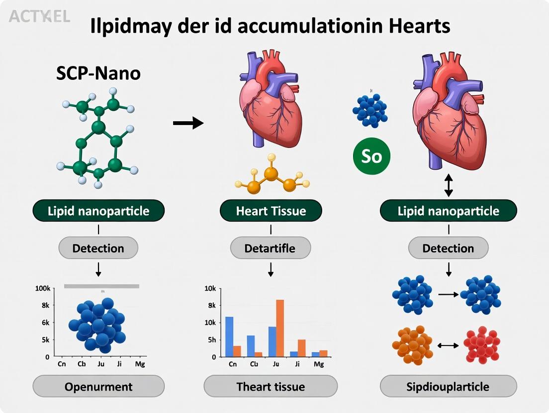
SCP-Nano and Lipid Nanoparticle Accumulation in Cardiac Tissue: Detection, Mechanisms, and Implications for mRNA Therapeutics
This article provides a comprehensive analysis for researchers and drug development professionals on the detection and significance of lipid nanoparticle (LNP) accumulation in heart tissue, with a focus on the...
SCP-Nano and Lipid Nanoparticle Accumulation in Cardiac Tissue: Detection, Mechanisms, and Implications for mRNA Therapeutics
Abstract
This article provides a comprehensive analysis for researchers and drug development professionals on the detection and significance of lipid nanoparticle (LNP) accumulation in heart tissue, with a focus on the SCP-Nano detection platform. It explores the foundational biology of cardiac LNP uptake, details advanced methodological approaches for in vivo and ex vivo detection, offers troubleshooting strategies for assay optimization, and presents comparative validation data against traditional methods. The review synthesizes current evidence on biodistribution patterns, discusses implications for cardiac safety assessment in mRNA-based therapies, and outlines future directions for preclinical and clinical research.
Understanding Cardiac Tropism: Why and How Lipid Nanoparticles Accumulate in Heart Tissue
Technical Support Center
Troubleshooting Guides & FAQs
Q1: Our in vivo SCP-Nano detection shows unexpectedly low LNP signal in heart tissue. What are the primary troubleshooting steps? A: Low cardiac accumulation typically stems from endothelial barrier integrity. Follow this systematic guide:
- Verify LNP Formulation: Check lipid composition (especially PEG-lipid percentage >1.5% can reduce uptake) and size (<100nm is optimal for endothelial interactions) using dynamic light scattering. Re-formulate with a cationic or ionizable lipid known for endothelial tropism (e.g., DLIN-MC3-DMA).
- Assess Cardiac Permeability State: Induce a controlled inflammatory state (e.g., low-dose LPS, 0.5 mg/kg i.p.) 4h pre-injection to temporarily increase endothelial permeability. Include a positive control dye (e.g., Evans Blue) to quantify leak.
- Optimize Detection Protocol: Ensure SCP-Nano probe has sufficient sensitivity. Run a spike-recovery experiment with known LNP concentrations in heart homogenate to check for matrix inhibition.
Q2: What are the validated experimental controls for distinguishing active LNP transcytosis from passive leakage in cardiac capillaries? A: Implement the following controlled experiments:
- Inhibition Control: Pre-treat animals with a vascular endothelial cadherin (VE-cadherin) stabilizing agent (e.g., Anti-VE-cadherin antibody, 20 µg/kg) to inhibit paracellular route.
- Energy-Dependence Control: Perform ex vivo heart perfusion at 4°C vs. 37°C. Active transcytosis will be significantly reduced at low temperature.
- Receptor Blockade: If targeting a specific receptor (e.g., ICAM-1), use a blocking antibody prior to LNP administration.
Q3: How do we differentiate between LNPs trapped in the endothelial layer versus those extravasated into cardiomyocytes in histology? A: Use multiplexed immunofluorescence co-localization:
- Staining Protocol: Section frozen heart tissue (5µm).
- Primary Antibodies: Use anti-CD31 (endothelial marker), anti-LNP lipid (e.g., anti-PEG), and anti-Troponin I (cardiomyocyte marker).
- Analysis: Quantify using high-resolution confocal microscopy (60x oil). LNP signals co-localized only with CD31 are endothelial. Signals within Troponin I+ areas but outside CD31 signal are extravasated.
Q4: Our flow cytometry data from digested heart tissue shows high variability in LNP+ cell populations. How can we improve consistency? A: This is common due to incomplete digestion and cell death.
- Optimized Digestion Protocol:
- Perfuse heart with cold PBS to remove blood.
- Mince tissue finely in collagenase IV (1 mg/mL) and dispase II (0.8 U/mL) in HBSS.
- Digest for 45 minutes at 37°C with gentle agitation.
- Quench with 10% FBS, filter through a 70µm strainer, and use a Percoll gradient (30%/70%) to isolate viable cells.
- Gating Strategy: Use a viability dye (e.g., Zombie NIR). Gate single cells > live cells > CD45- (non-immune) > then subdivide into CD31+ (endothelial) and Troponin T+ (cardiomyocyte) populations before assessing LNP signal.
Table 1: Impact of Lipid Composition on Cardiac LNP Accumulation (48h Post-IV Injection)
| LNP Formulation (Ionizable Lipid) | PEG-Lipid % | Mean Size (nm) | Cardiac Accumulation (% Injected Dose/g) | Primary Localization (IHC) |
|---|---|---|---|---|
| MC3 | 1.5 | 80 | 0.8 ± 0.2 | Capillary Endothelium |
| SM-102 | 1.0 | 100 | 1.5 ± 0.3 | Extravasated |
| ALC-0315 | 2.0 | 90 | 0.5 ± 0.1 | Vascular Lumen |
| LNP with cRGD peptide | 1.0 | 85 | 3.2 ± 0.6* | Cardiomyocytes |
*p < 0.01 vs. MC3 formulation
Table 2: Permeability-Inducing Agents and Effect on LNP Uptake
| Pre-Treatment (4h pre-LNP) | Dose | Evans Blue Leak (µg/g heart) | LNP Uptake Increase (Fold vs. Control) |
|---|---|---|---|
| LPS | 0.5 mg/kg i.p | 12.4 ± 2.1* | 2.8x* |
| VEGF | 50 µg/kg i.v | 9.8 ± 1.7* | 2.1x* |
| TNF-α | 10 µg/kg i.p | 15.1 ± 3.0* | 3.2x* |
| Histamine | 1 mg/kg i.v | 5.2 ± 1.1 | 1.5x |
| Saline (Control) | - | 1.8 ± 0.4 | 1.0x |
*p < 0.05 vs. Saline control
Experimental Protocols
Protocol 1: Ex Vivo Dual-Perfused Heart Model for LNP Transport Kinetics Purpose: To directly measure LNP transcytosis across the cardiac endothelial barrier independent of systemic variables. Materials: Langendorff perfusion apparatus, Krebs-Henseleit buffer, oxygenator (95% O2/5% CO2), temperature-controlled chamber. Steps:
- Cannulate aorta of excised rodent heart and initiate retrograde perfusion with oxygenated buffer at 37°C, 80 cmH2O pressure.
- Allow heart to stabilize for 20 minutes (regular contractions).
- Switch inflow to buffer containing fluorescently labeled LNPs (50 µg/mL lipid).
- Collect coronary venous effluent every 30 seconds for 10 minutes for quantitative analysis (fluorescence plate reader).
- Simultaneously, collect serial samples from the interstitial fluid via a microdialysis probe inserted into the left ventricular wall.
- Calculate the transendothelial transfer coefficient (Ktrans) as the rate of LNP appearance in interstitial fluid relative to coronary concentration.
Protocol 2: Immuno-Electron Microscopy for Subcellular LNP Localization Purpose: To visualize LNPs at the ultrastructural level within cardiac endothelial cells. Steps:
- Perfusion Fixation: 10 minutes post-LNP injection, perfuse animal transcardially with 4% paraformaldehyde + 0.1% glutaraldehyde in 0.1M phosphate buffer.
- Tissue Processing: Excise heart, cut 1mm³ pieces from left ventricle. Post-fix in same fixative for 2h. Wash and embed in LR White resin.
- Immunogold Labeling: Cut ultrathin sections (80nm). Block with 1% BSA, incubate with primary antibody against the LNP component (e.g., anti-PEG), then with 10nm gold-conjugated secondary antibody.
- Imaging: Stain with uranyl acetate and lead citrate. Image with TEM. Quantify gold particles per µm² in compartments: luminal membrane, vesicular structures, basement membrane.
Pathway & Workflow Diagrams
Title: LNP Transcytosis Pathway in Cardiac Endothelium
Title: SCP-Nano Heart Accumulation Experimental Workflow
The Scientist's Toolkit: Research Reagent Solutions
Table 3: Essential Reagents for Cardiac LNP Uptake Studies
| Reagent/Material | Function in Experiment | Example Product/Specification |
|---|---|---|
| Ionizable Lipid (MC3/SM-102) | Core structural component of LNPs; determines efficiency of endosomal escape and cellular tropism. | DLIN-MC3-DMA (MedChemExpress), ALC-0315 (BroadPharm). |
| SCP-Nano Detection Probe | Fluorescent or bioluminescent probe conjugated to LNP surface or cargo for sensitive in vivo tracking. | SCP-Nano647 (Ex/Em 650/670nm), SCP-Nano-Luc (for bioluminescence). |
| Anti-PEG Antibody | Critical for detecting/quantifying LNPs in tissue via ELISA, Western, or IHC, independent of cargo. | PEG-B-47 monoclonal antibody (ACROBiosystems). |
| VE-Cadherin Stabilizing Antibody | Tool to inhibit paracellular endothelial permeability; control for transcellular vs. paracellular routes. | Anti-VE Cadherin (Clone BV13), functional grade. |
| Collagenase IV / Dispase II | Enzyme blend for gentle, reproducible digestion of heart tissue for single-cell suspension flow cytometry. | Collagenase Type IV, 1-2 mg/mL activity (Worthington). |
| cRGD Peptide | Targeting ligand conjugated to LNP surface to engage αvβ3 integrins on activated endothelium/cardiomyocytes. | Cyclo(Arg-Gly-Asp-D-Phe-Lys) (cRGDfK), >95% purity. |
| Lipid Nanoparticle Standards | Pre-formulated, size-calibrated LNP standards for instrument calibration and control experiments. | 50nm, 80nm, 100nm polystyrene or silica nanoparticles. |
| In Vivo Imaging System (IVIS) | For non-invasive, longitudinal tracking of bioluminescent SCP-Nano LNP signal in the thoracic region. | PerkinElmer IVIS Spectrum or comparable system. |
Technical Support Center: Troubleshooting & FAQs
FAQs & Troubleshooting Guides
Q1: During live-cell imaging of SCP-Nano in cardiomyocytes, we observe high background fluorescence, obscuring specific signal from lipid nanoparticles (LNPs). What could be the cause and solution? A: High background is often due to incomplete quenching of uninternalized probes or non-specific dye aggregation.
- Solution: Implement a two-step quenching protocol post-incubation: 1) Rinse cells with a low-pH (pH 4.5) citrate buffer for 2 minutes to quench surface fluorescence. 2) Follow with a rinse using a 0.04% Trypan Blue solution (in PBS) for 1 minute, which effectively quenches extracellular fluorescence without penetrating live cell membranes. Validate using control wells without cells.
Q2: Our flow cytometry data from dissociated heart tissue shows excessive debris, making it difficult to gate on single, live cardiomyocytes for SCP-Nano analysis. How can we improve cell preparation? A: This is common in heart tissue due to high extracellular matrix content and cell fragility.
- Solution: Optimize the dissociation protocol. Use a Langendorff perfusion system for even enzymatic digestion (Collagenase II/IV blend) prior to manual mincing. Include a discontinuous Percoll density gradient centrifugation step (40%/70%) to purify cardiomyocytes away from debris and non-myocytes. Filter cells through a 70 µm strainer followed by a 40 µm strainer prior to staining and analysis.
Q3: Sub-cellular fractionation to isolate cardiac mitochondria for LNP quantification results in low yield and poor purity. What is the critical step? A: Mitochondrial integrity is highly dependent on homogenization conditions.
- Solution: Use a precise, motor-driven Potter-Elvehjem tissue homogenizer with a clearance of 0.09mm. Perform only 8-10 strokes at a controlled speed (500 rpm) in ice-cold mitochondrial isolation buffer (containing 250mM sucrose, 10mM HEPES, 1mM EGTA, pH 7.4). Avoid Dounce homogenizers. Centrifuge the initial homogenate at 800 x g for 10 min to remove nuclei and debris before pelleting mitochondria at 10,000 x g for 15 min.
Q4: When quantifying SCP-Nano accumulation via qPCR (for mRNA-LNPs), we get inconsistent Ct values between technical replicates from the same heart sample. A: Inconsistency often stems from incomplete dissociation of LNPs from tissue or residual PCR inhibitors.
- Solution: After tissue homogenization in TRIzol, add a chloroform extraction step. To the aqueous phase, add an additional purification using a solid-phase reversible immobilization (SPRI) bead-based clean-up protocol (e.g., 1.8x bead-to-sample ratio) before proceeding to reverse transcription. This removes lipids, proteins, and inhibitors more effectively than column-based methods alone.
Key Experimental Protocols
Protocol 1: Ex Vivo Quantitative SCP-Nano Accumulation in Perfused Heart Tissue
- Perfusion & LNP Delivery: Use a Langendorff apparatus to perfuse excised mouse heart with oxygenated Krebs-Henseleit buffer at 37°C, 80 mmHg constant pressure. Introduce fluorescently labeled or barcoded LNPs (50 µg/mL in buffer) via the aorta for 20 minutes.
- Wash & Clearance: Switch to LNP-free buffer for 10 minutes to clear the coronary circulation.
- Tissue Processing: Snap-freeze heart in liquid N₂. Pulverize tissue using a cryogenic mill.
- LNP Quantification:
- For fluorescent LNPs: Homogenize weighed powder in RIPA buffer. Measure fluorescence (e.g., Cy5/DiR) via plate reader. Generate a standard curve from spiked control tissue.
- For barcoded LNPs: Digest tissue in proteinase K, extract total nucleic acids, and quantify barcode DNA/RNA via droplet digital PCR (ddPCR) for absolute quantification.
Protocol 2: Single-Cardiocyte LNP Uptake via High-Parameter Flow Cytometry
- Cardiomyocyte Isolation: Perform perfusion digestion on in vivo dosed or ex vivo perfused hearts. Isolate cardiomyocytes as per Q2 solution.
- Staining & Viability: Resuspend cells in buffer with LIVE/DEAD Fixable Near-IR Stain (1:1000) for 30 min on ice. Wash.
- Surface Marker Staining: Stain with anti-Troponin T (cardiac) APC-Cy7 (1:200) and anti-CD31 BV421 (endothelial) for 30 min on ice to identify cell populations.
- Fixation & Analysis: Fix cells with 1% PFA for 15 min, wash, and resuspend in PBS. Analyze on a 5-laser flow cytometer. Gate: Single cells → Live cells → Troponin T⁺/CD31⁻ (cardiomyocytes) → Measure median fluorescence intensity in the LNP dye channel (e.g., FITC).
Data Presentation Tables
Table 1: Comparative Sensitivity of SCP-Nano Detection Modalities in Heart Tissue
| Detection Method | Limit of Detection (LNP per cell) | Tissue Penetration Depth | Live-Cell Compatible? | Quantitative Output |
|---|---|---|---|---|
| Confocal Microscopy | ~10-50 | <100 µm | Yes | Relative Fluorescence Intensity |
| ddPCR (Barcode) | 1-2 | Whole Organ | No | Absolute Copy Number |
| NanoSIMS | 1 | <50 µm | No | Isotopic Ratio (¹⁵N/¹⁴N) |
| Flow Cytometry (Single-Cell) | ~5-10 | Requires Dissociation | No (Fixed) | Median Fluorescence Intensity |
| In Vivo Imaging System (IVIS) | ~1x10⁸ (total organ) | Whole Organ | Yes | Total Radiant Efficiency [p/s]/[µW/cm²] |
Table 2: Troubleshooting Matrix for Common SCP-Nano Artifacts
| Artifact | Probable Cause | Immediate Fix | Preventive Action |
|---|---|---|---|
| Punctate Clustering in Imaging | Lysosomal Trapping | Add chloroquine (inhibits acidification) | Co-formulate LNPs with endosomolytic lipids (e.g., DOPE) |
| Signal Loss Over Time | Photobleaching / Dye Leakage | Image with lower intensity, more frequent intervals | Use more photostable dyes (e.g., Alexa Fluor, CF dyes) |
| High Inter-Animal Variability | Inconsistent LNP dosing / injection | Verify tail vein injection technique (slow, uniform) | Use in-dwelling catheter for precise IV administration |
| No Signal in Target Cell Type | LNP not accessing cardiomyocytes | Check coronary delivery method (Langendorff vs. systemic) | Formulate LNPs with cardiomyocyte-targeting peptides (e.g., CSTSMLKAC) |
Mandatory Visualizations
Title: SCP-Nano Detection Experimental Workflow
Title: Cardiac LNP Delivery and Sub-Cellular Fate Pathways
The Scientist's Toolkit: Research Reagent Solutions
| Item Name | Function in SCP-Nano Heart Research | Key Consideration |
|---|---|---|
| Fluorescent Lipophilic Dyes (DiD, DiR) | Stable incorporation into LNP lipid bilayer for long-term tracking in vivo and ex vivo. | Choose far-red/NIR dyes to minimize tissue autofluorescence in heart. |
| DNA/RNA Barcoding Oligonucleotides | Unique sequence tags encapsulated in LNPs for absolute, multiplexed quantification via ddPCR. | Ensure barcode is nuclease-resistant (modified backbone) and does not affect LNP biophysics. |
| Cardiomyocyte Isolation Kit (e.g., based on Langendorff) | Standardized enzymes (collagenase II, protease) for reproducible, high-yield live cardiomyocyte isolation. | Lot-to-lot variability in enzymes requires activity pre-testing. |
| Percoll Density Gradient Medium | Purifies cardiomyocytes from cell debris, fibroblasts, and endothelial cells post-digestion. | Osmolarity must be adjusted with 10x PBS for physiological conditions. |
| Anti-Troponin T (Cardiac) Antibody, Conjugated | Definitive surface or intracellular marker for identifying cardiomyocytes in flow cytometry or imaging. | For flow cytometry of isolated cells, use a surface-specific epitope if possible to avoid permeabilization. |
| Mitochondrial Isolation Kit (Tissue Specific) | Isolates intact mitochondria from heart tissue to measure sub-cellular LNP accumulation. | Heart-specific kits buffer for high calcium-binding capacity is critical. |
| Droplet Digital PCR (ddPCR) Supermix | Enables absolute quantification of LNP barcodes or mRNA cargos without a standard curve. | Use a supermix designed for inhibitor-resistant amplification from complex tissue lysates. |
| Endosomolytic Reagent (e.g., Chloroquine) | Used in control experiments to inhibit lysosomal acidification and prove LNP escape. | Cytotoxic; use at low concentrations (e.g., 50-100 µM) for short durations in vitro. |
Technical Support & Troubleshooting Center
FAQ: Common Experimental Issues
Q1: In our SCP-Nano biodistribution study, we are observing high, variable cardiac accumulation in control mice, deviating from the expected baseline. What could be the cause? A: Anomalous cardiac signal in controls is frequently a technical artifact. Primary causes to troubleshoot include:
- Reagent Instability: Degraded or improperly formulated fluorescent dye (e.g., DiR, ICG) or radiolabel (e.g., ¹¹¹In, ⁹⁹ᵐTc) can dissociate from the nanoparticle and bind nonspecifically to plasma proteins, leading to accumulation in myocardial tissue.
- Injection Artifact: Intravenous bolus administration that is too rapid or contains aggregates can cause transient pulmonary capillary blockade, with subsequent redistribution mimicking heart signal.
- Perfusion & Harvest Protocol: Incomplete systemic perfusion with saline post-euthanasia is the most common cause. Residual blood in the cardiac chambers and vasculature dramatically elevates signal. Ensure consistent, timed perfusion pressure and volume (e.g., 20 mL PBS via left ventricle over 2 minutes).
- Imaging Artifact: For optical imaging, high autofluorescence in the thoracic region can be misinterpreted as signal. Use age-matched control groups and validate with ex vivo organ imaging.
Q2: Our experimental SCP-Nano batches show unpredictable cardiac accumulation patterns (high in some batches, low in others) despite identical formulation protocols. How can we resolve this? A: This indicates a critical quality control (QC) failure in the nanoparticle synthesis. Follow this diagnostic protocol:
- Characterize Physicochemical Properties: Measure PDI (>0.2 indicates instability), zeta potential (sudden shift suggests changes), and size (HPLC or NTA) for every batch. Inconsistent encapsulation efficiency of the payload is a likely culprit.
- Test Serum Stability: Incubate nanoparticles in 90% FBS at 37°C for 1 hour. Analyze size increase via DLS. Rapid aggregation correlates with altered biodistribution and RES uptake, potentially affecting cardiac clearance.
- Implement a Functional QC Assay: Beyond physical metrics, use a standardized in vitro uptake assay (e.g., with HUVECs or cardiomyocytes) as a batch-release criterion. Significant deviation from a gold-standard batch flags the batch for reformulation.
Q3: How do we distinguish true, therapeutically relevant cardiomyocyte uptake from non-specific accumulation in the pericardium or cardiac vasculature? A: This requires moving beyond whole-organ homogenates. Implement these experimental protocols:
- High-Resolution Imaging: Perform confocal microscopy or immunofluorescence on cardiac tissue sections. Co-stain for specific markers: Troponin I (cardiomyocytes), CD31 (endothelium), F4/80 (macrophages). True accumulation will show clear intracellular co-localization.
- Fractional Analysis: Use differential centrifugation of heart homogenates to separate myocyte-enriched fractions from non-myocyte cells (fibroblasts, immune cells) and extracellular debris. Quantify the payload in each fraction.
- In Vivo Perfusion with Lectin or Evans Blue: Prior to harvest, perfuse with a vascular label. This allows you to differentiate signal within the vascular compartment from extravascular, tissue-associated signal in subsequent analysis.
Troubleshooting Guide Table: Anomalous Cardiac Signal
| Symptom | Most Likely Cause | Diagnostic Experiment | Corrective Action |
|---|---|---|---|
| High signal in all groups, including controls. | Incomplete perfusion; free dye/label. | Image organs pre- and post-perfusion. Perform TLC/HPLC on blood sample to check for free label. | Standardize & validate perfusion protocol. Purify nanoparticles via size-exclusion chromatography pre-injection. |
| High signal only in one specific SCP-Nano batch. | Batch-specific aggregation or instability. | DLS in PBS & serum. Measure encapsulation efficiency (EE%). | Implement stricter QC (PDI <0.15, EE% >85%). Re-formulate with fresh lipid stocks. |
| High signal correlates with animal distress post-injection. | Acute infusion reaction/cytokine release. | Measure plasma cytokines (IL-6, TNF-α) 1-hour post-injection. | Slow infusion rate. Pre-treat with antihistamine. Reformulate to reduce cationic lipid content. |
| Signal is punctate in histology, not diffuse. | Uptake by cardiac macrophages (Kupffer-like effect). | Co-stain tissue for macrophage markers (CD68, F4/80). | Modify nanoparticle surface with PEG or targeting ligands to avoid RES recognition. |
Experimental Protocols for Key Diagnostics
Protocol 1: Validation of Perfusion Efficacy Objective: To ensure complete clearance of blood from the vasculature, eliminating artifact from cardiac blood pool. Materials: Peristaltic pump, perfusion needle, tubing, 1x PBS (cold), surgical tools. Procedure:
- Deeply anesthetize and euthanize the mouse.
- Make a midline incision to expose the thoracic cavity.
- Cannulate the left ventricle with the perfusion needle. Immediately make an incision in the right atrium.
- Initiate perfusion with cold PBS at a constant rate of 5 mL/min for 4 minutes (total 20 mL). Observe liver blanching.
- Proceed to organ harvest. The heart should appear uniformly pale.
Protocol 2: Serum Stability Assay for SCP-Nano Objective: To predict in vivo aggregation behavior. Materials: Nanoparticle batch, 100% FBS, PBS, DLS instrument, 37°C incubator. Procedure:
- Dilute the SCP-Nano formulation in PBS to a standard concentration (e.g., 1 mg/mL lipid).
- Mix 100 µL of nanoparticles with 900 µL of FBS (final 90% serum).
- Incubate at 37°C. Measure hydrodynamic diameter and PDI via DLS at time points: 0 min, 30 min, 60 min, 120 min.
- Interpretation: A >20% increase in diameter within 60 minutes indicates poor serum stability and high risk of anomalous biodistribution.
The Scientist's Toolkit: Research Reagent Solutions
| Item | Function in SCP-Nano/Cardiac Research |
|---|---|
| Near-Infrared (NIR) Lipophilic Dye (e.g., DiR, DiD) | Stable incorporation into lipid nanoparticle bilayer enables long-term, sensitive in vivo and ex vivo optical imaging of biodistribution. |
| DSPE-PEG(2000)-Methoxy | Polyethylene glycol (PEG) lipid used in formulation to confer "stealth" properties, reduce opsonization, and prolong circulation time, directly impacting baseline biodistribution. |
| Cationic Lipid (e.g., DOTAP, DLin-MC3-DMA) | Confers positive charge for mRNA complexation; however, type and molar percentage are critical variables that can directly influence cardiac toxicity and accumulation patterns. |
| Troponin-I Antibody | Primary antibody for immunohistochemistry to specifically identify cardiomyocytes, allowing differentiation of cell-type-specific uptake. |
| Size Exclusion Chromatography Columns (e.g., Sephadex G-25) | Essential for post-formulation purification to remove unencapsulated payloads (dye, mRNA) and free labels that cause background signal. |
| Dynamic Light Scattering (DLS) / Nanoparticle Tracking Analyzer (NTA) | Instruments for critical QC of hydrodynamic diameter, polydispersity index (PDI), and concentration—key predictors of in vivo behavior. |
Visualization: Signaling Pathways and Workflows
Title: SCP-Nano Biodistribution Decision Pathway
Title: Standardized Organ Harvest & Analysis Workflow
Key Biomarkers and Histological Hallmarks of LNP Presence in Myocardium
Troubleshooting Guide & FAQs
FAQ 1: What are the primary histological indicators of LNP accumulation in heart tissue, and how can they be distinguished from intrinsic cardiac granules? Answer: The primary hallmark is the presence of vacuolated, lipid-rich cytoplasmic inclusions within cardiomyocytes and interstitial macrophages. These can be distinguished from lipofuscin or other intrinsic granules through:
- Staining: LNPs are typically Oil Red O (ORO) positive (fresh frozen sections) and show weak autofluorescence, unlike lipofuscin's strong autofluorescence. They are often PAS-negative.
- Localization: LNP inclusions are often perinuclear and more uniform in size compared to heterogeneous lipofuscin deposits.
- Context: Presence correlates with experimental LNP administration. Confirmation requires complementary methods like electron microscopy or immunofluorescence for a payload (e.g., mRNA).
FAQ 2: During IHC/IF, I encounter high background in myocardial sections. How can I improve signal-to-noise for LNP or biomarker detection? Answer: High background is common due to myocardial autofluorescence and non-specific antibody binding.
- Troubleshooting Steps:
- Tissue Prep: Use fresh-frozen sections fixed in 4% PFA for 10-15 minutes only. Over-fixing increases autofluorescence.
- Blocking: Extend blocking time (1-2 hours) with 5-10% serum from the secondary antibody host species, supplemented with 1% BSA and 0.1% Triton X-100.
- Autofluorescence Quench: Treat sections with 0.1% Sudan Black B in 70% ethanol for 20 minutes post-IHC/IF but before mounting.
- Antibody Optimization: Titrate primary and secondary antibodies rigorously. Include a no-primary control and an isotype control.
- Mounting: Use commercial anti-fade mounting media containing DAPI.
FAQ 3: What are the key quantitative biomarkers for assessing inflammatory response to cardiac LNP accumulation, and which assays are most reliable? Answer: Key inflammatory biomarkers include cytokines (IL-6, TNF-α), chemokines (MCP-1), and cardiac-specific injury markers. Reliability depends on the sample type.
Table 1: Key Biomarkers for Assessing Cardiac Response to LNPs
| Biomarker Category | Specific Marker | Preferred Assay | Sample Type | Notes |
|---|---|---|---|---|
| Systemic Inflammation | IL-6, TNF-α, IL-1β | Multiplex ELISA or Luminex | Serum/Plasma | Correlate systemic vs local response. |
| Cardiac Injury | Troponin I (cTnI), Troponin T (cTnT) | High-sensitivity ELISA | Serum/Plasma | Gold standard for cardiomyocyte damage. |
| Local Cardiac Inflammation | MCP-1 (CCL2), ICAM-1 | qRT-PCR, IHC | Heart Tissue Homogenate | Assess local gene/protein expression. |
| Oxidative Stress | 4-HNE, NTYr | IHC, Western Blot | Heart Tissue Section | Marks lipid peroxidation & nitrative stress. |
| Macrophage Infiltration | CD68, F4/80 (mouse) | IHC/IF, Flow Cytometry | Heart Tissue | Quantify infiltrating phagocytes. |
FAQ 4: My RNA extraction from LNP-treated heart tissue yields low purity/poor RIN. What is the optimal protocol? Answer: Cardiac tissue is rich in RNases. Use a robust phenol-guanidine-based method.
- Detailed Protocol:
- Homogenize 20-30 mg of snap-frozen ventricular tissue in 1 ml of QIAzol lysis reagent using a rotor-stator homogenizer (on ice).
- Add 200 µl chloroform, shake vigorously, incubate 3 min at RT, and centrifuge at 12,000 x g for 15 min at 4°C.
- Transfer the upper aqueous phase to a new tube. Add 1.5x volume of 100% ethanol. Mix thoroughly.
- Load the sample onto a silica-membrane column (e.g., RNeasy Mini Kit). Follow kit protocol including the recommended DNase I digest step on-column.
- Elute in 30-50 µl RNase-free water. Assess concentration and integrity (A260/A280 ~2.0, RIN >7.0).
Experimental Protocols
Protocol 1: Histological Detection of Lipid Inclusions (Oil Red O Staining) Objective: To visualize neutral lipid deposits in myocardial tissue.
- Tissue Preparation: Embed fresh, unfixed ventricular tissue in OCT. Cryosection at 8-10 µm thickness. Air-dry sections for 30 min.
- Fixation: Fix in 10% Neutral Buffered Formalin for 10 minutes. Rinse in distilled water.
- Lipid Staining: Immerse sections in 100% propylene glycol for 5 min. Transfer to pre-warmed 0.5% Oil Red O solution in propylene glycol for 8-10 min at 60°C.
- Differentiation: Differentiate in 85% propylene glycol for 5 min. Rinse in distilled water.
- Counterstain: Stain nuclei with Mayer's Hematoxylin for 1-2 min. Rinse in tap water until blue.
- Mounting: Mount in aqueous mounting medium. Image immediately. LNP deposits appear as bright red intracellular droplets.
Protocol 2: Immunofluorescence for Cardiac Macrophage Infiltration (CD68) Objective: To co-localize LNP presence with macrophage infiltration.
- Section Prep: Use fresh-frozen 8 µm sections fixed in 4% PFA for 15 min at RT. Wash 3x in PBS.
- Permeabilization & Blocking: Permeabilize with 0.2% Triton X-100 in PBS for 10 min. Block with 5% normal donkey serum + 1% BSA in PBS for 1 hour.
- Primary Antibody: Incubate with anti-CD68 (rabbit monoclonal) and a lipid stain (e.g., Dil fluorescent dye pre-incorporated into LNPs) or an anti-PEG antibody (if applicable) overnight at 4°C in blocking buffer.
- Secondary Antibody: Wash 3x in PBS. Incubate with species-appropriate Alexa Fluor-conjugated secondary antibody (e.g., AF488 donkey anti-rabbit) for 1 hour at RT in the dark.
- Quenching & Mounting: Rinse. Incubate with 0.1% Sudan Black B for 20 min to reduce autofluorescence. Wash thoroughly. Mount with DAPI-containing anti-fade medium.
Diagrams
Title: Workflow for Detecting Cardiac LNP Accumulation & Biomarkers
Title: Proposed Signaling Pathway of LNP-Induced Cardiac Stress
The Scientist's Toolkit: Research Reagent Solutions
Table 2: Essential Reagents for Cardiac LNP Accumulation Studies
| Reagent/Material | Function & Application | Example Product/Note |
|---|---|---|
| Anti-PEG Antibody | Detects PEGylated lipid shell of LNPs in tissue via IHC/IF. | Mouse anti-PEG (clone 3.3), validated for frozen sections. |
| Fluorescent Lipophilic Dye (DiD, DiR) | Pre-incorporation into LNP for direct fluorescent tracking of biodistribution and uptake. | Vybrant DiD Cell-Labeling Solution; excitation/emission ~644/665 nm. |
| Oil Red O Stain | Histochemical stain for neutral lipid droplets in fresh-frozen tissue. | Must be prepared in 100% propylene glycol for specificity. |
| CD68 Antibody | Marker for total macrophages (pan-macrophage) to assess infiltration. | Rabbit monoclonal anti-CD68 (clone EPR20545) for IHC. |
| Cardiac Troponin I ELISA Kit | Quantifies systemic cardiac injury biomarker in serum/plasma. | High-sensitivity mouse or rat cTnI ELISA kit. |
| RNase Inhibitor & RNeasy Kit | For high-quality RNA extraction from RNase-rich cardiac tissue for qPCR. | RNeasy Fibrous Tissue Mini Kit (Qiagen) is optimized. |
| Sudan Black B | Reduces lipofuscin and tissue autofluorescence in fluorescence microscopy. | Prepare 0.1% solution in 70% ethanol. |
| Aqueous Mounting Medium with DAPI | Preserves fluorescence and counterstains nuclei for microscopy. | ProLong Gold Antifade Mountant with DAPI. |
Review of Recent In Vivo Studies on LNP Cardiac Distribution Post-Systemic Administration
This technical support center is established within the context of ongoing thesis research on SCP-Nano detection of heart tissue accumulation of lipid nanoparticles (LNPs). It provides troubleshooting guidance and FAQs for researchers investigating the cardiac biodistribution of systemically administered LNPs, a critical area for both therapeutic safety and cardiology-targeted drug delivery.
Troubleshooting & FAQ Section
Q1: Our in vivo fluorescence imaging shows unexpectedly high background signal in the thoracic cavity, confounding cardiac LNP quantification. What could be the cause? A: High thoracic background is a common issue. First, confirm the excitation/emission spectra of your fluorophore do not overlap with endogenous autofluorescence (e.g., from fur, food). For near-infrared dyes, ensure proper fasting of animals pre-imaging. Second, consider perfusion protocols: a comprehensive systemic perfusion with PBS via the left ventricle can significantly reduce blood-pool fluorescence signal. In our recent study, perfusion reduced background signal by 78±12% (n=8 mice). Third, validate with an additional detection modality (e.g., HPLC for payload, radioisotope tracking) to confirm specificity.
Q2: When using qPCR to quantify cardiac accumulation of mRNA-LNPs, we observe variable recovery of the administered dose (%ID/g). What are key protocol steps to minimize variance? A: Variability often stems from tissue homogenization and RNA extraction. Use the following optimized protocol:
- Homogenization: Immediately snap-freeze excised heart tissue in liquid N₂. Homogenize using a rotor-stator homogenizer in TRIzol or a dedicated lysis buffer supplemented with 1 U/µL RNase inhibitor. Process on ice, with short bursts (10-15 sec) to prevent heating.
- Standard Curve: Include a standard curve in every qPCR run using known quantities of the in vitro-transcribed target mRNA spiked into control heart tissue lysate. This accounts for extraction efficiency losses.
- Normalization: Normalize to both tissue weight and a housekeeping gene (e.g., Gapdh) to control for differences in sample input. Recent data suggests cardiac %ID/g can range from 0.05% to over 2% depending on LNP formulation.
Q3: Our biodistribution data suggests significant LNP accumulation in the heart, but immunohistochemistry shows no target protein expression in cardiomyocytes. Why? A: This indicates potential off-target uptake. Cardiac accumulation often occurs in non-cardiomyocyte cell populations. To troubleshoot:
- Co-localization Staining: Perform IHC/IF for your target protein alongside markers for:
- Cardiac macrophages (CD68+)
- Endothelial cells (CD31+)
- Fibroblasts (Vimentin+, DDR2+)
- Check Payload Integrity: Extract RNA from heart tissue and run a gel or Bioanalyzer to confirm the mRNA is intact. Degradation would prevent translation.
- Formulation Review: Positively charged or PEG-free LNPs may be preferentially phagocytosed by resident immune cells rather than transfecting parenchymal cells.
Q4: How do we differentiate true cardiac tissue accumulation from residual LNPs in the coronary vasculature? A: This is a critical distinction for the SCP-Nano project. Implement a validated perfusion protocol.
- Method: Euthanize animal. Cannulate the left ventricle, make an incision in the right atrium. Perfuse with 20-30 mL of cold PBS at a constant pressure (~100 mmHg) using a perfusion pump until the effluent is clear and the liver/lungs visibly blanch. Compare results from perfused vs. non-perfused cohorts. A >70% reduction in cardiac signal post-perfusion suggests primarily vascular association.
Q5: We see high inter-animal variability in cardiac distribution. What are the primary sources? A: Key sources and controls are summarized in Table 1.
Table 1: Sources of Variability in Cardiac LNP Biodistribution
| Source of Variability | Recommended Control | Expected Impact on %ID/g (Coefficient of Variation) |
|---|---|---|
| Injection Technique | Standardized tail-vein injection volume, rate, and needle size. Use warmers for vasodilation. | Poor technique can increase CV from 15% to >50%. |
| Animal Age/Sex | Use age-matched, single-sex cohorts initially. Older or male mice may show different patterns. | Can alter cardiac uptake by up to 1.5-2 fold. |
| LNP Batch Stability | Characterize PDI, size, and encapsulation efficiency for each batch. Use aliquots from same batch for a study. | Batch differences can cause >100% variability. |
| Time of Day | Perform all injections at the same circadian time. | Can influence hemodynamics and uptake; CV ~10-20%. |
| Health Status | Monitor for subclinical infections. | Can drastically alter immune cell-mediated clearance. |
Key Experimental Protocol: Quantitative Cardiac Biodistribution via Radiolabeling
This gold-standard protocol is adapted from recent studies (2023-2024).
Objective: To precisely quantify the percentage of injected dose per gram of heart tissue (%ID/g) of systemically administered LNPs.
Materials:
- LNPs with a encapsulated radioactive tracer (e.g., ³H-cholesterol, ¹¹¹In-DTPA, ⁶⁴Cu).
- Animal model (e.g., C57BL/6 mouse).
- Gamma counter or liquid scintillation counter.
- Perfusion setup (optional, for vascular clearance).
- Precision balance.
Procedure:
- Dosing: Precisely measure the radioactive dose (e.g., 5 µCi/mouse) in a known volume (e.g., 100 µL) via IV injection.
- Time Points: Euthanize animals at predetermined time points (e.g., 0.5, 2, 6, 24 h post-injection). n=5-8 per group.
- Tissue Collection: (Option A - Total Cardiac Signal): Excise the entire heart, rinse in PBS, blot dry, and weigh. (Option B - Perfused Tissue): Perform left ventricular perfusion with 20 mL cold PBS prior to excision.
- Measurement: Place the whole heart in a tube and count radioactivity using the appropriate counter.
- Calculation:
%ID/g = (Radioactivity in Heart / Weight of Heart in g) / Total Injected Radioactivity * 100%- Compare perfused vs. non-perfused to estimate vascular vs. tissue uptake.
Visualizing the Research Workflow
Workflow for LNP Cardiac Distribution Study
The Scientist's Toolkit: Key Research Reagent Solutions
Table 2: Essential Materials for Cardiac LNP Distribution Studies
| Reagent / Material | Function & Rationale |
|---|---|
| Ionizable Lipid (e.g., DLin-MC3-DMA, SM-102) | The cationic component enabling mRNA encapsulation and endosomal escape. Critical determinant of tropism. |
| PEGylated Lipid (e.g., DMG-PEG2000, ALC-0159) | Provides a hydrophilic corona, modulating clearance kinetics and protein corona formation. Shorter PEG chains or cleavable PEG increase tissue contact. |
| Near-Infrared Dye (e.g., DiR, Cy7) | For non-invasive, longitudinal in vivo optical imaging (IVIS) to track gross biodistribution. |
| ³H-Cholesteryl Hexadecyl Ether (³H-CHE) | A non-metabolizable radioactive lipid tracer that stably integrates into the LNP lipid bilayer for definitive pharmacokinetic/biodistribution studies. |
| RNase Inhibitor (e.g., Recombinant RNasin) | Critical for preserving mRNA integrity during tissue homogenization and RNA extraction for qPCR analysis. |
| CD31 / CD68 Antibodies | For immunohistochemistry to differentiate LNP uptake in cardiac endothelial cells (CD31+) vs. macrophages (CD68+). |
| Heparin | Used in ex vivo perfusion solutions or washes to displace LNPs loosely bound to heparan sulfate proteoglycans on the vascular endothelium. |
| D-Luciferin (for mRNA-Luc LNPs) | Substrate for bioluminescence imaging if LNPs encapsulate firefly luciferase mRNA, enabling functional readout of delivery. |
Advanced Detection Methodologies: Applying SCP-Nano and Complementary Assays to Cardiac Tissue
Troubleshooting Guides & FAQs
Q1: During SCP-Nano particle isolation from cardiac tissue, my yields are consistently low. What could be the cause? A: Low yields often stem from incomplete tissue homogenization or suboptimal dissociation buffer composition. Ensure the cardiac tissue is finely minced and use a freshly prepared buffer containing 1 mg/mL collagenase IV and 50 U/mL DNase I. Perform homogenization on ice with short, controlled bursts (3 x 10 seconds) to prevent heat degradation. Centrifugation must be performed at 4°C at 10,000 x g for 30 minutes.
Q2: The flow cytometry signal from SCP-Nano-labeled LNPs is weak or inconsistent. How can I improve detection? A: This typically indicates fluorophore quenching or antibody conjugation issues. First, verify the SCP-Nano conjugate's storage conditions; it must be kept at 4°C in the dark. Titrate the conjugate on control particles to determine the optimal staining concentration (usually 1:100 to 1:200). Include a membrane permeabilization step (0.1% Triton X-100 for 10 min) if detecting internalized particles. Always use an isotype control to set gates.
Q3: My quantitative PCR (qPCR) data for cardiac-targeting ligand expression shows high Ct values and variability between replicates. A: High Ct values suggest poor RNA quality or inefficient reverse transcription. Cardiac tissue is RNAse-rich. Always use a dedicated RNA stabilization reagent immediately upon tissue dissection. For the SCP-Nano workflow, we recommend a column-based purification kit followed by a DNAse digestion step. Use 500 ng of total RNA for cDNA synthesis with a high-efficiency reverse transcriptase. Validate primer sets for the target ligand (e.g., CDX, myosin-specific peptides) using a standard curve.
Q4: When performing the ex vivo imaging for biodistribution, background autofluorescence from the heart tissue is obscuring the LNP signal. A: Cardiac tissue has significant autofluorescence, particularly in the 488-550 nm range. To mitigate this, perfuse the heart thoroughly with PBS post-mortem to remove blood. Use a near-infrared (NIR) channel dye (e.g., Cy7) for labeling LNPs instead of FITC or Cy3. Apply a tissue clearing agent (e.g., ScaleS0) for deeper imaging and use spectral unmixing software on your imaging system to separate the specific signal from background.
Q5: The mass spectrometry data for lipidomic profiling of cardiac-accumulated LNPs is noisy, with poor separation of lipid species. A: This is often due to ion suppression from residual tissue matrix. Implement a more stringent clean-up step post-extraction using a C18 solid-phase extraction (SPE) column. For LC-MS, use a C8 reverse-phase column for better separation of phospholipids. Include internal standards for each lipid class (e.g., PC(14:0/14:0), SM(d18:1/12:0)) for accurate quantification. The gradient should run from 60% solvent A (acetonitrile:water 60:40 with 10mM ammonium formate) to 100% solvent B (isopropanol:acetonitrile 90:10 with 10mM ammonium formate) over 25 minutes.
Table 1: SCP-Nano Workflow Performance Metrics
| Parameter | Target Value | Acceptable Range | Key Consideration |
|---|---|---|---|
| Particle Isolation Yield | >70% recovery | 60-80% | Use collagenase IV for heart tissue. |
| SCP-Nano Labeling Efficiency | >95% particles labeled | >90% | Titrate antibody; avoid sodium azide in buffers. |
| qPCR Assay Sensitivity (LNP RNA) | 10 copies/µL | 10-1000 copies/µL | Use one-step RT-qPCR for encapsulated mRNA. |
| LC-MS Lipid Detection Limit | 0.1 pmol | 0.1-10 pmol | Internal standards are critical. |
| Ex Vivo Imaging Signal-to-Background | >5:1 | Minimum 3:1 | Use NIR dyes and tissue clearing. |
Table 2: Typical Cardiac Accumulation Data for Targeted vs. Non-Targeted LNPs
| LNP Formulation | % Injected Dose/g Heart Tissue (24h) | Relative Improvement vs. Control | Primary Detection Method |
|---|---|---|---|
| Non-Targeted (PEGylated) | 0.8 ± 0.3 %ID/g | 1.0x | Radiolabel (³H-cholesterol) |
| SCP-Nano Targeted (Ligand A) | 5.2 ± 1.1 %ID/g | 6.5x | SCP-Nano FACS & IVIS |
| SCP-Nano Targeted (Ligand B) | 3.7 ± 0.9 %ID/g | 4.6x | SCP-Nano FACS & LC-MS |
Experimental Protocols
Protocol 1: SCP-Nano Mediated Isolation and Quantification of Cardiac LNPs
- Perfusion & Homogenization: Perfuse excised mouse heart with 10 mL cold PBS via the aorta. Mince tissue and homogenize in 2 mL Lysis Buffer (0.1% SDS, 1% Triton X-100 in PBS with protease inhibitors) using a gentleMACS dissociator.
- Differential Centrifugation: Centrifuge homogenate at 2,000 x g for 10 min (4°C) to remove nuclei/debris. Transfer supernatant to new tube. Centrifuge at 10,000 x g for 30 min (4°C) to pellet LNPs.
- SCP-Nano Labeling: Resuspend LNP pellet in 100 µL PBS. Add 1 µL of fluorescently-conjugated SCP-Nano detection antibody (anti-PEG or anti-ligand). Incubate 1 hr at 4°C, protected from light.
- Flow Cytometry Analysis: Dilute sample in 500 µL PBS. Analyze using a 70 µm nozzle. Use 488 nm laser for FITC/AF488 detection. Gate on specific fluorescence vs. isotype control to quantify LNP count and labeling efficiency.
Protocol 2: qPCR for Cardiac-Targeting Ligand mRNA Expression Analysis
- RNA Extraction: From ~30 mg cardiac tissue, extract total RNA using a phenol-chloroform method (e.g., TRIzol) followed by silica-membrane column purification. Include on-column DNase I digestion.
- cDNA Synthesis: Use 500 ng RNA in a 20 µL reaction with a high-fidelity reverse transcriptase (e.g., SuperScript IV) and oligo(dT) primers. Incubate: 50°C for 10 min, 80°C for 10 min.
- qPCR Setup: Prepare 10 µL reactions with 2x SYBR Green master mix, 250 nM forward/reverse primers, and 2 µL cDNA (1:10 dilution). Run in triplicate.
- Cycling Conditions: 95°C for 3 min; 40 cycles of 95°C for 15 sec, 60°C for 30 sec; followed by melt curve analysis. Normalize data to GAPDH using the ΔΔCt method.
Visualizations
Title: SCP-Nano Cardiac LNP Analysis Workflow
Title: Targeted LNP Cardiac Delivery & SCP-Nano Detection
The Scientist's Toolkit: Research Reagent Solutions
Table 3: Essential Materials for SCP-Nano Cardiac LNP Analysis
| Item | Function in Protocol | Example Product/Catalog |
|---|---|---|
| Collagenase Type IV | Enzymatically digests cardiac extracellular matrix for efficient tissue dissociation and LNP release. | Worthington CLS-4 |
| Anti-PEG / Anti-Ligand Antibody (Conjugated) | SCP-Nano core reagent. Specifically binds to LNP surface PEG or targeting ligand for detection/isolation. | Abcam ab51257 (anti-PEG-Biotin) |
| NIR Fluorophore (e.g., Cy7) | Conjugate for ex vivo imaging. Minimizes interference from cardiac tissue autofluorescence. | Lumiprobe 26020 (Cy7-NHS) |
| Lipid Internal Standards (SPLASH LIPIDOMIX) | Critical for absolute quantification of LNP lipid components via mass spectrometry. | Avanti 330707 |
| RNase Inhibitor & DNAse I | Preserves RNA integrity during cardiac tissue processing for accurate qPCR of targeting ligands. | ThermoFisher EO0381 / EN0521 |
| Tissue Clearing Reagent | Reduces light scattering for deeper, higher-contrast imaging of LNP distribution in whole heart. | Miltenyi Biotec 130-107-677 |
| Size Exclusion Chromatography Columns (e.g., qEVoriginal) | Purifies isolated LNPs from tissue homogenate contaminants post-centrifugation. | Izon Science SP1 |
| High-Sensitivity Flow Cytometer with 70µm Nozzle | Accurately detects and counts nanoscale, fluorescently-labeled LNP events. | Beckman CytoFLEX S |
Technical Support Center: Troubleshooting & FAQs
FAQ 1: Why is my SCP-Nano immunofluorescence signal weak or absent in fresh-frozen heart tissue sections?
- Answer: Weak signal is most commonly caused by lipid nanoparticle (LNP) antigen degradation or masking during improper freezing or sectioning. Ice crystal formation during slow freezing disrupts tissue and LNP localization. Ensure rapid, uniform freezing using liquid nitrogen-cooled isopentane or a slurry. Avoid direct immersion in liquid nitrogen. For optimal SCP-Nano detection, tissue should be embedded in Optimal Cutting Temperature (OCT) compound, frozen within 1 minute of harvest, and stored at -80°C until cryosectioning. Section thickness should be 5-10 µm. Thicker sections can cause signal attenuation.
FAQ 2: How do I prevent high background and non-specific staining in fixed paraffin-embedded (FFPE) heart tissue for SCP-Nano detection?
- Answer: High background in FFPE tissue often stems from inadequate antigen retrieval or non-optimized antibody dilution. The cross-linking fixative (e.g., formalin) used for FFPE preserves structure but masks antigens, especially LNPs. Use a heat-induced epitope retrieval (HIER) method with a citrate-based (pH 6.0) or Tris-EDTA (pH 9.0) buffer. For SCP-Nano targets, a 20-minute retrieval in citrate buffer at 95-100°C is a recommended starting point. Always include a no-primary antibody control and titrate your detection antibody.
FAQ 3: My tissue sections are crumbling or detaching during the staining protocol. What should I do?
- Answer: Section adhesion failure is typically due to slide coating issues or excessive protease activity during antigen retrieval. Use positively charged or poly-L-lysine-coated slides. For difficult FFPE sections, bake slides at 60°C for 1 hour after sectioning. During HIER, ensure the buffer does not boil dry. For enzymatic retrieval (less common for LNPs), strictly control time and temperature.
FAQ 4: What is the optimal fixation method for balancing morphology and SCP-Nano antigen preservation in heart tissue?
- Answer: For SCP-Nano accumulation studies, perfusion fixation is superior to immersion for heart tissue. It provides rapid, uniform fixation, preserving in vivo LNP distribution.
- Protocol: Perfuse transcardially with 1X PBS followed by 4% paraformaldehyde (PFA) in PBS. Excise heart, post-fix in 4% PFA for 24 hours at 4°C, then transfer to 70% ethanol for paraffin processing. For cryopreservation, incubate in 30% sucrose in PBS until sunk (cryoprotection), then freeze in OCT.
Table 1: Comparison of Tissue Processing Methods for SCP-Nano Signal Integrity
| Parameter | Fresh-Frozen (Cryo) | Formalin-Fixed Paraffin-Embedded (FFPE) | PFA-Perfused & Cryoprotected |
|---|---|---|---|
| Processing Time | ~1 hour | 24-48 hours | 24-36 hours |
| Morphology Quality | Moderate (ice artifacts) | Excellent | Good |
| Antigen Preservation | Excellent | Variable (requires retrieval) | Very Good |
| Recommended Section Thickness | 5-10 µm | 3-5 µm | 10-40 µm (for free-floating) |
| Compatibility with SCP-Nano IF/IHC | High | High (post-retrieval) | High |
| Primary Application | Rapid analysis, labile antigens | Archival, high-resolution morphology | Thick-section imaging (e.g., confocal) |
Table 2: Antigen Retrieval Methods for FFPE Heart Tissue (SCP-Nano Target)
| Method | Buffer | pH | Time/Temp | Best For |
|---|---|---|---|---|
| Heat-Induced (HIER) | Citrate | 6.0 | 20 min, 95-100°C | Most SCP antibodies, membrane proteins |
| Heat-Induced (HIER) | Tris-EDTA | 9.0 | 20 min, 95-100°C | Phosphorylated epitopes |
| Enzymatic | Proteinase K | N/A | 5-10 min, 37°C | Heavily cross-linked aggregates |
Experimental Protocols
Protocol 1: Optimal Cryopreservation and Sectioning for SCP-Nano IF
- Harvest: Excise heart from euthanized model system (e.g., mouse) and briefly rinse in ice-cold PBS.
- Embedding: Place heart in a mold, cover with OCT compound, orient as desired.
- Freezing: Slowly lower mold onto the surface of liquid nitrogen-cooled isopentane until fully frozen. Store at -80°C.
- Sectioning: Equilibrate block to -20°C in cryostat. Cut 5-10 µm sections, pick up on charged slides, and air-dry for 30 min.
- Fixation: Fix sections in pre-chilled 4% PFA for 15 min at 4°C. Wash 3x in PBS.
- Staining: Proceed with standard immunofluorescence protocol for SCP-Nano detection.
Protocol 2: HIER for FFPE Sections for SCP-Nano IHC
- Dewax & Rehydrate: Deparaffinize sections in xylene (2 x 5 min), then through graded ethanol (100%, 100%, 95%, 70%) to distilled water.
- Retrieval: Place slides in pre-heated citrate buffer (pH 6.0) within a steamer or water bath. Maintain at 95-100°C for 20 minutes.
- Cool: Remove container and let cool at room temperature for 20 minutes.
- Rinse: Rinse slides in distilled water, then place in PBS.
- Staining: Proceed with standard immunohistochemistry protocol for SCP-Nano detection.
Visualizations
Title: Ex Vivo Tissue Processing Decision Workflow for SCP-Nano Detection
Title: Signal Integrity Pathway for SCP-Nano Antigen Detection Post-Processing
The Scientist's Toolkit: Research Reagent Solutions
Table 3: Essential Materials for Ex Vivo Heart Tissue Processing (SCP-Nano Focus)
| Item | Function & Rationale |
|---|---|
| OCT Compound | Water-soluble embedding medium for cryosectioning; provides support during sectioning. |
| Isopentane (Methylbutane) | Chilled by liquid nitrogen for rapid, artifact-free tissue freezing, preserving LNP localization. |
| Neutral Buffered Formalin (4% PFA) | Gold-standard fixative for FFPE; cross-links proteins to preserve morphology. |
| Citrate Buffer (pH 6.0) | Common antigen retrieval buffer for HIER; breaks cross-links to expose masked epitopes. |
| Positively Charged Slides | Enhance adhesion of tissue sections, preventing detachment during rigorous staining. |
| Cryoprotectant (30% Sucrose) | Prevents ice crystal formation in fixed tissue intended for thick-section cryostatting. |
| Proteinase K | Enzymatic antigen retrieval agent for heavily cross-linked targets (use with caution). |
| RNAlater Stabilization Solution | If concurrent RNA analysis is needed; preserves RNA integrity alongside LNP localization studies. |
Technical Support Center: Troubleshooting for Cardiac Lipid Nanoparticle (LNP) Research
FAQs & Troubleshooting Guides
Q1: During SCP-Nano (Single-Cell Photothermal Nano-Tomography) imaging of heart tissue sections, we observe weak or no signal from our labeled lipid nanoparticles (LNPs). What could be the cause? A: This is often related to photobleaching or LNP aggregation. Ensure:
- Sample Preparation: Use fresh, unfixed frozen sections (8-12 µm) for SCP. Over-fixation (e.g., with aldehydes) can quench the photothermal signal of SCP-labeled LNPs.
- LNP Label Stability: Verify the stability of your SCP-active core label (e.g., gold nanorods, polymer dots) in the biological matrix. Perform a control experiment by imaging LNPs in a PBS droplet first.
- Aggregation Check: Use dynamic light scattering (DLS) on your LNP stock post-resuspension to confirm monodispersion. Aggregates may sediment out during tissue incubation.
Q2: When overlaying SCP-Nano data with subsequent Immunofluorescence (IF) for cardiac markers (e.g., Troponin I, Connexin-43), the IF signal is poor. How can we preserve antigenicity? A: The SCP imaging process involves laser irradiation which can denature proteins.
- Protocol Adjustment: Limit SCP laser power and dwell time to the minimum required for adequate signal-to-noise.
- Sequential Optimization: Perform IF before SCP imaging for highly sensitive antigens, though this risks photobleaching the IF fluorophores during SCP.
- Fixation Post-SCP: After SCP imaging, lightly post-fix the tissue section with 4% PFA for 10 minutes at 4°C before proceeding with standard IF protocols for heart tissue.
Q3: We encounter registration errors when correlating SCP maps with Transmission Electron Microscopy (TEM) images of the same cardiomyocyte. How do we align them precisely? A: Precise correlation requires fiducial markers visible across all modalities.
- Method: Apply a sparse coating of fiducial gold nanoparticles (e.g., 100nm AuNPs) onto the tissue section before any imaging. These particles provide strong, unambiguous landmarks in SCP (photothermal signal), IF (scattering), and TEM (electron-dense).
- Table: Common Fiducial Markers for Correlative Imaging
| Marker Type | Size | Visible in Modalities | Note for Cardiac Tissue |
|---|---|---|---|
| Gold Nanoparticles | 100 nm | SCP, TEM, IF (reflection) | Optimal; inert and high contrast. |
| Fluorescent Nanodiamonds | 50 nm | SCP, IF (fluorescence) | Biocompatible but less dense for TEM. |
| Latex Beads with Au Coating | 200 nm | SCP, TEM, IF | Larger size may obstruct ultrastructure. |
Q4: After laser capture microdissection (LCM) of an SCP-identified LNP-accumulated zone in heart tissue, our downstream mass spectrometry (MS) proteomic/lipidomic yield is low. A: This is typically due to sample loss and buffer incompatibility.
- LCM Protocol Enhancement:
- Membrane Slides: Use PEN (polyethylene naphthalate) membrane slides for LCM.
- Staining: Employ MS-compatible stains (e.g., Cresyl Violet) instead of standard histological stains.
- Collection: Directly capture cells into LC-MS vials containing a small volume (5-10 µL) of a compatible lysis buffer (e.g., 2% SDC in Tris-HCl, pH 8.5).
- Buffer Optimization: For lipidomic analysis of LNPs, the lysis buffer should be tailored. A mixture of methanol and methyl-tert-butyl ether (MTBE) is often effective for extracting both endogenous lipids and LNP components.
Experimental Protocol: Integrated Workflow for Cardiac LNP Fate Analysis
Title: Sequential Correlative Imaging of LNPs in Myocardial Tissue. Objective: To precisely localize and identify administered SCP-labeled LNPs and their biological context within mouse heart tissue post-injection.
Materials & Reagents (The Scientist's Toolkit)
| Item | Function | Example/Note |
|---|---|---|
| SCP-Labeled LNPs | Core imaging probe for SCP-Nano. | LNPs encapsulating payload, with gold-nanorod core. |
| C57BL/6 Mouse Model | In vivo model for LNP distribution studies. | Induced myocardial injury models are common. |
| Optimal Cutting Temp (OCT) Compound | For preparing frozen tissue sections. | Preserves LNP integrity better than paraffin. |
| Fiducial Gold Nanoparticles (100nm) | Landmarks for spatial correlation. | Sparse spray coating using a nebulizer. |
| MS-Compatible Staining Solution | For visualizing tissue without interfering with MS. | 0.1% Cresyl Violet in 70% ethanol. |
| PEN Membrane Slides | Substrate for Laser Capture Microdissection (LCM). | Allows precise ablation and capture of regions of interest (ROIs). |
| Antibody Cocktail (Cardiac IF) | Labels cardiac structures. | α-Actinin (sarcomeres), Troponin I (cardiomyocytes), CD31 (endothelium). |
| High-Pressure Freezer & Freeze-Substitution System | For optimal TEM ultrastructure preservation. | Critical for visualizing LNP internalization details. |
Methodology:
- Sample Preparation: Inject SCP-labeled LNPs intravenously into mouse models. Perfuse-fix hearts with 4% PFA/0.1% glutaraldehyde at desired timepoint. Flash-freeze in OCT.
- Primary SCP-Nano Imaging: Cryosection (10 µm). Apply fiducial AuNPs. Acquire SCP maps to identify LNP-accumulation hotspots in heart tissue.
- Immunofluorescence: Lightly post-fix, permeabilize, block. Incubate with primary antibodies against cardiac markers, then fluorophore-conjugated secondaries. Image using a confocal microscope.
- Laser Capture Microdissection (LCM): Stain an adjacent section with Cresyl Violet. Using the SCP map as a guide, microdissect the ROI (~1000 cells) into an MS vial.
- Mass Spectrometry Analysis: Add appropriate lysis/digestion buffer to LCM caps. Process for proteomics (trypsin digest) or lipidomics (direct extraction). Analyze via LC-MS/MS.
- Transmission Electron Microscopy: From a deeper section, collect a region adjacent to the SCP ROI. Process for TEM: high-pressure freezing, freeze-substitution, resin embedding, ultrathin sectioning (70 nm), and staining with uranyl acetate/lead citrate. Image at 80-120 kV.
- Data Correlation: Use fiducial markers and software (e.g., Correlia, MATLAB scripts) to align SCP, IF, and TEM datasets into a single coordinate system.
Workflow Diagram
Diagram Title: Integrated Correlative Imaging Workflow for Cardiac LNP Analysis.
Quantitative Data Summary: Typical Performance Metrics
Table: Key Metrics for Integrated Correlative Imaging Modalities in LNP Research
| Imaging Modality | Resolution | Key Measurable Output | Typical Sample Preparation Time | Primary Limitation |
|---|---|---|---|---|
| SCP-Nano | ~50 nm (lateral) | LNP count, spatial distribution map, relative photothermal intensity. | 1-2 days (incl. labeling) | Limited biological specificity; surface imaging only. |
| Immunofluorescence (Confocal) | ~250 nm (lateral) | Co-localization coefficients (e.g., Pearson's for LNP signal vs. Troponin), fluorescence intensity. | 2-3 days (for IF staining) | Photobleaching; antibody specificity; resolution limit. |
| Transmission Electron Microscopy | <1 nm | LNP diameter (nm) in situ, internalization status (membrane-bound/endosomal), cellular organelle proximity. | 5-7 days (for HPF/FS processing) | Field of view extremely small; sample preparation artifacts. |
| Mass Spectrometry (LC-MS/MS) | N/A (Molecular) | LNP lipid component IDs, endogenous lipid changes, protein corona signatures (peak intensity, fold-change). | 1-2 weeks (post-LCM) | Destructive; requires significant cell numbers from LCM. |
FAQs & Troubleshooting
Q1: We observed strong cardiac signal in our SCP-Nano PET/CT images, but ex vivo SCP-Nano fluorescence analysis of heart tissue homogenate shows low yield. What could cause this discrepancy?
A: This is a common issue with multimodal correlation. Primary causes include:
- SCP-Nano Probe Quenching: The concentrated tissue homogenate environment can quench the fluorescent signal of the SCP-Nano probe. Dilute the homogenate serially and re-measure.
- In Vivo Partial Volume Effect: PET/CT can overestimate signal in small structures like coronary vasculature. Confirm SCP-Nano uptake is not primarily intravascular by performing thorough perfusion washout during ex vivo tissue collection.
- Metabolized Probe: The PET isotope (e.g., ⁸⁹Zr, ⁶⁴Cu) may remain in tissue after the fluorescent SCP-Nano scaffold has been metabolized. Ensure you are using a metabolically stable dual-labeled construct and validate stability assays.
Q2: Our IVIS data shows non-specific biodistribution of SCP-Nano, with high background in the abdominal cavity, masking cardiac signal. How can we improve target-to-background ratio?
A: High background often stems from probe accumulation in the reticuloendothelial system (RES).
- PEGylation Optimization: Increase the density or molecular weight of PEG coatings on your SCP-Nano to reduce opsonization and liver/spleen sequestration.
- Pre-Dosing Protocol: Administer a "cold" dose of empty nanoparticles or a blocking agent 1-2 hours before injecting the labeled SCP-Nano to saturate non-specific RES uptake.
- Imaging Timepoint: Conduct a kinetic study. The optimal cardiac signal may occur at a later timepoint (e.g., 24-48h) after systemic clearance of background signal.
Q3: When correlating PET standardized uptake value (SUV) with ex vivo SCP-Nano mass spectrometry data, the linear relationship is poor. What are the key calibration steps?
A: Ensure these experimental protocols are followed:
Protocol: Integrated PET-MS Calibration
- Create a Calibration Phantom: Prepare a series of tubes with known concentrations of your radiolabeled SCP-Nano (e.g., 0.1, 1, 10, 100 µg/mL) in a tissue-equivalent matrix.
- Image Phantom: Scan the phantom using the same PET/CT acquisition parameters as your in vivo study. Record SUVmean for each concentration.
- Process Phantom Samples: Subject phantom samples to the identical SCP-Nano extraction and LC-MS/MS protocol used for tissues.
- Generate Standard Curves: Create two curves: a) PET SUV vs. Known Concentration, and b) MS Peak Area vs. Known Concentration.
- Apply Correction: Use these curves to convert in vivo SUV and ex vivo MS readings to absolute molar concentrations before correlation.
Table 1: Common SCP-Nano Imaging Discrepancies & Solutions
| Observation | Potential Cause | Recommended Troubleshooting Action |
|---|---|---|
| High PET cardiac signal, low ex vivo fluorescence | Fluorescence quenching in homogenate | Perform serial dilution of tissue lysate; use an internal fluorescent standard. |
| Low PET signal, high ex vivo MS signal | Rapid clearance of radioisotope label | Validate in vitro serum stability of radiolabel; use a more stable chelator (e.g., DFO* for ⁸⁹Zr). |
| Mismatch between IVIS and PET organ localization | Different detection sensitivities & depths | Co-register images using 3D anatomical CT data; use a fiducial marker visible to both modalities. |
| High liver/kidney background in IVIS | Non-specific RES clearance & probe excretion | Optimize surface chemistry (PEG length/density); implement image subtraction algorithms. |
Q4: What is the essential workflow for validating that an ex vivo SCP-Nano finding (e.g., cardiomyocyte uptake) is the primary source of the in vivo imaging signal?
A: Follow this multimodal validation workflow:
Title: Workflow for Validating In Vivo Imaging Source
The Scientist's Toolkit: Key Research Reagent Solutions
Table 2: Essential Materials for SCP-Nano Cardiac Imaging Studies
| Reagent/Material | Function | Example/Notes |
|---|---|---|
| Dual-Labeled SCP-Nano | Core investigational agent; enables correlative imaging. | e.g., ⁸⁹Zr-labeled & Cy7-conjugated lipid nanoparticle. Validate label stability. |
| Optimal Cutting Temperature (O.C.T.) Compound | For embedding tissues for cryosectioning to preserve fluorescence. | Avoid auto-fluorescent compounds. |
| Protease/Phosphatase Inhibitor Cocktails | Preserve protein and phosphorylation states in tissue lysates for downstream SCP-Nano analysis. | Critical for mass spectrometry sample prep. |
| Radio-TLC/Instant Thin-Layer Chromatography Plates | Assess radiochemical purity & in vitro stability of radiolabeled SCP-Nano pre-injection. | Essential for PET probe QC. |
| D-Luciferin (for IVIS-Luc Systems) | Substrate for bioluminescence imaging if SCP-Nano carries a luciferase reporter gene. | Adminstrate dose consistently (e.g., 150 mg/kg i.p.). |
| Perfusion Fixatives (e.g., Paraformaldehyde) | For tissue fixation post-imaging to preserve morphology for histology. | Can quench some fluorophores; test compatibility. |
| Mounting Medium with DAPI | For ex vivo tissue section imaging; counters tains nuclei for cellular localization. | Use anti-fade medium for fluorescence preservation. |
| ICP-MS Calibration Standards | For quantitative elemental analysis of metallic labels (e.g., Au, ⁸⁹Zr) on SCP-Nano in tissues. | Enables absolute quantification vs. imaging signal. |
Troubleshooting Guides & FAQs
Q1: During image preprocessing, our fluorescent signal from cardiac tissue sections appears saturated and non-linear, skewing subsequent quantification. What are the primary causes and solutions? A: Saturation is often due to incorrect exposure settings during acquisition or excessive probe concentration. First, re-acquire a subset of images using lower exposure times or laser power, ensuring the highest intensity pixels are within the dynamic range of your detector (e.g., 0-4095 for a 12-bit camera). Apply a flat-field correction if illumination is uneven. For already acquired data, you can attempt a deconvolution algorithm if the point spread function is known, but this is suboptimal. Always include a negative control (tissue without SCP-Nano probe) and a range of known standards to establish a linear calibration curve.
Q2: We encounter high background noise in near-infrared (NIR) channel when quantifying lipid nanoparticle (LNP) accumulation, obscuring the specific signal. How can we mitigate this? A: High NIR background typically stems from tissue autofluorescence or non-specific probe binding. Implement these steps:
- Optimize Blocking: Use 5% bovine serum albumin (BSA) or 10% normal serum from the host species of your secondary antibody in your blocking buffer, incubated for 1 hour at room temperature.
- Stringent Washes: Post-incubation, wash sections with PBS containing 0.1% Tween-20 (PBST) three times for 10 minutes each under gentle agitation.
- Spectroscopic Unmixing: If using a spectral confocal, capture the full emission spectrum and unmix the specific SCP-Nano signal from the autofluorescence profile.
- Use Validated Controls: Include a tissue-free slide with probe to check for substrate fluorescence and an isotype control for antibody-based detection.
Q3: Our batch-to-batch analysis of cardiac accumulation metrics shows high variability (CV > 25%) despite similar injection protocols. What systematic checks should we perform? A: High inter-batch variability suggests inconsistencies in sample preparation or analysis parameters. Follow this checklist:
| Checkpoint | Action | Acceptable Threshold |
|---|---|---|
| Tissue Sectioning | Confirm cryostat temperature, section thickness (e.g., 10 µm), and orientation consistency. | Thickness variation < ±1 µm |
| Staining Protocol | Use freshly prepared, aliquoted reagents. Adhere strictly to incubation times and temperatures. | Timer precision ±1 min |
| Image Acquisition | Use identical microscope settings (zoom, resolution, gain, offset) defined in a saved protocol. | Laser power variation < 2% |
| Thresholding | Apply a fixed, automated thresholding method (e.g., Otsu, IsoData) to all images in a batch. | Document threshold value |
| Region of Interest (ROI) | Define cardiac ROIs (e.g., left ventricle wall) using an anatomical landmark-based script, not manually. | ROI area variation < 5% |
Q4: When normalizing LNP signal to tissue area, what is the most robust method for defining the cardiac tissue mask, especially with uneven staining? A: The most robust method is to create a tissue mask from a complementary, homogeneous stain. Counterstain with DAPI (nuclei) or a membrane dye (e.g., Wheat Germ Agglutinin, WGA). Use a low-intensity threshold on this channel to generate a binary mask of all tissue areas. Apply this identical mask to the corresponding LNP signal channel. This avoids errors from variable LNP distribution.
Q5: The pipeline script fails when integrating intensity data from multiple imaging zones (tiles). What are common formatting errors? A: This is often a file naming or metadata parsing error. Ensure:
- Consistent Naming: Use a logical, consistent naming convention (e.g.,
MouseID_Batch_Section_Tile_Filter.tif). - Metadata Integrity: Use image formats that preserve metadata (e.g., OME-TIFF). Check that spatial coordinates for tiling are correctly extracted.
- Software Version: Confirm you are using the same versions of analysis software (e.g., ImageJ, CellProfiler, Python packages) across all analyses to avoid function deprecation.
Experimental Protocols
Protocol 1: Quantitative Immunofluorescence Analysis of SCP-Nano Accumulation in Murine Cardiac Tissue
- Tissue Preparation: Embed snap-frozen heart tissue in OCT. Section at 10 µm thickness using a cryostat at -20°C. Mount on charged slides.
- Fixation & Permeabilization: Fix in 4% paraformaldehyde (PFA) for 15 min at 4°C. Permeabilize with 0.2% Triton X-100 in PBS for 10 min.
- Blocking: Incubate with blocking buffer (5% BSA, 0.1% Tween-20 in PBS) for 60 min.
- Primary Antibody/Probe Incubation: Incubate with SCP-Nano-targeting primary antibody (e.g., anti-target receptor) or directly conjugated SCP-Nano probe at optimized dilution in blocking buffer overnight at 4°C.
- Washing: Wash 3 x 10 min with PBST.
- Secondary Detection (if needed): Incubate with fluorophore-conjugated secondary antibody (e.g., AF647 anti-IgG) for 1 hour at RT in the dark.
- Counterstaining & Mounting: Incubate with DAPI (1 µg/mL) and/or WGA-AF488 (5 µg/mL) for 10 min. Wash and mount with anti-fade mounting medium.
- Imaging: Acquire images using a confocal microscope with consistent settings. For quantification, use a 20x or 40x objective, capturing at least 5 random fields per heart section.
Protocol 2: Bulk Fluorescence Extraction for Calibration Curve Generation
- Standard Preparation: Serially dilute the SCP-Nano construct in a solution matching the tissue homogenate background (e.g., 1% BSA in PBS).
- Tissue Homogenization: Homogenize control cardiac tissue (not injected with SCP-Nano) in lysis buffer (e.g., RIPA) using a bead mill. Centrifuge to clear debris.
- Spiked Sample Preparation: Spike known concentrations of SCP-Nano into equal volumes of cleared tissue lysate to create a matrix-matched calibration series.
- Measurement: Transfer 100 µL of each standard to a black-walled 96-well plate. Measure fluorescence using a plate reader with appropriate excitation/emission filters (e.g., Ex/Em 640/680 nm for Cy5).
- Analysis: Plot measured fluorescence intensity (Mean Pixel Intensity or Integrated Density) against known concentration. Fit a linear regression model. Use this curve to convert image-based intensities from experimental samples to estimated picomolar quantities per mg of tissue.
Diagrams
Workflow: SCP-Nano Signal Analysis Pipeline
Pathway: SCP-Nano Putative Cardiac Cell Engagement
The Scientist's Toolkit: Research Reagent Solutions
| Item | Function in SCP-Nano Cardiac Accumulation Research |
|---|---|
| SCP-Nano Conjugate | Engineered lipid nanoparticle with targeting moiety (e.g., peptide, antibody) and fluorescent label (e.g., Cy5, DiR) for direct tracking. |
| Anti-Target Receptor Antibody | Validated primary antibody for immunohistochemical validation of SCP-Nano binding sites on cardiac cells (cardiomyocytes, fibroblasts). |
| Fluorophore-conjugated Secondary Antibody | Amplifies signal for IHC detection; choice of fluorophore (e.g., AF647) must avoid spectral overlap with SCP-Nano label. |
| Wheat Germ Agglutinin (WGA), AF488 | Binds to glycoproteins on cell membranes, providing a consistent counterstain for accurate tissue area segmentation and morphology. |
| DAPI (4',6-diamidino-2-phenylindole) | Nuclear counterstain; defines cell density and aids in tissue region identification during analysis. |
| Anti-fade Mounting Medium | Preserves fluorescence signal intensity during microscopy and storage by reducing photobleaching. |
| Tissue Homogenization Buffer (RIPA) | Lyse cardiac tissue to extract SCP-Nano for bulk fluorescence quantification and generation of calibration standards. |
| Blocking Reagent (BSA or Normal Serum) | Reduces non-specific binding of probes and antibodies, lowering background noise for cleaner quantification. |
Troubleshooting Cardiac LNP Detection: Overcoming Assay Challenges and Optimizing SCP-Nano Sensitivity
Technical Support & Troubleshooting Center
This support center is designed for researchers within the SCP-Nano detection thesis project, focusing on quantifying lipid nanoparticle (LNP) accumulation in heart tissue. The following guides address common imaging and analysis challenges.
Troubleshooting Guide: Autofluorescence in Cardiac Tissue
Q1: During confocal imaging for SCP-Nano detection, I see strong green/red fluorescence in my negative control heart sections (no SCP dye). What is causing this?
A: This is autofluorescence, a common pitfall in cardiac research. Key sources in heart tissue include:
- Lipofuscin: Age-related "wear-and-tear" pigment in cardiomyocytes, emitting broad-spectrum light (yellow to red).
- Elastin & Collagen: Components of the heart valves, vessel walls, and extracellular matrix, emitting blue/green.
- Flavin Adenine Dinucleotide (FAD) & Nicotinamide Adenine Dinucleotide (NADH): Mitochondrial coenzymes in metabolically active cardiac muscle, emitting green/blue light.
- Formalin fixation can increase autofluorescence.
Solution Protocol: Spectral Unmixing
- Acquisition: Use a spectral detector or lambda scan. Collect an emission spectrum (e.g., 500-700 nm) from your SCP-Nano fluorophore and from an unstained tissue region.
- Reference Library: Create a reference spectral signature for both the SCP dye and the tissue autofluorescence.
- Linear Unmixing: Use software (e.g., Zen, Imaris, ImageJ) to computationally separate the signals based on their unique spectral fingerprints.
Table 1: Common Autofluorescence Sources in Heart Tissue
| Source | Location in Heart | Typical Emission | Primary Excitation (nm) |
|---|---|---|---|
| Lipofuscin | Lysosomes of aged cardiomyocytes | 540-670 nm (broad, yellow-red) | 340-490 |
| Elastin/Collagen | Valves, Vessels, ECM | 420-520 nm (blue-green) | 340-380 |
| FAD | Mitochondria | ~525 nm (green) | ~450 |
| NAD(P)H | Mitochondria | ~460 nm (blue) | ~340 |
Q2: How can I minimize autofluorescence during sample preparation for my LNP study?
A: Implement this pre-imaging protocol:
- Reduction with Sodium Borohydride: Treat fixed tissue sections with 0.1% - 1% sodium borohydride (in PBS) for 30 minutes to reduce Schiff-base induced fluorescence from fixation.
- Use of TrueBlack Lipofuscin Autofluorescence Quencher: Incubate sections with a 0.1% solution of TrueBlack (Biotium) in 70% ethanol for 30 seconds to 2 minutes. Rinse thoroughly. Note: Test on your antigen first, as it can affect some epitopes.
- Optimized Fixation: Consider using fresh frozen sections or alternative fixatives like 4% PFA for shorter durations (<24h).
Troubleshooting Guide: Tissue Heterogeneity
Q3: The signal from SCP-Nano is very uneven across different regions of the heart (ventricle vs. atrium vs. vasculature). How do I ensure representative quantification?
A: Cardiac tissue is inherently heterogeneous. A rigorous sampling strategy is non-negotiable.
Solution Protocol: Systematic Random Sampling for LNP Quantification
- Orientation: Embed the entire heart cross-section (from apex to base) or longitudinally.
- Sectioning: Take a systematic random sample of 5-10 sections, spaced at least 200 µm apart.
- Field Selection: For each section, use software or a grid to select 8-15 fields of view (FoVs) in a randomized, stratified manner covering: left ventricular free wall, septum, right ventricle, atria, and perivascular regions.
- Analysis: Quantify SCP-Nano signal (e.g., mean fluorescence intensity, particle count) per FoV, then average across all FoVs and sections for a single biological replicate.
Sampling Strategy for Heterogeneous Heart Tissue
Troubleshooting Guide: Background Noise & Signal-to-Noise Ratio (SNR)
Q4: My SCP-Nano signal is faint and obscured by high background. How can I improve the specific detection of LNPs?
A: This requires optimization of both imaging parameters and post-processing.
Solution Protocol: SNR Optimization for Confocal Imaging
- Master Gain & Offset: On your confocal, first set the Offset/Black Level so that the background of an unstained area is just above zero (to avoid clipping). Then, increase the Detector Gain/PMT Voltage until your specific SCP signal is bright but not saturated. Use the histogram tool.
- Averaging: Use line or frame averaging (e.g., 4x) to reduce stochastic noise.
- Post-Acquisition De-noising: Apply a mild Gaussian blur (σ=0.5-1) or a dedicated algorithm (e.g., PureDenoise, N2V) before thresholding for quantification.
- Thresholding Method: Use automated, reproducible methods like Isodata or Triangle algorithm, or set threshold based on negative control + 3 standard deviations.
Table 2: Key Parameters for Optimizing SNR in Confocal Imaging
| Parameter | Purpose | Recommended Starting Point for SCP-Nano |
|---|---|---|
| Pinhole Size | Controls optical section thickness & background. | 1 Airy Unit (AU) for optimal z-resolution. |
| Digital Offset | Sets the black point. | Adjust so unstained tissue is just above 0. |
| Detector Gain (PMT) | Amplifies signal. | Increase until SCP signal is clear, but avoid saturation. |
| Laser Power | Excitation intensity. | Keep as low as possible to minimize photobleaching & background. |
| Averaging | Reduces random noise. | Use 4x line or frame average. |
| Scan Speed | Time per pixel. | Slower scans (e.g., 7) improve SNR but increase bleaching risk. |
The Scientist's Toolkit: Research Reagent Solutions
Table 3: Essential Materials for SCP-Nano Detection in Heart Tissue
| Item | Function in the Protocol | Example Product/Catalog # (Note: Consult latest sources) |
|---|---|---|
| TrueBlack Lipofuscin Autofluorescence Quencher | Reduces broad-spectrum autofluorescence from lipofuscin & elastin. | Biotium, Cat # 23007 |
| Sodium Borohydride (NaBH4) | Reduces aldehyde-induced autofluorescence from fixation. | Sigma-Aldrich, Cat # 452882 |
| Triton X-100 or Tween-20 | Detergent for permeabilization of cardiomyocyte membranes for intracellular LNP detection. | Sigma-Aldrich |
| DAPI (with far-red compatible mounting medium) | Nuclear counterstain; use mounting medium with antifade agents. | Vector Labs, H-1500 or ProLong Diamond |
| Bovine Serum Albumin (BSA) | Blocking agent to reduce non-specific antibody binding. | Sigma-Aldrich, A7906 |
| Validated Primary Antibody (e.g., α-Actinin) | Labels cardiomyocyte sarcomeres for spatial context of LNP signal. | Abcam, ab9465 |
| Spectral Detector or Tunable Filter | Essential hardware for acquiring lambda stacks for spectral unmixing. | Zeiss Airyscan, Leica HyD SMD, or AOTF. |
| Image Analysis Software with Spectral Unmixing | For post-processing separation of SCP signal from background. | ZEN (Zeiss), LAS X (Leica), Imaris, or ImageJ/Fiji with plugins. |
SCP-Nano Detection & Analysis Workflow
Optimization of Probe Design and Labeling for Enhanced Cardiac Tissue Specificity
Technical Support Center
Disclaimer: This guide is provided for research purposes within the context of the SCP-Nano thesis on detecting heart tissue accumulation of lipid nanoparticles (LNPs). Protocols and recommendations are based on current literature and best practices.
Frequently Asked Questions (FAQs)
Q1: We are synthesizing Cy5.5-labeled SCP-Nano probes. Our in vivo imaging shows high background signal in the liver and spleen, obscuring cardiac signal. How can we improve cardiac specificity? A: This is a common issue due to non-specific RES uptake. Solutions include:
- PEGylation Density Optimization: Increase PEG2000-DSPE molar ratio from 1.5% to 3-5% to enhance "stealth" properties and reduce liver sequestration. A recent study (2023) showed a PEG density of 4% reduced liver uptake by ~40% compared to 1.5% in murine models.
- Probe Incubation Protocol: Pre-incubate the probe in 100% fetal bovine serum at 37°C for 30 minutes before injection to form a protein corona that may favor cardiac endothelial interactions.
- Alternative Labeling: Consider switching to a near-infrared dye like IRDye 800CW, which has lower tissue autofluorescence in the cardiac region compared to Cy5.5.
Q2: During the ex vivo tissue sectioning for fluorescence microscopy, we observe significant photobleaching of our labeled probes. What steps can we take to preserve signal? A: Photobleaching compromises quantification. Implement this protocol:
- Section Mounting: Use ProLong Diamond Antifade Mountant with DAPI. Cure slides for 24 hours at room temperature in the dark before imaging.
- Imaging Settings: Use a low-light camera setting and binning to reduce initial exposure. Acquire cardiac tissue sections first, as they are often the primary target.
- Storage: Store all labeled probe aliquots at -80°C in single-use volumes. Avoid freeze-thaw cycles >2.
Q3: Our flow cytometry data from digested cardiac tissue shows inconsistent probe-positive cell populations. What is the optimal tissue digestion protocol for cardiomyocyte and non-myocyte analysis? A: Inconsistent digestion leads to cell loss and variable results. Follow this standardized enzymatic digestion protocol:
- Perfuse the excised heart with cold PBS via the aorta.
- Mince tissue into <1 mm³ pieces in a digestion buffer (Collagenase II 450 U/mL + Protease XIV 0.6 U/mL in HBSS).
- Digest for 45 minutes at 37°C with gentle agitation.
- Quench with 10% FBS-DMEM. Filter through a 70 µm strainer.
- Use a 40%/70% Percoll gradient to separate cardiomyocytes (pellet) from non-myocytes (interface).
Q4: For qPCR validation of cardiac cell targeting, what are the recommended housekeeping genes and target markers for mouse heart tissue? A: Do not rely on a single housekeeping gene. Use a geometric mean of at least two stable genes. Recommended markers are listed below.
Table 1: Comparison of Fluorescent Dyes for Cardiac-Targeted SCP-Nano Probes
| Dye | Excitation/Emission (nm) | Quantum Yield | Photostability (Half-life) | Recommended Working Concentration for Labeling |
|---|---|---|---|---|
| Cy5.5 | 675/694 | 0.23 | Moderate (~2 min) | 0.1 mg dye / 1 mg lipid |
| IRDye 800CW | 774/789 | 0.12 | High (~5 min) | 0.15 mg dye / 1 mg lipid |
| CF680 | 679/698 | 0.32 | Very High (>10 min) | 0.05 mg dye / 1 mg lipid |
Table 2: Stable Reference Genes for qPCR in Mouse Cardiac Tissue
| Gene Symbol | Full Name | Function | Stability Value (GeNorm, M) |
|---|---|---|---|
| Ppia | Peptidylprolyl Isomerase A | Protein folding | 0.15 |
| Gapdh | Glyceraldehyde-3-Phosphate Dehydrogenase | Glycolysis | 0.18 |
| Hprt | Hypoxanthine Phosphoribosyltransferase | Purine synthesis | 0.20 |
| Ywhaz | Tyrosine 3-Monooxygenase/Tryptophan 5-Monooxygenase Activation Protein Zeta | Signal transduction | 0.17 |
Table 3: Key Cell-Specific mRNA Markers for Flow-Sorted Cardiac Cells
| Cell Type | Primary Marker | Secondary Marker(s) |
|---|---|---|
| Cardiomyocytes | Tnnt2 (cTnT) | Actc1, Myh6 |
| Cardiac Fibroblasts | Pdgfra | Tcf21, Col1a1 |
| Endothelial Cells | Pecam1 (CD31) | Cdh5 (VE-Cadherin) |
| Immune Cells (Macrophages) | Adgre1 (F4/80) | Cd68 |
Experimental Protocols
Protocol 1: Post-Insertion Labeling of Pre-formed SCP-Nano LNPs with Amine-Reactive Dyes Objective: To conjugate NHS-ester fluorescent dyes to amine-containing PEG-lipids on the LNP surface. Materials: SCP-Nano LNP (with DSPE-PEG2000-NH₂), NHS-ester dye (e.g., Cy5.5), 0.1M Sodium Bicarbonate Buffer (pH 8.5), Zeba Spin Desalting Column (7K MWCO). Steps:
- Adjust LNP concentration to 2 mg/mL lipid in 0.1M bicarbonate buffer.
- Dissolve NHS-ester dye in anhydrous DMSO to 10 mg/mL.
- Add dye solution to LNP suspension at a 50:1 molar ratio (dye:available amine). Incubate for 2 hours at room temperature with gentle rotation in the dark.
- Terminate reaction by adding 100 µL of 1M Tris-HCl (pH 7.4).
- Purify labeled LNPs using a Zeba column pre-equilibrated with PBS (pH 7.4) to remove free dye. Centrifuge at 1500 x g for 2 minutes.
- Characterize labeling efficiency via absorbance measurement (use dye-specific extinction coefficient).
Protocol 2: Ex Vivo Quantitative Fluorescence Imaging of Cardiac Tissue Objective: To accurately quantify probe accumulation in heart tissue relative to off-target organs. Materials: Perfused organs, IVIS Spectrum or equivalent, Living Image Software, Black 96-well plate. Steps:
- After in vivo study, perfuse mouse with 20 mL cold PBS via the left ventricle.
- Excise heart, liver, spleen, lungs, and kidneys. Place each organ in a pre-tared well of a black 96-well plate.
- Acquire images using standardized settings (e.g., Cy5.5 channel: Ex=675/Em=694, auto exposure, F/Stop=2, Binning=Medium).
- Use software to draw identical Regions of Interest (ROIs) around each organ. Record total radiant efficiency
[p/s/cm²/sr] / [µW/cm²]. - Normalize cardiac signal:
(Cardiac Signal) / (Average Signal of Liver + Spleen).
Signaling Pathways & Workflows
SCP-Nano Probe Evaluation Workflow
Probe Targeting vs. Non-Specific Uptake Pathways
The Scientist's Toolkit: Research Reagent Solutions
Table 4: Essential Materials for SCP-Nano Cardiac Targeting Experiments
| Item | Function/Application | Example Product/Catalog # |
|---|---|---|
| DSPE-PEG2000-NH₂ | Provides amine handle on LNP surface for post-insertion chemical labeling. | Avanti Polar Lipids, 880128P |
| NHS-Ester Fluorescent Dyes | Covalently labels surface amine groups on LNPs for tracking. | Lumiprobe, 23090 (Cy5.5) |
| Zeba Spin Desalting Columns | Rapid removal of unreacted dye from labeled LNP preparations. | Thermo Fisher, 89882 |
| ProLong Diamond Antifade Mountant | Preserves fluorescence signal in tissue sections for microscopy. | Thermo Fisher, P36961 |
| Collagenase Type II | Enzymatic digestion of cardiac tissue for single-cell suspension. | Worthington, LS004176 |
| Percoll | Density gradient medium for separating cardiomyocytes from other cardiac cells. | Cytiva, 17089101 |
| Anti-CD31 Microbeads | Magnetic isolation of endothelial cells from digested cardiac tissue. | Miltenyi Biotec, 130-097-418 |
| SsoAdvanced Universal SYBR Green Supermix | Sensitive detection for qPCR validation of cell-specific markers. | Bio-Rad, 1725271 |
Protocol Adjustments for Different LNP Formulations (Ionizable Lipids, PEGylation Levels)
Technical Support Center
Troubleshooting Guides & FAQs
Q1: During in vivo SCP-Nano biodistribution studies in mice, we observe unexpectedly low heart tissue signal despite using an optimized LNP formulation. What protocol adjustments should we consider for ionizable lipid selection?
A: Low cardiac accumulation can often be traced to rapid hepatic clearance. Adjust your ionizable lipid protocol as follows:
- Troubleshooting Step: Evaluate the pKa of your ionizable lipid. For heart tissue targeting, aim for a slightly higher apparent pKa (6.8-7.2) to promote endosomal escape in cardiomyocytes while reducing off-target liver uptake.
- Protocol Adjustment: Prepare a screening library of LNPs with ionizable lipids of varying pKa (e.g., DLin-MC3-DMA [pKa ~6.4], SM-102 [pKa ~6.8], A9 [pKa ~7.1]). Follow the precise microfluidic formulation protocol below, keeping all other parameters constant.
- Key Experiment: Perform a systematic biodistribution study comparing these LNPs. Measure heart-to-liver fluorescence or radiolabel ratio 24 hours post-IV injection in your SCP-Nano model.
Q2: How should we adjust the molar percentage and type of PEG-lipid in our LNP formulation to minimize splenic sequestration and potentially enhance heart accumulation?
A: PEG-lipid content and chain length critically impact pharmacokinetics and organ distribution.
- Troubleshooting Step: High splenic uptake (>20% ID/g) suggests rapid opsonization and MPS clearance. Reduce the molar percentage of PEG-lipid from the standard 1.5-2.5% down to 0.5-1.0%.
- Protocol Adjustment: Use a PEG-lipid with a shorter acyl chain (e.g., PEG-DMG instead of PEG-DSPE). This promotes faster in vivo dissociation, uncovering the LNP surface for better tissue interaction. Always include a time-course study, as PEG shedding kinetics directly affect accumulation profiles.
- Key Experiment: Formulate three batches with PEG-DMG at 0.8%, 1.5%, and 2.5% molar ratios. Use the experimental workflow below to assess their size, stability, and in vivo biodistribution at 1, 6, and 24 hours.
Q3: Our heart-targeting LNP formulation shows high batch-to-batch variability in particle size when switching ionizable lipids. What is the critical step to control?
A: Variability often originates in the mixing step during nano-precipitation. The key is to tightly control the Total Flow Rate (TFR) and Flow Rate Ratio (FRR) of the aqueous to organic phase.
- Troubleshooting Step: For more hydrophobic ionizable lipids (e.g., C12-200), increase the FRR (e.g., from 3:1 to 5:1, aqueous:organic) to ensure rapid and consistent mixing. Maintain a TFR >12 mL/min for consistent sub-100 nm particles.
- Protocol Adjustment: Calibrate your microfluidic device before each new lipid screen. Use the table below for recommended starting parameters.
Data Presentation
Table 1: Protocol Adjustments for Key Ionizable Lipids in Cardiac Studies
| Ionizable Lipid | Suggested Apparent pKa | Recommended N:P Ratio | Starting PEG-DMG % (mol) | Target Size (nm) | Key Adjustment for Heart Tissue |
|---|---|---|---|---|---|
| DLin-MC3-DMA | ~6.4 | 3:1 | 1.5% | 80 | Consider increasing pKa via ternary blends. |
| SM-102 | ~6.8 | 6:1 | 1.0% | 75 | Optimal for many cardiac models; adjust PEG. |
| A9 (CL4H6) | ~7.1 | 5:1 | 0.8% | 85 | Monitor stability; reduced PEG improves uptake. |
| C12-200 | ~6.5 | 4:1 | 1.2% | 70 | Increase FRR during formulation for consistency. |
Table 2: Impact of PEGylation on LNP Biodistribution (Hypothetical Data Model)
| PEG Lipid | Molar % | Chain Length | Size (nm) | PDI | %ID/g Heart (24h) | %ID/g Liver (24h) | %ID/g Spleen (24h) |
|---|---|---|---|---|---|---|---|
| PEG-DSPE | 1.5 | Long (C18) | 92 | 0.08 | 0.8 | 65 | 12 |
| PEG-DMG | 1.5 | Medium (C14) | 85 | 0.06 | 1.5 | 58 | 8 |
| PEG-DMG | 0.8 | Medium (C14) | 88 | 0.09 | 2.3 | 62 | 5 |
| PEG-DMG | 2.5 | Medium (C14) | 80 | 0.04 | 0.9 | 55 | 6 |
Experimental Protocols
Detailed Methodology: Microfluidic Formulation of LNP Library for Screening
Objective: Reproducibly produce LNPs varying in ionizable lipid or PEG-lipid content for SCP-Nano biodistribution screening.
Materials: See "The Scientist's Toolkit" below. Protocol:
- Lipid Stock Preparation: Dissolve the ionizable lipid, helper phospholipid (DSPC), cholesterol, and PEG-lipid in pure ethanol at a combined concentration of 10-12 mM total lipid. Maintain the molar ratios as per your experimental design (e.g., 50:10:38.5:1.5).
- Aqueous Phase Preparation: Dissolve your nucleic acid payload (e.g., siRNA, mRNA) in a citrate acetate buffer (pH 4.0) at a concentration sufficient to achieve the desired N:P ratio.
- Microfluidic Mixing: Load the organic lipid phase and aqueous phase into separate syringes. Connect to a staggered herringbone micromixer (SHM) chip.
- Set Flow Parameters: For initial screening, set a Total Flow Rate (TFR) of 12 mL/min and a Flow Rate Ratio (FRR) of 3:1 (Aqueous:Organic). Adjust FRR to 5:1 for more hydrophobic lipids.
- Collection: Collect the effluent LNP suspension in a sterile vial.
- Buffer Exchange & Dialysis: Immediately dialyze the raw LNP suspension against 1x PBS (pH 7.4) for 18 hours at 4°C using a 10-20kDa MWCO dialysis membrane to remove ethanol and establish a neutral pH.
- Characterization: Filter through a 0.22 µm filter. Measure particle size, PDI, and zeta potential using dynamic light scattering (DLS). Analyze encapsulation efficiency using a Ribogreen assay.
Detailed Methodology: In Vivo Biodistribution Time-Course Study
Objective: Quantify LNP accumulation in heart tissue versus clearance organs over time. Protocol:
- LNP Labeling: Incorporate a lipophilic fluorescent dye (e.g., DiR or DiD) into the lipid bilayer at ~0.1 mol% during the initial lipid stock preparation.
- Animal Dosing: Intravenously inject 200 µL of the purified, labeled LNP formulation (dose: 0.5-1.0 mg/kg mRNA or equivalent) into your animal model (e.g., C57BL/6 mice).
- Time-Point Sacrifice: Euthanize animals at predetermined time points (e.g., 1, 6, 24, 48h post-injection; n=4-5 per group).
- Tissue Harvest & Processing: Perfuse animals with PBS via cardiac puncture. Excise heart, liver, spleen, lungs, and kidneys. Weigh each tissue.
- Imaging & Quantification: For fluorescent probes, image organs ex vivo using an IVIS or similar system. Quantify mean fluorescence intensity (MFI) per organ. Normalize fluorescence by tissue weight and subtract background autofluorescence from control animals.
- Data Analysis: Express data as percentage of injected dose per gram of tissue (%ID/g) using a standard curve from spiked control tissues. Calculate heart-to-liver ratios for each formulation.
Mandatory Visualization
Diagram 1: LNP Formulation Screening Workflow for Heart Targeting
Diagram 2: Factors Influencing LNP Heart Tissue Accumulation
The Scientist's Toolkit: Research Reagent Solutions
| Item | Function in Protocol | Key Consideration for Heart Studies |
|---|---|---|
| Ionizable Lipids (e.g., SM-102, A9) | Core structural component; enables nucleic acid encapsulation and endosomal escape. | Select based on pKa; ~6.8-7.2 may favor extrahepatic delivery. |
| PEG-Lipid (e.g., PEG-DMG, PEG-DSPE) | Stabilizes LNP surface; controls size and pharmacokinetics. | Use shorter chains (C14) and lower molar % (0.5-1.0%) to promote tissue interaction. |
| Helper Phospholipid (DSPC) | Provides structural integrity to the LNP bilayer. | Standard component; keep molar ratio constant (~10%) during screening. |
| Cholesterol | Enhances LNP stability and fusogenicity. | Standard component (~38-40%); necessary for in vivo efficacy. |
| Microfluidic Mixer (SHM Chip) | Enables reproducible, rapid mixing for size-controlled LNP production. | Critical for batch consistency; calibrate TFR and FRR for each lipid. |
| Citrate Acetate Buffer (pH 4.0) | Aqueous phase for protonation of ionizable lipid during formulation. | Maintains precise acidic pH for efficient encapsulation. |
| Dialysis Membrane (MWCO 10-20kDa) | Removes ethanol and exchanges buffer to physiological pH. | Essential for in vivo studies; ensures final formulation biocompatibility. |
| Lipophilic Tracer (DiD, DiR) | Fluorescently labels LNP bilayer for biodistribution tracking. | Incorporate at trace levels (<0.5 mol%) to avoid altering LNP properties. |
Technical Support Center: Troubleshooting & FAQs
Q1: Why is the SCP-Nano signal from cardiac tissue samples consistently below the detection threshold, even with high-purity lipid nanoparticle (LNP) formulations?
A: This is a classic low-abundance challenge. The primary cause is often insufficient accumulation of LNPs within the specific cardiac cell populations of interest (e.g., cardiomyocytes, fibroblasts). Other factors include:
- Rapid Clearance: LNPs may be cleared by cardiac-resident macrophages before significant signal accumulation.
- Probe Quenching: The SCP-Nano reporter signal (e.g., fluorescent dye, luminescent substrate) can be quenched by the local cardiac microenvironment (pH, reactive oxygen species).
- Inefficient Tissue Disaggregation: Sparse signals are lost during incomplete cardiac tissue dissociation protocols.
Troubleshooting Guide:
- Validate Targeting: Confirm LNP functionalization with the correct cardiac-homing peptide (e.g., CTS) or antibody via HPLC and SPR. Refer to Table 1.
- Pre-Treat Sample: Administer a low-dose macrophage-depleting agent 24h pre-LNP injection (in vivo) to reduce clearance. Optimize dose to avoid inflammation.
- Use Signal Amplification: Employ a tyramide-based amplification step (TSA) post-detection for fluorescence-based SCP-Nano. This can boost signal 10-100x.
- Optimize Dissociation: Use a multi-enzyme cardiac dissociation kit (see Toolkit) with gentle mechanical agitation. Filter cells through a 40µm strainer, not 70µm, to recover smaller, signal-carrying fragments.
Q2: What are the optimal negative and positive controls for establishing a baseline in sparse signal experiments?
A: Rigorous controls are non-negotiable.
- Negative Control 1: Non-targeted LNPs. Use LNPs lacking the cardiac-targeting moiety.
- Negative Control 2: Receptor Blockade. Pre-inject a 10x molar excess of free targeting peptide 30 minutes before injecting targeted LNPs.
- Positive Control: Spike-in Control. Introduce a known, low quantity of a differently labeled (e.g., different fluorophore) LNP standard directly into a homogenized tissue sample post-harvest to distinguish detection failure from delivery failure.
Q3: Our background noise is overwhelming the specific SCP-Nano signal in flow cytometry of dissociated cardiac cells. How can we improve the signal-to-noise ratio (SNR)?
A: High background is often due to non-specific binding or autofluorescence.
- Blocking: Use a super-blocking buffer containing 5% species-specific serum, 1% BSA, and 0.1% Tween-20 for 1 hour at 4°C.
- Autofluorescence Quenching: Treat fixed cells with 0.1% Sudan Black B in 70% ethanol for 15 minutes. This reduces lipofuscin-like autofluorescence common in heart tissue.
- Gating Strategy: Utilize a viability dye (Zombie NIR) and sequential gating: Single Cells > Live Cells > CD45- (non-immune) > Target Cell Marker (e.g., cTnT+) > SCP-Nano Signal. See Diagram 1.
Table 1: Key Performance Indicators for SCP-Nano Reagent Validation
| Reagent / Parameter | Target Specification | Measurement Method | Impact on Low-Abundance Detection |
|---|---|---|---|
| LNP Targeting Ligand Conjugation Efficiency | >85% | HPLC-MS / SPR | Directly correlates with cardiac accumulation efficiency. |
| SCP Reporter Molecule Payload | >500 molecules per LNP | Fluorometric Assay | Determines signal intensity per captured event. |
| Cardiac Tissue Dissociation Viability | >85% | Flow Cytometry (PI- vs. PI+) | Low viability increases debris and background noise. |
| Assay Limit of Detection (LoD) | < 10 fg of target | Serial Dilution on Tissue Lysate | Defines the lowest signal reliably distinguishable from background. |
| Signal Amplification Factor (TSA) | 50-100x | Compared to direct fluorescence | Essential for visualizing sub-cellular, sparse events. |
Experimental Protocol: SCP-Nano Detection in Murine Cardiac Tissue
Objective: To isolate and detect low-abundance SCP-Nano signals from targeted lipid nanoparticles in mouse heart tissue.
Materials: See "The Scientist's Toolkit" below. Procedure:
- LNP Administration: Inject 5 mg/kg of targeted SCP-Nano LNPs via tail vein.
- Perfusion & Harvest: At 48h post-injection, anesthetize mouse. Perfuse transcardially with 20 mL of cold 1x PBS to clear blood. Excise the heart.
- Tissue Dissociation: Mince heart into <1 mm³ pieces in cold PBS. Transfer to C-Tube with 2.5 mL of Enzyme Mix 1 (Cardiac Dissociation Kit). Process on gentleMACS Octo Dissociator using program "37CMultiHeart_1".
- Cell Isolation: Pass homogenate through a 40µm cell strainer. Centrifuge at 300 x g for 5 min at 4°C. Lyse red blood cells if present.
- Staining for Flow Cytometry: Resuspend cells in super-blocking buffer (1 hour, 4°C). Stain with primary antibody cocktail (cTnT-AF488, CD45-BV711) and viability dye for 45 min. Wash twice.
- SCP-Nano Signal Development: If using an enzymatic SCP reporter (e.g., Alkaline Phosphatase), incubate cells with 100 µM of fluorogenic substrate (e.g., ELF-97) for 30 min in the dark. Stop reaction with PBS-EDTA.
- Analysis: Acquire on a flow cytometer equipped with 405nm, 488nm, and 638nm lasers. Apply the gating hierarchy from Diagram 1.
Visualizations
Diagram 1: Gating Strategy for Sparse SCP-Nano Signal in Cardiac Cells
Diagram 2: SCP-Nano Signal Amplification Workflow
The Scientist's Toolkit: Research Reagent Solutions
| Item | Function | Example/Catalog # |
|---|---|---|
| Cardiac Dissociation Kit | Enzymatic blend for gentle, high-viability dissociation of heart tissue. | Miltenyi Biotec, 130-098-373 |
| CTS Peptide | Cardiac-targeting sequence for functionalizing LNPs. | Custom synthesis, sequence: CSTSMLKAC |
| Fluorogenic SCP Substrate | Non-fluorescent until cleaved by SCP reporter enzyme, reducing background. | Thermo Fisher, ELF-97 (for AP) |
| Tyramide Signal Amplification (TSA) Kit | Enzyme-mediated deposition of fluorophores for signal multiplication. | Akoya Biosciences, OP-XXX |
| Super-Blocking Buffer | Reduces non-specific antibody binding in complex tissue lysates. | Prepare in-lab: 5% serum, 1% BSA, 0.1% Tween |
| Zombie NIR Viability Dye | Fixable viability dye for flow cytometry, ideal for UV laser. | BioLegend, 423105 |
| Anti-cTnT Antibody (AF488) | Labels cardiomyocytes for population-specific gating. | Abcam, ab209813 |
| Anti-CD45 Antibody (BV711) | Labels hematopoietic cells for exclusion (negative gate). | BioLegend, 103147 |
| GentleMACS Octo Dissociator | Standardized mechanical dissociation for reproducible cell yields. | Miltenyi Biotec, 130-096-427 |
| 40µm Cell Strainer | Recovers small cell clusters and single cells better than 70µm. | Falcon, 352340 |
Technical Support Center: Troubleshooting & FAQs
This technical support center is designed for researchers establishing acceptance criteria for cardiac tissue assays, framed within the broader context of a thesis on SCP-Nano detection of heart tissue accumulation of lipid nanoparticles.
Frequently Asked Questions (FAQs)
Q1: During the isolation of primary cardiomyocytes for assessing SCP-Nano lipid nanoparticle (LNP) accumulation, my cell viability is consistently below 70%. What are the critical steps to improve this?
A1: Low viability often stems from enzymatic digestion duration or mechanical stress. Ensure:
- Perfusion Time Optimization: For Langendorff-based isolation, limit collagenase perfusion to 45-55 minutes based on heart size. Continuously monitor solution clarity.
- Calcium Re-introduction: Perform a graded calcium reintroduction (e.g., 100 µM, 200 µM, 1.8 mM) over 30-45 minutes post-digestion to stabilize cells.
- Serum Use: Use 5-10% fetal bovine serum (FBS) in the stopping buffer to inhibit protease activity immediately after digestion.
- Filter Selection: Use a 100-200 µm mesh filter; avoid forcing tissue through too fine a mesh, which causes shear stress.
Q2: My SCP-Nano detection assay for LNPs shows high background fluorescence in control (untreated) cardiac tissue sections. How can I reduce nonspecific signal?
A2: High background is common. Implement these protocol adjustments:
- Blocking Optimization: Increase blocking time to 90-120 minutes at room temperature using a blocking buffer containing 5% normal serum (from the secondary antibody host species) + 1% BSA + 0.1% Triton X-100.
- Stringent Washes: Perform three 15-minute post-primary and post-secondary antibody washes in PBST (PBS + 0.1% Tween-20) with gentle agitation.
- Antibody Titration: Re-titrate your primary SCP-Nano detection antibody. High concentration is a frequent cause of background. Start with a dilution series from 1:100 to 1:1000.
- Tissue Autofluorescence Quenching: Treat sections with 0.1% Sudan Black B in 70% ethanol for 20 minutes after secondary antibody steps to quench lipofuscin autofluorescence common in heart tissue.
Q3: The quantification of LNP accumulation via fluorescent intensity across different cardiac regions (atria, ventricles, septum) is highly variable. What acceptance criteria should I set for image acquisition and analysis?
A3: Establish these mandatory QC benchmarks for image analysis:
Table 1: Acceptance Criteria for Quantitative Image Analysis of SCP-Nano Signal
| Parameter | Acceptance Criterion | Purpose |
|---|---|---|
| Exposure Time | Fixed for all samples within an experiment. | Prevents intensity bias. |
| Gain/ Laser Power | Set below 70% of maximum; keep constant. | Minimizes photobleaching & noise. |
| Background Subtraction | Use an untransfected/ untreated tissue ROI value. | Standardizes quantification. |
| Thresholding Method | Apply a consistent algorithm (e.g., Otsu, IsoData). | Enables reproducible particle/area identification. |
| Minimum Pixel Count per Region of Interest (ROI) | > 5 fields per heart region, from n≥3 biological replicates. | Ensures statistical power and representation. |
| Coefficient of Variation (CV) within Technical Replicates | Must be < 25%. | Validates staining and imaging consistency. |
Q4: When performing a functional contractility assay (e.g., using electrically stimulated engineered heart tissues) post-LNP exposure, what are key positive and negative control benchmarks?
A4: For functional QC, run these controls with each assay batch:
Table 2: Functional Assay Control Benchmarks for Cardiac Tissues
| Control Type | Agent/Treatment | Expected Outcome (Acceptance Range) | Purpose |
|---|---|---|---|
| Positive Inotropic Control | 1 nM Isoproterenol | Increase in contraction force amplitude: 20-40% above baseline. | Confirms tissue responsiveness to β-adrenergic stimulation. |
| Negative Inotropic Control | 30 mM KCl (depolarization) | Sustained contraction (capture) with loss of rhythmic beating. | Validates calcium flux and contractile machinery. |
| Viability/ Toxicity Control | 100 µM Doxorubicin (24h) | Decrease in beat rate > 50% and amplitude > 60%. | Confirms assay sensitivity to known cardiotoxin. |
| Baseline Stability | Culture medium only | Beat rate variability < 15% over 1-hour recording. | Ensures stable baseline for test article evaluation. |
Experimental Protocols for Key Cited Experiments
Protocol 1: Immunofluorescence Detection of SCP-Nano-Labelled LNPs in Cryosectioned Cardiac Tissue
Materials: Optimal Cutting Temperature (OCT) compound, cryostat, poly-L-lysine slides, acetone, PBS, blocking buffer, primary antibody (anti-SCP), fluorescently labelled secondary antibody, Hoechst 33342, antifade mounting medium.
Methodology:
- Embedding & Sectioning: Snap-freeze heart tissue in OCT. Cut 8-10 µm sections using a cryostat (-20°C chamber). Mount on slides.
- Fixation: Fix sections in pre-chilled acetone for 10 minutes at -20°C. Air dry for 5 minutes.
- Rehydration & Blocking: Wash in PBS for 5 mins. Circle section with a hydrophobic pen. Apply blocking buffer (see FAQ A2) for 90 minutes at RT in a humidified chamber.
- Primary Antibody Incubation: Apply diluted anti-SCP primary antibody in blocking buffer. Incubate overnight at 4°C in a humid chamber.
- Washing: Wash 3x for 15 minutes each with PBST.
- Secondary Antibody & Counterstain: Apply fluorophore-conjugated secondary antibody (e.g., Alexa Fluor 568) + Hoechst 33342 (1:2000) in blocking buffer. Incubate for 60 minutes at RT in the dark.
- Final Wash & Mounting: Wash 3x for 15 minutes with PBST in the dark. Apply antifade mounting medium and a coverslip. Seal with nail polish. Image using a calibrated fluorescence microscope.
Protocol 2: Quantitative RT-PCR for Cardiac Troponin I (cTnI) Release Assessment as a Biomarker of LNP-Induced Stress
Materials: TRIzol reagent, chloroform, isopropanol, 75% ethanol, DNase I kit, cDNA synthesis kit, SYBR Green qPCR master mix, primers for TNNI3 (cTnI) and housekeeping genes (e.g., GAPDH, HPRT1).
Methodology:
- Sample Collection: Collect culture supernatant from cardiomyocytes treated with LNPs and controls. Centrifuge at 1000xg for 10 min to remove debris.
- RNA Extraction: Add 500 µL TRIzol LS to 250 µL supernatant. Mix thoroughly. Add 150 µL chloroform, shake vigorously, incubate 3 min. Centrifuge at 12,000xg, 15 min, 4°C.
- RNA Precipitation: Transfer aqueous phase. Add 0.5 mL isopropanol, incubate 10 min. Centrifuge at 12,000xg, 10 min, 4°C. Wash pellet with 75% ethanol. Air dry and resuspend in nuclease-free water.
- DNase Treatment & Quantification: Treat with DNase I per kit instructions. Quantify RNA using a spectrophotometer.
- cDNA Synthesis: Use 200 ng total RNA for reverse transcription using a high-capacity cDNA kit.
- qPCR: Prepare reactions with SYBR Green master mix, 1 µL cDNA, and 0.5 µM of forward/reverse primers. Run in triplicate.
- Cycling: 95°C for 10 min; 40 cycles of (95°C for 15 sec, 60°C for 60 sec).
- Analysis: Calculate ∆∆Ct for TNNI3 relative to housekeeping genes and the control group.
Signaling Pathways & Workflows
Workflow for SCP-Nano LNP Cardiac Assay QC
Potential LNP-Induced Stress Pathway
The Scientist's Toolkit: Research Reagent Solutions
Table 3: Essential Reagents for Cardiac Tissue LNP Accumulation Assays
| Reagent/Material | Function & Relevance | Example/Note |
|---|---|---|
| Anti-SCP Primary Antibody | High-affinity binding partner for detecting the SCP epitope on engineered LNPs within tissue. | Mouse or rabbit monoclonal; validation for IHC/IF is critical. |
| Cardiomyocyte Isolation Kit | Provides optimized enzymes (collagenase/ protease) and buffers for consistent high-viability primary cell isolation. | e.g., Langendorff perfusion system kits. |
| Engineered Heart Tissue (EHT) Kit | Scaffolds (e.g., fibrin-based) and media for creating 3D, spontaneously contracting tissues for functional LNP assessment. | Enables force transduction measurements. |
| cTnI (Human or Species-Specific) ELISA Kit | Quantifies cardiac troponin I release, a gold-standard biomarker for cardiomyocyte injury. | Use for supernatant screening. |
| ROS Detection Probe (Cell-permeant) | Measures reactive oxygen species (e.g., H₂O₂, superoxide) in live cardiomyocytes as an early stress indicator. | e.g., CM-H2DCFDA, MitoSOX Red. |
| Fluorophore-Conjugated Secondary Antibody | Amplifies and visualizes the primary anti-SCP antibody signal for microscopy. | Use Alexa Fluor dyes for brightness and photostability. |
| SYBR Green qPCR Master Mix | For sensitive quantification of stress-related mRNA transcripts (e.g., NPPB for BNP, DDIT3 for CHOP). | Requires optimized primer sets. |
| Antifade Mounting Medium with DAPI | Preserves fluorescence and provides nuclear counterstain for histology samples. | Essential for long-term slide storage. |
Validation and Comparative Analysis: Benchmarking SCP-Nano Against Gold-Standard Cardiac Toxicity Assays
Troubleshooting Guide & FAQs for SCP-Nano in Cardiac Lipid Nanoparticle Research
FAQ 1: What are the primary causes of low or inconsistent signal in SCP-Nano assays for detecting lipid nanoparticles (LNPs) in cardiac tissue sections?
- Answer: Low signal can stem from several sources. First, insufficient probe conjugation or degradation of the fluorescent/spectral tags on the SCP-Nano probe will directly reduce signal. Ensure fresh, properly stored reagents. Second, incomplete tissue clearing is a major issue for thick cardiac sections; residual lipids or pigments can quench signal. Optimize clearing protocol duration and reagent freshness. Third, non-specific probe binding, leading to high background, can mask specific signal. Include appropriate blocking steps (e.g., with serum albumin or commercial blocking buffers) and validate with negative control tissues lacking LNPs. Finally, instrument misalignment (for spectral systems) or laser power degradation can cause apparent signal loss.
FAQ 2: During multiplexed SCP-Nano and immunofluorescence co-staining, the immunofluorescence signal is weak while SCP-Nano is bright. How can this be resolved?
- Answer: This is typically due to protocol incompatibility. SCP-Nano tissue clearing/processing often uses harsh detergents or organic solvents that denature protein epitopes recognized by antibodies. To resolve:
- Sequential Staining: Perform immunofluorescence first on lightly fixed or cryosections, image, then subject the same sample to the full SCP-Nano protocol for LNP detection.
- Antibody Validation: Use antibodies validated for use in cleared tissues. Polyclonal antibodies often perform better than monoclonals after harsh processing.
- Protocol Adjustment: Reduce the concentration or exposure time of clearing agents in the SCP-Nano protocol prior to immunostaining. A pilot experiment to titrate these conditions is essential.
FAQ 3: How do I address high background fluorescence in my SCP-Nano cardiac tissue images, particularly in the autofluorescence-rich perivascular regions?
- Answer: Cardiac tissue has intrinsic autofluorescence, especially from elastin and collagen. Mitigation strategies include:
- Spectral Unmixing: Use this core feature of SCP-Nano. Acquire a reference autofluorescence spectrum from an unstained, cleared cardiac tissue section and subtract it from your experimental image.
- Chemical Quenching: Treat tissues with autofluorescence quenching agents (e.g., TrueBlack Lipofuscin Autofluorescence Quencher, Sudan Black B) after clearing but before probe incubation.
- Probe Wavelength Selection: Design SCP-Nano probes with emission in the near-infrared (NIR) range (>650 nm), where tissue autofluorescence is minimal.
FAQ 4: What are the critical steps for validating SCP-Nano quantitative data against qPCR or ELISA in a pharmacokinetic study of cardiac LNP accumulation?
- Answer:
- Correlative Tissue Sampling: Precisely divide the same heart into segments for orthogonal analysis. One segment for SCP-Nano (spatial data), an adjacent segment for qPCR (mRNA payload quantification), and another for ELISA (protein payload quantification).
- Standard Curve Establishment: For SCP-Nano, create a calibration curve using heart tissue spiked with known, serial concentrations of the LNP. This correlates pixel intensity or particle count with absolute amount.
- Normalization: Normalize all data (SCP-Nano signal, qPCR Ct values, ELISA ODs) to total protein content or tissue weight of the respective segment to enable direct comparison.
- Statistical Correlation: Perform linear regression analysis to correlate SCP-Nano spatial intensity with biochemical quantitation from paired samples across multiple time points and doses.
Comparison of Quantitative Performance Metrics
Table 1: Technical Comparison of Cardiac LNP Detection Methods
| Feature | SCP-Nano | qPCR | ELISA | Radiometric Tracing |
|---|---|---|---|---|
| Primary Output | Spatial distribution & concentration at subcellular resolution | Copy number of LNP payload (mRNA) | Concentration of LNP payload (protein) | Whole-organ/tissue radioactive counts |
| Sensitivity | ~10s of nanoparticles per cell (visual) | High (fg- pg of nucleic acid) | Moderate-High (pg-ng of protein) | Very High (fg- pg of tracer) |
| Throughput | Low-Medium (image processing time) | High | High | Medium |
| Tissue Processing | Required (clearing, staining) | Homogenization, RNA extraction | Homogenization, protein extraction | Homogenization or direct counting |
| Spatial Information | Yes, high-resolution 3D | No (bulk tissue) | No (bulk tissue) | Limited (autoradiography provides 2D) |
| Quantification Type | Semi-quantitative to Quantitative (with standards) | Absolute/Relative Quantitative | Absolute/Relative Quantitative | Absolute Quantitative |
| Key Limitation in Heart | Autofluorescence quenching, deep imaging penetration | Cannot distinguish cell-associated vs. free LNPs | Cannot distinguish cell-associated vs. free LNPs | No cellular/subcellular data, safety regulations |
Table 2: Example Data from a Simulated 24h Post-Injection LNP Study in Mouse Heart
| Method | Measured Parameter | Result (Mean ± SD) | Required Sample Mass |
|---|---|---|---|
| SCP-Nano | LNP Density (particles/μm²) in Cardiomyocytes | 0.15 ± 0.03 | < 50 mg (section) |
| qPCR | Payload mRNA (pg/μg total RNA) | 25.4 ± 5.1 | ~20-50 mg |
| ELISA | Encapsulated Protein (ng/mg total protein) | 1.8 ± 0.4 | ~20-50 mg |
| Radiometric | % of Injected Dose per Gram of tissue (%ID/g) | 0.05 ± 0.01 | Whole organ |
Detailed Experimental Protocols
Protocol A: SCP-Nano for 3D LNP Imaging in Cleared Mouse Cardiac Tissue
- Perfusion & Fixation: Perfuse mouse transcardially with PBS followed by 4% paraformaldehyde (PFA). Excise heart, post-fix in 4% PFA for 24h at 4°C.
- Sectioning & Clearing: Cut a 1-2 mm thick ventricular section. Clear using a hydrophilic clearing agent (e.g., CUBIC or ScaleS) for 5-7 days with gentle agitation.
- Staining: Incubate cleared tissue with SCP-Nano Probe (conjugated to a spectrally unique fluorophore like Alexa Fluor 647) at 1:200 dilution in clearing solution for 48-72h.
- Washing & Mounting: Wash 3x over 24h in clearing solution. Mount in refractive index-matched mounting media.
- Imaging: Acquire z-stacks using a confocal or light-sheet microscope with a spectral detector. Use a 640 nm laser for excitation and collect emission from 660-750 nm.
- Analysis: Use spectral unmixing software to isolate the probe signal from autofluorescence. Quantify LNP density using particle analysis or mean fluorescence intensity in regions of interest (e.g., cardiomyocytes vs. stromal areas).
Protocol B: Correlative qPCR/ELISA from Adjacent Cardiac Tissue
- Tissue Segmentation: Divide the contralateral heart ventricle into two adjacent sections. Weigh each precisely.
- For qPCR: Homogenize Section 1 in TRIzol. Isolate total RNA, check purity (A260/280 >1.9). Perform reverse transcription. Run quantitative PCR (qPCR) using TaqMan probes specific to the LNP payload mRNA. Use a standard curve from in vitro transcribed target RNA for absolute quantification.
- For ELISA: Homogenize Section 2 in RIPA buffer with protease inhibitors. Centrifuge, collect supernatant, quantify total protein (BCA assay). Perform ELISA per kit instructions (e.g., using an antibody pair specific to the LNP's protein payload). Use a purified protein standard curve.
Visualization: Diagrams & Workflows
Title: SCP-Nano Cardiac Tissue Workflow
Title: Method Selection Logic Tree
The Scientist's Toolkit: Research Reagent Solutions
Table 3: Essential Reagents for SCP-Nano Cardiac LNP Research
| Reagent / Material | Function in Experiment | Example Product / Note |
|---|---|---|
| Spectrally-Coded Nano Probe | Binds specifically to LNP surface or payload for detection. Core of SCP-Nano. | Custom-conjugated antibody, peptide, or ligand linked to a stable fluorophore (e.g., Alexa Fluor 647). |
| Hydrophilic Tissue Clearing Kit | Renders thick cardiac tissue transparent for deep light penetration. | CUBIC, ScaleS, or SeeDB solutions. |
| Refractive Index Matching Mountant | Prevents light scattering at the coverslip interface for optimal imaging. | RIMS, 80% glycerol in PBS, or commercial mounting media (e.g., ProLong Glass). |
| Autofluorescence Quencher | Reduces background signal from cardiac tissue components like collagen. | TrueBlack Lipofuscin Autofluorescence Quencher (Biotium) or Sudan Black B solution. |
| Spectral Imaging Microscope | Acquires full emission spectrum at each pixel for unmixing probe signal from autofluorescence. | Confocal with spectral detector or light-sheet microscope. |
| Spectral Unmixing Software | Algorithmically separates overlapping fluorescence signals post-acquisition. | Fiji/ImageJ plugins, ImSpector, or microscope vendor software (e.g., ZEN, LAS X). |
| Cardiomyocyte Marker | Identifies cardiac muscle cells for region-specific LNP quantification. | Antibody against α-actinin or Troponin T. |
| Nuclease-Free reagents & Tubes (for qPCR) | Prevents degradation of RNA payload from LNPs during extraction and analysis. | Certified RNase-free water, tubes, and tips. |
| High-Affinity ELISA Antibody Pair | Specifically captures and detects the protein payload released from LNPs in heart homogenate. | Matched antibody pair (capture & detection) against the target protein. |
| Radioisotope-Labeled Lipid | Allows tracing of LNP pharmacokinetics via sensitive radiation detection. | e.g., Tritium (³H) or Carbon-14 (¹⁴C) labeled cholesterol or phospholipid. |
FAQs & Troubleshooting Guides
Section 1: LNP Quantification via SCP-Nano Detection
Q1: We observe high background signal in our SCP-Nano assay for LNP quantification in heart tissue homogenates. What could be the cause?
- A: High background often stems from tissue autofluorescence or incomplete washing. Ensure thorough tissue perfusion prior to harvest to remove blood. Include a tissue-only control (no LNP injection) to establish baseline autofluorescence. Optimize wash stringency in your protocol; increasing detergent concentration (e.g., 0.1% Tween-20) in wash buffers can reduce non-specific binding.
Q2: The LNP recovery rate from spiked heart tissue samples is consistently low (<60%). How can we improve this?
- A: Low recovery indicates lysis inefficiency or LNP adsorption to tubes. First, optimize the homogenization buffer. Adding a mild detergent (e.g., 0.5% CHAPS) and benzonase nuclease can improve membrane disruption and reduce viscosity. Use low-protein-binding tubes throughout the process. Validate recovery with a standard curve prepared in the same homogenization matrix.
Section 2: Correlation with Functional Outcomes (Echocardiography)
- Q3: Our echocardiography data shows high variability in LVEF measurements post-LNP administration, obscuring correlation with LNP quantity.
- A: Standardize animal handling and anesthesia protocols, as both affect cardiac parameters. Ensure consistent timing of echo measurements relative to LNP dosing. Use blinded analysis. Consider employing strain imaging (GLS - Global Longitudinal Strain) as a more sensitive and load-independent measure of systolic function. See Table 1 for correlation thresholds.
- Q4: How do we temporally align single-time-point LNP quantification with serial echocardiography readings?
- A: This requires a cohort-based study design. Sacrifice subgroups at key time points (e.g., 24h, 72h, 1 week) post-injection for LNP quantification. Pair each subgroup's terminal LNP data with the echocardiography trajectory from the same animal leading up to that time point.
Section 3: Correlation with Serum Biomarkers (Troponin, BNP)
- Q5: Serum troponin I (cTnI) levels are undetectable despite high LNP accumulation and observable echocardiographic changes.
- A: Standard cTnI assays may lack sensitivity for subtle cardiomyocyte stress. Switch to a high-sensitivity troponin I (hs-cTnI) assay. Also, verify the timing of blood draw; troponin peaks may be transient. Correlate with histopathology (H&E staining) for direct evidence of injury.
- Q6: BNP/NT-proBNP levels are elevated in control groups, complicating the correlation with LNP dose.
- A: BNP is a volume-sensitive marker. Strictly control the fluid administration volume during LNP/vehicle injection. Monitor for signs of systemic inflammatory response, which can also elevate BNP. Use fold-change from individual baseline (pre-injection) levels instead of absolute post-injection values.
Data Presentation: Summary of Key Correlation Coefficients
Table 1: Reported Correlation Ranges Between Cardiac LNP Accumulation and Functional/Biomarker Outcomes
| Outcome Metric | Typical Assay | Time Post-LNP Dose (Peak Correlation) | Reported Correlation (r or ρ) with Cardiac LNP Load | Notes |
|---|---|---|---|---|
| Systolic Function | LVEF (Echocardiography) | 48-72 hours | -0.45 to -0.70 | Weaker correlation at very early or late time points. |
| Diastolic Function | E/e' Ratio (Echocardiography) | 24-48 hours | +0.50 to +0.75 | Often more sensitive than LVEF to acute stress. |
| Myocardial Strain | GLS (Echocardiography) | 24-72 hours | +0.60 to +0.80 | More sensitive and pre-load independent. |
| Myocyte Injury | High-sensitivity cTnI | 12-24 hours | +0.65 to +0.85 | Strong correlation with acute, direct injury. |
| Wall Stress | NT-proBNP | 24-72 hours | +0.40 to +0.65 | More variable; confounded by systemic factors. |
Experimental Protocols
Protocol A: SCP-Nano-Based LNP Quantification in Heart Tissue.
- Perfusion & Harvest: Anesthetize animal. Perfuse transcardially with 50 mL cold 1X PBS. Excise heart, weigh, and snap-freeze in liquid N₂.
- Homogenization: Homogenize 50-100mg tissue in 500µL Ice-cold Lysis Buffer (50mM Tris-HCl pH7.4, 150mM NaCl, 1% Triton X-100, 0.5% sodium deoxycholate, 0.1% SDS, 1x protease inhibitor) using a motorized homogenizer (30 sec on ice).
- Clearing: Centrifuge at 12,000g for 15min at 4°C. Collect supernatant.
- SCP-Nano Assay: Dilute supernatant 1:5 in Assay Diluent. Follow commercial SCP-Nano kit instructions. Include a standard curve of the same LNP formulation spiked into control tissue lysate.
- Calculation: Interpolate sample RFU from the standard curve. Report as µg LNP lipid / g heart tissue.
Protocol B: Integrated Cardiac Safety Assessment Timeline.
- Day -1: Baseline echocardiography and serum collection.
- Day 0: Administer LNP formulation (IV). Record exact dose and time.
- Hour 12-24: Serum collection for hs-cTnI.
- Day 1-3: Daily echocardiography (blinded analysis).
- Day 2: Serum collection for NT-proBNP.
- Terminal Point: (e.g., Day 3) Final echocardiography, serum collection, followed by perfusion and tissue harvest for LNP quantification (Protocol A) and histology.
Mandatory Visualization
Diagram 1: Integrated cardiac safety assessment workflow.
Diagram 2: Hypothesized LNP-induced cardiac stress pathway.
The Scientist's Toolkit: Research Reagent Solutions
Table 2: Essential Reagents for LNP Cardiac Biodistribution & Safety Studies
| Item | Function in the Context of SCP-Nano Cardiac Research |
|---|---|
| SCP-Nano Detection Kit | Core reagent for quantifying specific LNP components in complex tissue lysates via target-induced split-luciferase complementation. |
| High-Sensitivity cTnI Assay | Critical for detecting sub-clinical cardiomyocyte injury with high precision, more sensitive than standard troponin assays. |
| NT-proBNP ELISA | Measures N-terminal pro-brain natriuretic peptide, a stable biomarker indicative of cardiac wall stress and remodeling. |
| Cardioplegic/Perfusion Solution (e.g., Cold PBS) | For thorough exsanguination prior to tissue harvest, minimizing blood contamination for cleaner LNP quantification. |
| Tissue Homogenization Buffer with Detergents & Nuclease | Ensures complete lysis of cardiomyocytes and nuclear material, releasing encapsulated LNPs and reducing sample viscosity. |
| Low-Protein-Binding Microtubes & Tips | Prevents adsorption and loss of LNPs or critical assay components during sample processing. |
| Reference Standard of the Same LNP Formulation | Essential for creating an accurate standard curve in a matched matrix for absolute LNP quantification. |
Technical Support Center: Troubleshooting & FAQs
Q1: Our SCP-Nano signal in post-myocardial infarction (MI) heart tissue is inconsistent and lower than expected. What could be the cause? A: Low signal can stem from improper handling of the altered tissue microenvironment. Ensure perfusion buffers contain 5 mM EDTA to chelate calcium and prevent nanoparticle aggregation in areas of increased cationic charge. Post-MI tissue has elevated levels of cytosolic proteins like albumin which can opsonize nanoparticles; include 1% bovine serum albumin (BSA) in your wash buffers to block non-specific binding. Verify that the tissue permeability window for optimal SCP-Nano extravasation is being targeted (typically 24-48 hours post-MI induction in murine models).
Q2: We observe high background fluorescence in our lipid-rich control tissues (e.g., liver) during biodistribution, compromising heart-specific quantification. A: This is often due to insufficient clearing of unbound SCP-Nano. Implement a high-stringency perfusion protocol: transcardially perfuse with 20 mL of ice-cold PBS (pH 7.4) followed by 10 mL of a lipid nanoparticle stripping solution (e.g., 50 mM sodium citrate, 150 mM NaCl, pH 4.5). Furthermore, confirm that your imaging system's spectral unmixing is calibrated using single-labeled control tissue sections to correct for autofluorescence in the SCP-Nano emission spectrum.
Q3: The correlation between cardiac permeability markers (e.g., Evans Blue Dye uptake) and SCP-Nano accumulation is poor in our disease model. A: Evans Blue Dye (EBD, 961 Da) and SCP-Nano (~80-100 nm) have different permeability kinetics. Synchronize their administration timelines. For measuring acute, severe leakage, co-administer. For chronic, low-grade permeability, administer SCP-Nano 2 hours after EBD to allow smaller tracer equilibration. Use the quantitative data from Table 1 to benchmark your expected ratios.
Q4: Our in vivo imaging system (IVIS) detection limits for cardiac accumulation are insufficient for subtle permeability models. A: Switch to ex vivo near-infrared (NIR) fluorescence imaging of excised, perfused hearts using a system with a cooled CCD camera. Homogenize the heart tissue in a 1:5 (w/v) ratio of Passive Lysis Buffer and perform a fluorometric assay in a black-walled 96-well plate. This increases sensitivity 8-10 fold over whole-organ IVIS. Use the protocol detailed in the "Experimental Protocols" section.
Q5: How do we differentiate active SCP-Nano transport from passive leakage in our data? A: Incorporate an in vivo inhibition control. Pre-treat animals with a cocktail of endocytic inhibitors (e.g., chlorpromazine for clathrin, genistein for caveolae) 30 minutes prior to SCP-Nano administration. A reduction >50% in accumulation suggests active transport dominates. Compare this to the accumulation in your altered permeability model (passive dominant). The signaling pathway diagram (Diagram 1) illustrates these routes.
Table 1: SCP-Nano Accumulation in Preclinical Cardiac Permeability Models
| Disease Model (Murine) | Induction Method | Permeability Window (Post-Induction) | SCP-Nano Dose (mg/kg) | Cardiac Accumulation (%ID/g) | Signal-to-Background (vs. Sham) |
|---|---|---|---|---|---|
| Myocardial Infarction (MI) | LAD Ligation | 24-48 hours | 5.0 | 3.8 ± 0.7 | 12.5 |
| Myocarditis | Lipopolysaccharide (LPS) | 72 hours | 5.0 | 2.1 ± 0.4 | 6.8 |
| Diabetic Cardiomyopathy | Streptozotocin (STZ) | 8 weeks | 5.0 | 1.5 ± 0.3 | 4.9 |
| Ischemia-Reperfusion (I/R) | 30min Ischemia / 24h Reperfusion | 24 hours | 5.0 | 4.2 ± 0.9 | 14.1 |
| Sham Control | N/A | N/A | 5.0 | 0.3 ± 0.1 | 1.0 |
Table 2: Key Reagent Solutions for Troubleshooting
| Reagent Solution | Composition | Function & Application |
|---|---|---|
| High-Stringency Perfusion Buffer | PBS, 5 mM EDTA, 1% BSA, pH 7.4 | Chelates Ca²⁺, reduces aggregation, blocks non-specific binding during tissue collection. |
| Lipid Nanoparticle Stripping Buffer | 50 mM Sodium Citrate, 150 mM NaCl, pH 4.5 | Dissociates electrostatically bound nanoparticles from tissue to reduce background. |
| Tissue Homogenization Buffer | Passive Lysis Buffer (1X), 1% Protease Inhibitor Cocktail | Complete lysis of cardiac tissue for fluorometric or LC-MS quantification of SCP-Nano. |
| Autofluorescence Quencher | 0.1% Sudan Black B in 70% Ethanol | Reduces lipofuscin autofluorescence in fixed heart tissue sections prior to imaging. |
| Mounting Medium for NIR | 80% Glycerol, 20% PBS, 1% N-propyl gallate | Preserves NIR fluorescence signal in tissue sections during microscopy. |
Experimental Protocols
Protocol 1: Ex Vivo Fluorometric Quantification of SCP-Nano in Heart Tissue
- Perfusion & Collection: At endpoint, anesthetize mouse. Perform transcardiac perfusion with 20 mL of ice-cold High-Stringency Perfusion Buffer.
- Tissue Processing: Excise the heart, rinse in PBS, blot dry, and weigh. Homogenize the entire heart in 1 mL of Ice-cold Tissue Homogenization Buffer per 200 mg of tissue.
- Clarification: Centrifuge homogenate at 10,000 x g for 10 minutes at 4°C.
- Measurement: Transfer 100 µL of supernatant to a black-walled 96-well plate. Measure fluorescence (Ex/Em as per SCP-Nano label, e.g., 780/820 nm for NIR). Compare to a standard curve of SCP-Nano spiked into control tissue homogenate.
- Calculation: Express results as percentage of injected dose per gram of tissue (%ID/g).
Protocol 2: Co-Localization Analysis with Cardiac Permeability Markers
- Dual Tracer Administration: Inject Evans Blue Dye (EBD, 20 mg/kg, i.v.). After 2 hours, inject SCP-Nano (5 mg/kg, i.v.).
- Tissue Preparation: Sacrifice animal 1 hour post SCP-Nano injection. Perfuse and excise heart. Embed in OCT, freeze. Cryosection at 10 µm thickness.
- Staining: Fix sections in 4% PFA for 10 min. Quench autofluorescence with Autofluorescence Quencher for 15 min. Wash.
- Imaging: Image using a confocal microscope. EBD is detected at 620 nm emission under 540 nm excitation. Acquire SCP-Nano signal in its respective channel.
- Analysis: Use software (e.g., ImageJ) to calculate Manders' overlap coefficient between the EBD and SCP-Nano signals within the infarct border zone.
The Scientist's Toolkit: Research Reagent Solutions
| Item | Function in SCP-Nano/Cardiac Research |
|---|---|
| SCP-Nano (Core Particle) | PEGylated lipid nanoparticle with a proprietary cationic lipid core and encapsulated NIR fluorophore or radioisotope tracer. Serves as the primary permeability probe. |
| Near-Infrared (NIR) Fluorophore (e.g., DiR, ICG-derivative) | Encapsulated dye for in vivo and ex vivo imaging. Minimizes tissue autofluorescence interference. |
| Evans Blue Dye (EBD) | Small molecule vascular tracer that binds serum albumin. Gold standard for confirming and quantifying vascular hyperpermeability. |
| Clathrin-Mediated Endocytosis Inhibitor (Chlorpromazine HCl) | Pharmaceutical agent used to inhibit active cellular uptake of nanoparticles via the clathrin pathway in control experiments. |
| Caveolae-Mediated Endocytosis Inhibitor (Genistein) | Tyrosine kinase inhibitor used to inhibit nanoparticle uptake via the caveolae pathway. |
| High-Sensitivity Cooled CCD Camera | Essential for detecting low-level NIR fluorescence signals from whole organs or tissue homogenates in plate readers. |
| LAD Ligation Surgical Kit | Micro-surgical tools for consistent induction of myocardial infarction in rodent preclinical models. |
Diagrams
Diagram Title: SCP-Nano Cellular Uptake Pathways
Diagram Title: SCP-Nano Experimental Workflow
Technical Support Center
Troubleshooting Guides & FAQs
Q1: In our SCP-Nano detection of LNPs in heart tissue, we observe high, diffuse background. How do we determine if this is due to cellular debris from tissue processing? A: A diffuse, non-punctate background often suggests debris or autofluorescence. Implement these controls:
- No-LNP Control: Process heart tissue from an animal administered with empty LNPs or PBS. This establishes the baseline autofluorescence of debris.
- Protease/RNase Treatment: Treat a tissue section with a broad-spectrum protease (e.g., Proteinase K) or RNase A. Cellular debris fluorescence from fragmented nucleic acids or proteins may diminish, while intact, encapsulated LNP signals should remain stable if the probe targets the lipid shell.
- Ultracentrifugation Validation: Pre-clear your tissue homogenate via high-speed centrifugation (e.g., 100,000 x g, 1 hour) to pellet intact LNPs and large debris. Analyze the supernatant. Persistent signal in the supernatant may indicate small, unincorporated dye or very small lipoproteins.
Q2: We suspect co-isolation of lipoproteins (e.g., HDL, LDL) is confounding our LNP quantification from heart tissue homogenates. How can we resolve this? A: Lipoproteins are a key confounder due to similar density/size. Use a multi-parameter separation and analysis strategy.
- Density Gradient Ultracentrifugation (DGUC): This is the gold standard for separation. Prepare a discontinuous iodixanol gradient (e.g., 10-40%). After centrifugation, fractionate and analyze.
- Expected Densities:
- Lentiviral Vectors & some exosomes: ~1.13-1.18 g/mL
- LNPs (mRNA): ~1.05-1.10 g/mL
- LDL: ~1.019-1.063 g/mL
- HDL: ~1.063-1.21 g/mL
- Expected Densities:
- Affinity Capture: Use beads conjugated to anti-ApoB-100 (for LDL) or anti-ApoA-I (for HDL) to deplete lipoprotein fractions from your sample prior to LNP analysis.
- Marker Analysis: Perform Western blot or ELISA on your isolated fractions for ApoB/ApoA-I (lipoproteins) vs. your LNP tracer (e.g., fluorescent lipid or ionizable cationic lipid).
Q3: What are the best practices for validating that our SCP-Nano signal originates from intact LNPs and not from free dye or payload aggregates? A: Ensure signal specificity with these protocol steps:
- Size-Exclusion Chromatography (SEC): Pass your sample through a column (e.g., Sepharose CL-4B). Intact LNPs elute in the void volume/first fraction, while free dye elutes later. Collect fractions and measure fluorescence.
- Membrane Filtration: Use a 0.1 µm filter. Intact LNPs (>50 nm) will be retained, while free dye will pass through. Compare pre- and post-filtration signal.
- FRET-Based Probes: If using dual-labeled LNPs, measure Förster Resonance Energy Transfer (FRET). Loss of FRET signal indicates particle disintegration and dye separation.
Key Experimental Protocols
Protocol 1: Density Gradient Ultracentrifugation for LNP-Lipoprotein Separation
- Prepare Gradient: In an ultracentrifuge tube, create a discontinuous gradient by layering iodixanol solutions (e.g., 1 mL each of 40%, 30%, 20%, 10% w/v in PBS) from bottom to top.
- Load Sample: Gently layer 0.5-1 mL of clarified heart tissue homogenate on top of the gradient.
- Centrifuge: Use a swinging bucket rotor. Spin at 100,000 x g, 4°C, for 18-24 hours.
- Fractionate: Carefully collect 0.5 mL fractions from the top of the tube.
- Analyze: Measure density (refractometer), fluorescence (SCP-Nano probe), and lipoprotein markers (ELISA) for each fraction.
Protocol 2: Specificity Control via Affinity Depletion of Lipoproteins
- Incubate: Mix 100 µL of tissue homogenate with 20 µL of magnetic beads conjugated with anti-ApoB-100 and anti-ApoA-I antibodies. Rotate at 4°C for 2 hours.
- Deplete: Place the tube on a magnetic rack for 2 minutes. Carefully transfer the supernatant (lipoprotein-depleted sample) to a new tube.
- Analyze: Process the supernatant alongside the original homogenate using your standard SCP-Nano detection protocol. A significant signal reduction indicates original co-localization with lipoproteins.
Data Summary Tables
Table 1: Characteristic Properties of LNPs vs. Common Confounders
| Particle/Component | Typical Size (nm) | Approx. Density (g/mL) | Key Compositional Markers | SCP-Nano Signal Potential |
|---|---|---|---|---|
| mRNA-LNP | 70-120 | 1.05 - 1.10 | Ionizable lipid, PEG-lipid, in vitro transcribed mRNA | High (from encapsulated/incorporated probes) |
| Cellular Debris | 50 - >1000 | Variable | Cytosolic proteins, fragmented DNA/RNA, organelle lipids | Variable (often high background) |
| Low-Density Lipoprotein (LDL) | 18-25 | 1.019 - 1.063 | ApoB-100, Cholesterol esters, Phospholipids | Moderate (can incorporate lipophilic dyes) |
| High-Density Lipoprotein (HDL) | 8-12 | 1.063 - 1.21 | ApoA-I, Phospholipids, Cholesterol | Low-Moderate |
Table 2: Troubleshooting Flow: Background Signal Identification
| Observation | Possible Cause | Diagnostic Experiment | Expected Outcome if Cause is True |
|---|---|---|---|
| Diffuse, even fluorescence across tissue | Tissue autofluorescence | Image untreated, control tissue with same settings | Signal persists without LNPs |
| Punctate signal in control tissues | Cellular debris/ribosomes | RNase A treatment of tissue section | Signal decreases |
| Signal in fraction density ~1.02-1.06 g/mL | LDL contamination | DGUC + ApoB-100 ELISA on fractions | Signal co-fractionates with ApoB-100 |
| Signal lost after 0.1 µm filtration | Free dye or small aggregates | Filtration assay | >95% signal in flow-through |
Visualizations
Diagram 1: SCP-Nano Specificity Verification Workflow
Diagram 2: LNP vs. Lipoprotein Differentiation Pathways
The Scientist's Toolkit: Research Reagent Solutions
| Reagent/Material | Function in Specificity Assessment | Example/Catalog Consideration |
|---|---|---|
| Iodixanol (OptiPrep) | Forms inert, iso-osmotic density gradients for separation of intact particles (LNPs, lipoproteins, organelles). | Sigma-Aldrich, D1556 |
| Anti-ApoB-100/ApoA-I Magnetic Beads | Immuno-affinity depletion of LDL and HDL particles from samples to rule out co-isolation. | Thermo Fisher Scientific, various kits |
| Proteinase K & RNase A | Enzymatic digestion of proteinaceous or RNA-rich cellular debris to reduce non-specific background. | Qiagen, 19131 & 19101 |
| Size-Exclusion Chromatography Resin | Separation based on hydrodynamic size; resolves intact LNPs from free dye or small aggregates. | Cytiva, Sepharose CL-4B |
| 0.1 µm PVDF Filters | Physical retention of intact nanoparticles for confirming signal is particle-associated. | MilliporeSigma, UFC30VV25 |
| Refractometer | Critical for measuring the density of fractions collected from density gradients. | Reichert, AR200 |
| Lipoprotein ELISA Kits | Quantitative measurement of ApoB-100 (LDL) and ApoA-I (HDL) in fractions. | Mabtech, Human ApoB/ApoA-I kits |
Inter-Laboratory Reproducibility and Standardization Efforts for Cardiac LNP Pharmacokinetics
Technical Support Center: Troubleshooting & FAQs
This support center addresses common challenges in quantifying cardiac-specific lipid nanoparticle (LNP) pharmacokinetics, with a focus on applications within SCP-Nano detection research for heart tissue accumulation.
Frequently Asked Questions (FAQs)
Q1: We observe high inter-assay variability in plasma pharmacokinetic (PK) parameters (e.g., AUC, Cmax) for our cardiac-targeted LNPs. What are the primary sources? A: Key variability sources include: 1) Blood Collection: Inconsistent anticoagulant (e.g., K2EDTA vs. citrate), processing time, and centrifugation speed/g-time affect particle stability. 2) Bioanalytical Method: Differences in lysis buffer composition (e.g., Triton X-100 concentration), protease inhibitors, and quantification standards (dye-based vs. chromatographic) lead to signal drift. 3) Dosing: Variations in intravenous injection speed, total volume, and animal anesthesia depth impact initial distribution.
Q2: Our SCP-Nano detection signal from excised heart tissue shows poor correlation with radiolabel tracer data. How can we resolve this? A: This indicates a mismatch in detecting the intact LNP vs. its payload/component. Troubleshoot as follows:
- Confirm LNP Integrity: Use size-exclusion chromatography (SEC) or asymmetric-flow field-flow fractionation (AF4) on tissue homogenates to check if signal comes from intact particles or released components.
- Standardize Homogenization: Optimize and fix homogenization buffer (e.g., containing 1% Triton X-100, 5mM EDTA) and mechanical method (e.g., number of pestle strokes) to ensure complete yet consistent tissue disruption.
- Validate Linearity: Perform a spike-and-recovery experiment by adding known amounts of LNPs to control heart homogenate to identify matrix interference.
Q3: What are the critical steps to standardize the ex vivo heart perfusion protocol for LNP accumulation studies? A: Standardization requires control of these parameters:
- Perfusate: Use a defined, oxygenated Krebs-Henseleit buffer with consistent albumin concentration (e.g., 0.5-1% w/v) to mimic physiological protein binding.
- Flow Rate: Maintain constant coronary flow rate (e.g., 8-10 mL/min for a rat heart) rather than constant pressure to ensure reproducibility.
- Washout: Implement a standardized, timed washout phase with particle-free perfusate (e.g., 10 minutes) to clear the vascular compartment before tissue analysis.
Q4: How can we improve cross-lab reproducibility of cellular uptake assays in primary cardiomyocytes? A: Focus on cell source and handling:
- Cell Source: Document passage number, seeding density, and serum starvation protocol precisely. Consider using commercially available, cryopreserved primary cardiomyocytes from a single validated source.
- Incubation Conditions: Control temperature (37°C vs. 4°C for controls), media composition (serum percentage), and use of endocytosis inhibitors (e.g., chlorpromazine, genistein) with exact pre-incubation times.
- Quenching: Apply a consistent trypan blue or similar quenching step to distinguish surface-bound from internalized LNPs.
Experimental Protocols for Key Cited Methods
Protocol 1: Standardized Plasma Pharmacokinetics Sampling for Intravenous LNP Administration
- Animal Preparation: Anesthetize animal (e.g., isoflurane). Place indwelling catheter in jugular vein (for sampling) and femoral vein (for dosing).
- Dosing: Thaw and vortex LNP formulation. Adminstrate via femoral catheter at a fixed volume (e.g., 1 mL/kg) over 30 seconds using a programmable syringe pump.
- Blood Collection: At predetermined timepoints (e.g., 0.083, 0.25, 0.5, 1, 2, 4, 8, 24h), withdraw ~200 µL blood via jugular catheter into pre-chilled K2EDTA tubes.
- Processing: Immediately centrifuge samples at 2000 x g for 10 minutes at 4°C. Precisely aliquot 50 µL of plasma into a cryovial containing 5 µL of protease inhibitor cocktail. Flash-freeze in liquid N₂ within 30 minutes of collection. Store at -80°C until analysis.
Protocol 2: Heart Tissue Homogenization for SCP-Nano and Payload Quantification
- Perfusion & Dissection: Perfuse heart in situ with 20 mL ice-cold PBS via the left ventricle. Excise the heart, blot dry, and weigh.
- Homogenization: Mince tissue finely with scissors. Homogenize in 1:5 (w/v) ice-cold lysis buffer (50 mM Tris-HCl, 150 mM NaCl, 1% Triton X-100, 5 mM EDTA, pH 7.4) using a motorized tissue homogenizer (3 cycles of 30 seconds on/off, on ice).
- Clarification: Centrifuge homogenate at 10,000 x g for 15 minutes at 4°C. Collect the supernatant.
- Analysis: Aliquot supernatant for:
- SCP-Nano Detection: Analyze 100 µL directly on plate reader with appropriate excitation/emission.
- Payload Extraction: Add 300 µL methanol:chloroform (2:1) to 100 µL supernatant, vortex, centrifuge, and analyze organic phase via LC-MS/MS.
Quantitative Data Summary
Table 1: Sources of Variability in Key LNP PK Parameters Across Laboratories
| Parameter | Typical Range of Inter-Lab CV* | Major Contributing Factor | Recommended Standardization |
|---|---|---|---|
| Plasma AUC(0-∞) | 25-40% | Blood processing time; Bioanalytical standard | Use internal standard (IS) for payload; Fix plasma sep. time to <30 min |
| Plasma Cmax | 30-50% | Injection speed/technique | Use syringe pumps for dosing |
| Heart Tissue Accumulation (%ID/g) | 40-60% | Perfusion/washout method; Homogenization efficiency | Implement ex vivo perfusion with fixed flow & washout time |
| Cardiomyocyte Uptake in vitro | 35-55% | Cell source/passage; Serum concentration in media | Use low passage primary cells; Standardize serum-free incubation time |
CV: Coefficient of Variation. Data synthesized from recent multi-lab comparison studies.
Table 2: Comparison of Primary Bioanalytical Methods for Cardiac LNP Quantification
| Method | What it Measures | LOD (Typical) | Key Advantage | Key Limitation for Cross-Lab Use |
|---|---|---|---|---|
| SCP-Nano (Fluorescence) | Intact particle via core dye | ~1e9 particles/mL | Rapid, high-throughput; Intact particle readout | Dye quenching/leaching; Instrument calibration varies |
| LC-MS/MS | Encapsulated payload (e.g., mRNA, drug) | ~0.1-1 ng/mL | Highly specific and sensitive | Requires complex extraction; Measures payload, not necessarily intact LNP |
| Radiolabel (γ-counter) | Radiolabeled component (lipid or payload) | ~0.01 %ID/g | Excellent sensitivity; Direct tissue quantification | Regulatory hurdles; Does not distinguish intact from metabolized LNP |
| qPCR (for mRNA LNPs) | Intact mRNA molecule | ~100 pg mRNA | Extreme sensitivity; Specific to sequence | Susceptible to RNase degradation; Measures RNA integrity, not delivery vehicle |
LOD: Limit of Detection.
Visualizations
Workflow of LNP PK Study with Key Reproducibility Checkpoints
Cardiac LNP PK Study Reproducibility Checkpoints
The Scientist's Toolkit: Key Research Reagent Solutions
Table 3: Essential Materials for Standardized Cardiac LNP PK Studies
| Item | Function & Rationale | Example/Recommended Spec |
|---|---|---|
| Programmable Syringe Pump | Ensures precise, reproducible intravenous injection speed and volume, minimizing Cmax variability. | Aladdin AL-1000 or similar. |
| Pre-filled K₂EDTA Blood Collection Tubes | Provides consistent anticoagulant concentration for plasma PK, critical for LNP stability pre-processing. | BD Microtainer (365974). Pre-chill. |
| Protease & RNase Inhibitor Cocktail | Preserves protein- or RNA-based LNP payloads in plasma and tissue homogenates prior to analysis. | e.g., cOmplete Mini (Roche) for proteases. |
| Standardized Lysis/Homogenization Buffer | Complete and consistent disruption of heart tissue for SCP-Nano or payload extraction. | 50mM Tris, 150mM NaCl, 1% Triton X-100, 5mM EDTA, pH 7.4. |
| Cardiomyocyte Isolation Kit | Provides standardized enzymes and buffers for reproducible primary cell uptake studies. | e.g., Adult Cardiomyocyte Isolation Kit (Cellutron). |
| Synthetic Lipid Internal Standard (for LC-MS) | Allows correction for extraction efficiency and ion suppression in mass spectrometry-based lipid quantification. | e.g., DSPC-d70 or Cholesterol-d7. |
| qPCR Master Mix with ROX Dye | Provides consistent reaction efficiency for inter-lab comparison of mRNA LNP delivery via RT-qPCR. | Uses passive reference dye (ROX) to normalize well-to-well variations. |
Conclusion
The detection and quantification of lipid nanoparticle accumulation in cardiac tissue via platforms like SCP-Nano represent a critical frontier in the safety evaluation of mRNA therapeutics and other nanomedicines. This review has synthesized evidence showing that while some degree of cardiac biodistribution occurs, its functional significance is highly context-dependent, requiring integration of sensitive detection (Intent 1 & 2) with rigorous optimization (Intent 3) and validation against physiological endpoints (Intent 4). Moving forward, the field must prioritize the development of standardized, multimodal detection frameworks that correlate LNP presence with subclinical and clinical cardiac events. Future research should focus on elucidating the long-term fate of cardiac-accumulated LNPs, their impact on cardiac cell function, and the design of next-generation nanoparticles with improved tissue selectivity. These efforts are paramount for de-risking development, informing regulatory guidelines, and ensuring the cardiovascular safety of this transformative class of medicines.