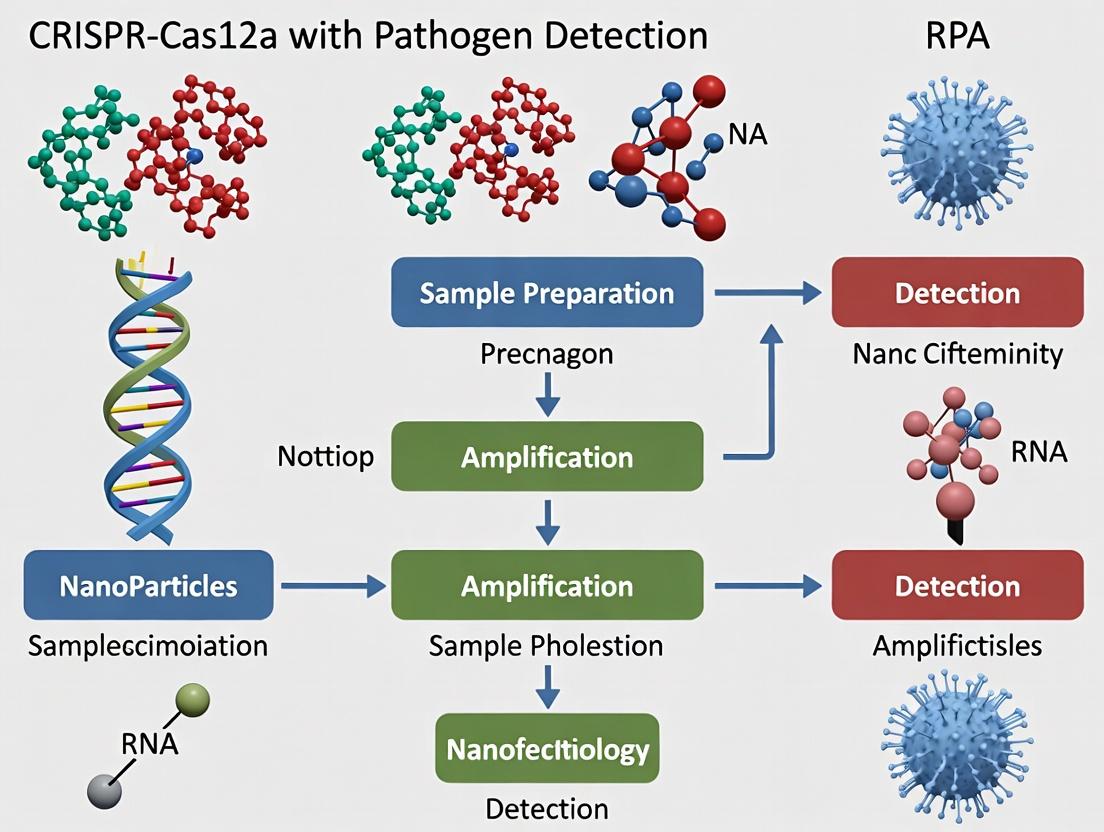
RPA-CRISPR/Cas12a: A Game-Changing, Isothermal Nucleic Acid Detection Platform for Rapid Pathogen Identification in Biomedical Research
This article provides a comprehensive guide for researchers and drug development professionals on the integrated RPA-CRISPR/Cas12a platform for pathogen detection.
RPA-CRISPR/Cas12a: A Game-Changing, Isothermal Nucleic Acid Detection Platform for Rapid Pathogen Identification in Biomedical Research
Abstract
This article provides a comprehensive guide for researchers and drug development professionals on the integrated RPA-CRISPR/Cas12a platform for pathogen detection. We explore the foundational principles of Cas12a's trans-cleavage activity and its synergy with Recombinase Polymerase Amplification (RPA). A detailed methodological framework covers assay design, workflow optimization, and specific applications for viral, bacterial, and fungal targets. We address common troubleshooting challenges and optimization strategies for sensitivity, specificity, and reaction kinetics. Finally, the article presents validation protocols and comparative analyses against gold-standard methods (qPCR) and other CRISPR-Cas systems, evaluating performance metrics, cost, and suitability for point-of-care and high-throughput settings. This resource aims to empower scientists to develop robust, rapid, and field-deployable diagnostic tools.
Demystifying the RPA-CRISPR/Cas12a Synergy: Core Principles and Advantages for Molecular Diagnostics
Within the broader thesis on developing CRISPR-Cas12a coupled with Recombinase Polymerase Amplification (RPA) for rapid, field-deployable pathogen detection, a foundational understanding of the Cas12a enzyme's intrinsic mechanics is critical. Unlike the more commonly known Cas9, Cas12a possesses distinct features in its target recognition and, most importantly, a potent nonspecific trans-cleavage activity upon target binding. This application note details the biochemical mechanisms and provides protocols for harnessing these properties in diagnostic assays.
Mechanism of Target Recognition
Cas12a is a Class 2, Type V-A CRISPR-associated nuclease. Its recognition and cleavage of target DNA proceed via a defined sequence of events.
Key Steps:
- Guide RNA (crRNA) Loading: The Cas12a protein is pre-loaded with a single CRISPR RNA (crRNA). The crRNA contains a ~20-24 nt spacer sequence complementary to the target DNA and a conserved stem-loop structure recognized by Cas12a.
- PAM Recognition: Cas12a scans double-stranded DNA (dsDNA) for a specific Protospacer Adjacent Motif (PAM). For most commonly used Cas12a orthologs (e.g., Lachnospiraceae bacterium ND2006, LbCas12a), the PAM is a 5'-TTTV-3' (where V is A, C, or G), located upstream of the target sequence.
- DNA Melting & R-loop Formation: Upon PAM binding, the enzyme locally unwinds the DNA. The crRNA spacer hybridizes to the target strand (complementary to the spacer), forming an R-loop structure.
- Target Strand Cleavage: The nuclease domains (RuvC) of Cas12a cleave the target DNA strand within the protospacer.
- Non-Target Strand Cleavage & Displacement: The non-target strand is displaced and subsequently cleaved by the same RuvC domain, completing double-stranded DNA cleavage. This is referred to as cis-cleavage.
Mechanism of Trans-Cleavage Activity
The defining diagnostic feature of Cas12a is its collateral, trans-cleavage activity. Upon successful formation of the Cas12a:crRNA:target DNA ternary complex, the enzyme undergoes a conformational change that activates its nonspecific single-stranded DNA (ssDNA) nuclease activity. This activated state cleaves any accessible ssDNA molecule in solution indiscriminately. This activity is repurposed in detection assays by including a fluorescent-quenched ssDNA reporter; cleavage of this reporter yields a fluorescent signal.
Table 1: Quantitative Comparison of Common Cas12a Orthologs
| Ortholog | PAM Sequence (5'→3') | cis-Cleavage (Target DNA) | Trans-Cleavage (Collateral) Activity | Optimal Temp (°C) | Typical crRNA Length (nt) |
|---|---|---|---|---|---|
| LbCas12a | TTTV (V=A/C/G) | Double-stranded break | High | 37 | 41-44 |
| AsCas12a | TTTV (V=A/C/G) | Double-stranded break | High | 37 | 40 |
| FnCas12a | YTTV (Y=C/T) | Double-stranded break | Moderate | 37 | 43 |
Experimental Protocol: Cas12a Trans-Cleavage Assay for Pathogen Detection (Post-RPA)
This protocol outlines the detection step following isothermal amplification (e.g., RPA) of a target pathogen gene.
A. Materials & Reagent Setup
- Nuclease-Free Water
- 10X Cas12a Reaction Buffer: 200 mM Tris-HCl, 100 mM MgCl₂, 500 mM NaCl, pH 7.5 at 25°C.
- Purified Cas12a Nuclease (LbCas12a): 100 nM working stock.
- Target-Specific crRNA: 1 µM stock in nuclease-free water. Design note: Spacer must be complementary to the amplified target region, immediately downstream of the PAM.
- Fluorescent ssDNA Reporter: 5'-FAM-TTATT-BHQ1-3' (or HEX/Iowa Black FQ), 10 µM stock.
- Amplified Target DNA: RPA product, diluted 1:10 in nuclease-free water.
- Real-time PCR Instrument or Plate Reader (for fluorescence measurement).
B. Procedure
- Prepare the reaction mix on ice:
- Nuclease-Free Water: to 20 µL final volume.
- 10X Cas12a Reaction Buffer: 2 µL.
- Cas12a Nuclease (100 nM): 1 µL (5 nM final).
- crRNA (1 µM): 2 µL (100 nM final).
- ssDNA Reporter (10 µM): 2 µL (1 µM final).
- Total Master Mix Volume: 17 µL.
- Aliquot 17 µL of Master Mix into each reaction well/tube.
- Add 3 µL of template:
- Test: 3 µL of diluted RPA product.
- No-Target Control (NTC): 3 µL nuclease-free water.
- Immediately place the plate/tube in a pre-warmed (37°C) real-time PCR instrument or plate reader.
- Measure fluorescence (FAM channel: Ex 485/Em 520) every 30 seconds for 60 minutes.
- Data Analysis: A positive detection is indicated by a rapid increase in fluorescence signal over time compared to the flat baseline of the NTC.
Visualizing the Cas12a Detection Workflow
Diagram 1: Cas12a-RPA Pathogen Detection Workflow (78 chars)
Diagram 2: Cas12a Target Binding & Trans-Cleavage Mechanism (94 chars)
The Scientist's Toolkit: Key Research Reagent Solutions
Table 2: Essential Materials for Cas12a-based Detection Assays
| Reagent / Material | Function & Role in Experiment | Key Considerations for Selection |
|---|---|---|
| Cas12a Nuclease (Purified) | The core enzyme for both specific target cleavage and nonspecific collateral activity. | Ortholog choice (Lb, As), high specific activity, low endotoxin levels, commercial availability as recombinant protein. |
| crRNA (Synthetic) | Guides Cas12a to the specific DNA target sequence via complementarity. | Requires careful design downstream of PAM (TTTV). Chemical modifications can enhance stability. Must be HPLC-purified. |
| Fluorescent-Quenched ssDNA Reporter | Substrate for trans-cleavage activity; cleavage yields detectable fluorescence. | Common sequence: 5'-6-FAM-TTATT-BHQ1-3'. Choice of fluorophore/quencher pair must match detection equipment. |
| Isothermal Amplification Mix (RPA) | Amplifies target pathogen DNA to detectable levels for Cas12a at constant temperature. | Kit includes recombinase, polymerase, proteins. Must be compatible with downstream Cas12a buffer (Mg²⁺ concentration is critical). |
| Nuclease-Free Buffers & Water | Provides optimal ionic conditions for Cas12a activity and prevents RNase/DNase degradation. | MgCl₂ concentration is a key variable affecting both RPA and Cas12a kinetics. Must be certified nuclease-free. |
| Real-Time Fluorescence Detector | Enables kinetic measurement of reporter cleavage, providing time-to-positive data. | Can be a real-time PCR machine, plate reader, or portable field device. Requires appropriate optical filters for chosen fluorophore. |
Recombinase Polymerase Amplification (RPA) is an isothermal nucleic acid amplification technique that operates at 37-42°C, mimicking in vitro the natural process of DNA replication and recombination. Within the context of CRISPR-Cas12a-based pathogen detection, RPA serves as a rapid, sensitive, and equipment-free front-end amplification step, enabling the detection of attomolar levels of target DNA from pathogens without the need for thermal cycling.
The core principles revolve around three key enzymatic activities:
- Recombinase: T4 uvsX recombinase forms filaments with primers and scans double-stranded DNA for homologous sequences, facilitating strand invasion and displacement loop (D-loop) formation.
- Single-Stranded DNA Binding Protein (SSB): T4 gp32 stabilizes the displaced strand, preventing primer displacement and reannealing of the template.
- Strand-Displacing DNA Polymerase: Bacillus subtilis Pol I (Bsu) extends the primer from the 3’ end, synthesizing new DNA.
This synergy allows for exponential amplification of the target sequence in under 20 minutes.
Quantitative Performance Data: RPA vs. Other Isothermal Methods
Table 1: Comparison of Key Isothermal Amplification Techniques for Pathogen Detection
| Parameter | Recombinase Polymerase Amplification (RPA) | Loop-Mediated Isothermal Amplification (LAMP) | Helicase-Dependent Amplification (HDA) | Nicking Enzyme Amplification Reaction (NEAR) |
|---|---|---|---|---|
| Typical Temp. | 37-42°C | 60-65°C | 60-65°C | 55-60°C |
| Time to Result | 10-20 min | 15-60 min | 60-120 min | 5-15 min |
| Typical Limit of Detection | 1-10 copies/reaction | 10-100 copies/reaction | 10-100 copies/reaction | 10-50 copies/reaction |
| Primer Design Complexity | Low (2 primers) | High (4-6 primers) | Low (2 primers) | Medium (2 primers) |
| Key Enzymes | Recombinase, SSB, Polymerase | Bst Polymerase | Helicase, Polymerase | Nicking Enzyme, Polymerase |
| Compatibility w/ CRISPR | High (Low temp, fast) | Medium (High temp, Mg²⁺ interfer.) | Medium | High (Fast) |
Table 2: Performance Metrics of RPA-CRISPR-Cas12a for Select Pathogens (Representative Data)
| Target Pathogen | Gene Target | RPA Time | Cas12a Guide RNA | Total Assay Time | Limit of Detection (LoD) | Clinical Sensitivity/Specificity |
|---|---|---|---|---|---|---|
| SARS-CoV-2 | N gene, E gene | 15 min | Specific to target | ~40 min | 5-10 copies/µL | >95% / >98% |
| Mycobacterium tuberculosis | IS6110 | 20 min | Specific to target | ~50 min | 1-2 copies/reaction | >90% / 100% |
| Pseudomonas aeruginosa | gyrB | 15 min | Specific to target | ~40 min | 10 CFU/mL | >95% / >95% |
| HPV 16/18 | E6/E7 | 20 min | Specific to target | ~50 min | 10 copies/reaction | >97% / >99% |
The Scientist's Toolkit: Essential Reagents and Materials
Table 3: Key Research Reagent Solutions for RPA-CRISPR-Cas12a Assays
| Item | Function & Description | Example Vendor/Product |
|---|---|---|
| Lyophilized RPA Pellet | Contains core enzymes (recombinase, SSB, polymerase), nucleotides, and buffer; enables room-temperature storage and rapid setup. | TwistAmp Basic (TwistDx) |
| Magnesium Acetate (MgOAc) Solution | Critical cofactor; initiates the RPA reaction upon addition. Typically 280mM stock. | Included in TwistAmp kits |
| Forward/Reverse RPA Primers | Target-specific oligonucleotides (30-35 nt) designed per standard guidelines. Reconstituted in nuclease-free water or TE buffer. | IDT, Thermo Fisher |
| Cas12a Nuclease (cpf1) | CRISPR effector; upon target recognition by gRNA, exhibits collateral ssDNA cleavage activity. | Alt-R A.s. Cas12a (IDT), EnGen Lba Cas12a (NEB) |
| crRNA / gRNA | Single guide RNA complementary to the RPA-amplified target; directs Cas12a for specific binding and trans-cleavage. | Synthesized chemically (IDT) or transcribed in vitro. |
| Fluorescent ssDNA Reporter | Dual-labeled (FAM-BHQ1) short single-stranded DNA oligonucleotide; cleavage by activated Cas12a generates fluorescent signal. | F-Q Reporter (IDT), Custom Synthesis |
| Lateral Flow Readout Strips | Alternative detection; uses labeled reporters (FAM/Biotin) captured on test/control lines. | Milenia HybriDetect, Ustar Biotech |
| Nuclease-Free Water | Solvent for primer/resgent reconstitution; essential to prevent enzymatic degradation. | Invitrogen, Sigma-Aldrich |
Detailed Experimental Protocols
Protocol 1: Basic RPA Amplification for CRISPR-Cas12a Input
Objective: To amplify target DNA from purified pathogen genomic DNA or lysate. Materials: TwistAmp Basic kit, target-specific RPA primers (10µM each), 280mM magnesium acetate, template DNA, nuclease-free water. Workflow:
- Prepare Master Mix (on ice):
- Nuclease-free water: to a final volume of 50µL
- Rehydration buffer (from kit): 29.5 µL
- Forward primer (10µM): 2.4 µL
- Reverse primer (10µM): 2.4 µL
- Template DNA (1-10 ng, or lysate): 2 µL
- Vortex gently and briefly spin down.
- Transfer the entire master mix (47.5 µL) into a lyophilized RPA pellet. Pipette up and down to fully resuspend.
- Initiate Reaction: Add 2.5 µL of 280 mM magnesium acetate (MgOAc) to the tube lid. Briefly centrifuge to mix MgOAc into the reaction.
- Incubate at 37-42°C for 15-20 minutes. A standard heat block or dry bath is sufficient.
- Proceed directly to CRISPR-Cas12a detection or store amplified product at 4°C for short-term use.
Protocol 2: Integrated RPA-CRISPR-Cas12a Fluorescence Detection
Objective: To detect RPA-amplified pathogen DNA via Cas12a collateral cleavage and real-time fluorescence. Materials: RPA product (from Protocol 1), LbCas12a nuclease (10µM), target-specific crRNA (10µM), fluorescent ssDNA reporter (10µM, e.g., FAM-TTATT-BHQ1), NEBuffer 2.1 or equivalent, plate reader/qPCR instrument. Workflow:
- Prepare Cas12a Detection Mix:
- Nuclease-free water: to final 20µL
- 2x Reaction Buffer (e.g., NEBuffer 2.1): 10 µL
- LbCas12a (10µM): 1 µL (final 500nM)
- crRNA (10µM): 1 µL (final 500nM)
- Fluorescent ssDNA Reporter (10µM): 1 µL (final 500nM)
- Mix gently.
- Combine: Add 2 µL of the RPA amplification product (from Protocol 1) to 18 µL of the Cas12a Detection Mix. Final reaction volume = 20 µL.
- Incubate & Read Fluorescence:
- Transfer to a 96-well PCR plate or microcuvette.
- Place in a real-time PCR instrument or fluorescence plate reader.
- Set temperature to 37°C.
- Measure fluorescence (FAM: Ex/Em ~485/535 nm) every minute for 30-60 minutes.
- Data Analysis: A positive result is indicated by a sharp, exponential increase in fluorescence over time. Negative samples show baseline drift.
Visualization of Workflows and Mechanisms
Title: RPA Enzymatic Mechanism and Amplification Cycle
Title: Integrated RPA-CRISPR-Cas12a Assay Workflow
Title: Cas12a Target Recognition and Trans-Cleavage Signaling
Within the broader research thesis on CRISPR-Cas12a combined with Recombinase Polymerase Amplification (RPA) for pathogen detection, this document details the integrated workflow that enables ultrasensitive, specific, and rapid diagnostic assays. This synergy leverages RPA’s isothermal amplification speed with Cas12a’s sequence-specific recognition and trans-cleavage activity, creating a powerful tool for researchers and drug development professionals targeting viral, bacterial, and other pathogenic nucleic acids.
Core Principles and Signaling Pathway
The fundamental signaling pathway relies on two sequential biomolecular events: Target Amplification followed by Cas12a-mediated Detection & Signal Generation.
Diagram 1: RPA-Cas12a Detection Signaling Pathway (82 chars)
The integrated RPA-Cas12a workflow demonstrates superior performance characteristics compared to standalone amplification or traditional PCR-based detection methods, particularly for point-of-care applications.
Table 1: Performance Comparison of Pathogen Detection Methods
| Parameter | qPCR (Gold Standard) | RPA Alone | RPA-Cas12a Integrated Workflow |
|---|---|---|---|
| Assay Time | 60 - 120 min | 15 - 30 min (amp only) | 35 - 60 min (total) |
| Temperature | Thermocycling (95°C, 50-60°C) | Isothermal (37-42°C) | Isothermal (37-42°C) |
| Limit of Detection (LoD) | 10 - 100 copies/µL | 1 - 10 copies/µL | 1 - 10 copies/µL |
| Specificity | High | Moderate | Very High (CRISPR-guided) |
| Signal-to-Noise Ratio | High | Moderate | >10:1 (with optimized reporter) |
| Instrument Simplicity | Complex (thermocycler) | Simple (heat block) | Simple (heat block, fluorimeter) |
| Multiplexing Potential | High (dye-based) | Low | Emerging (multiple Cas enzymes/reporters) |
Table 2: Optimized Reaction Conditions for Integrated Workflow
| Component | Recommended Concentration | Function & Notes |
|---|---|---|
| RPA Amplification | Pre-incubation: 39°C for 20-40 min | |
| Forward/Reverse Primer | 400 nM each | Target-specific amplification. Design for ~200-500 bp amplicon. |
| RPA Enzyme Pellets/Kit | As per mfr. | Contains recombinase, polymerase, etc. |
| MgOAc | 14 mM (final) | Essential cofactor, added last to initiate. |
| Cas12a Detection | Incubation: 37°C for 10-15 min | |
| Cas12a Enzyme (LbCas12a) | 50 - 100 nM | Effector nuclease. A.s. Cas12a also common. |
| crRNA | 50 - 100 nM | Designed to target amplified sequence. |
| ssDNA FQ Reporter | 200 - 500 nM | e.g., FAM-TTATT-BHQ1, poly-T backbone preferred. |
| Reaction Buffer | 1X NEBuffer 2.1 or 3.1 | Provides optimal ionic conditions. |
Detailed Experimental Protocols
Protocol 4.1: One-Pot RPA-Cas12a Fluorescent Detection of DNA Targets
Principle: Amplification and detection occur sequentially in a single, sealed tube to minimize contamination.
Materials: See "The Scientist's Toolkit" below. Procedure:
- Master Mix Preparation (on ice): In a 1.5 mL microcentrifuge tube, combine the following in order:
- Nuclease-free water: to a final volume of 50 µL.
- 2X RPA Reaction Buffer: 25 µL.
- Forward Primer (10 µM): 2 µL.
- Reverse Primer (10 µM): 2 µL.
- crRNA (10 µM): 0.5 µL.
- ssDNA FQ Reporter (10 µM): 1 µL.
- Cas12a Enzyme (1 µM): 0.5 µL.
- RPA Enzyme Pellets: 1 pellet (or liquid enzymes as per kit).
- Mix thoroughly by pipetting. Do not vortex enzyme-containing mixes.
- Aliquot 49.5 µL of the master mix into each 0.2 mL PCR tube or reaction strip.
- Initiate Reaction: Add 0.5 µL of template DNA (or nuclease-free water for NTC) to the side of the tube, then cap.
- Briefly centrifuge to combine reagents.
- Add Magnesium: Using a fine pipette, add 2.5 µL of 280 mM MgOAc (14 mM final) directly into the liquid. Cap and centrifuge immediately.
- Incubate: Place tubes in a pre-warmed real-time fluorimeter or heat block at 39°C. Collect fluorescence measurements (FAM channel, 1-min intervals) for 60 minutes.
- Data Analysis: A positive reaction is indicated by a sharp increase in fluorescence signal over a predetermined threshold (typically 5 standard deviations above the mean of the NTC). Time-to-positive (TTP) can be correlated with initial template concentration.
Protocol 4.2: Lateral Flow Readout for RPA-Cas12a (Two-Step)
Principle: Amplicon-activated Cas12a cleaves a labeled reporter, altering its migration on a lateral flow strip.
Procedure:
- Perform RPA: Carry out a standard 50 µL RPA reaction (Protocol 4.1 steps 1-6, excluding Cas12a, crRNA, and FQ reporter) at 39°C for 30 min.
- Cas12a Cleavage Reaction: Prepare a separate detection mix:
- Nuclease-free water: 15 µL
- Cas12a Reaction Buffer (10X): 2 µL
- crRNA (10 µM): 1 µL
- Cas12a (1 µM): 1 µL
- Biotin-ssDNA-FAM Reporter (10 µM): 1 µL
- Combine 5 µL of the completed RPA product with the 20 µL detection mix. Incubate at 37°C for 15 min.
- Lateral Flow Detection: Apply 50-80 µL of running buffer to the sample pad of a sandwich-style lateral flow strip (e.g., Milenia HybriDetect). Immediately pipette 10 µL of the cleavage reaction onto the sample pad.
- Allow the strip to develop for 5-10 minutes.
- Interpretation: Positive: Control line (C) and Test line (T) visible. The intact reporter is captured at T. Negative: Only the Control line (C) is visible. Cleavage destroys the T-line epitope.
Diagram 2: Two-Step RPA-Cas12a Lateral Flow Protocol (71 chars)
The Scientist's Toolkit: Key Research Reagent Solutions
| Item | Supplier Examples | Function in Workflow |
|---|---|---|
| RPA Kit (TwistAmp Basic/basic/fast) | TwistDx/Twist Bioscience | Provides all enzymes, buffers, and pellets for isothermal amplification. Essential for speed. |
| Purified Cas12a (LbCas12a, AsCas12a) | New England Biolabs, IDT, Thermo Fisher | The CRISPR effector protein. Requires high purity and nuclease-free buffers for consistent activity. |
| Custom crRNA | IDT, Synthego, Thermo Fisher | Guide RNA specific to the target amplicon. Critical for specificity. HPLC purification recommended. |
| Fluorescent ssDNA Reporters (e.g., FAM-TTATT-BHQ1) | Biosearch Technologies, IDT | The trans-cleavage substrate. Quenched fluorescence pre-cleavage, signal post-cleavage. |
| Lateral Flow Strips (HybriDetect) | Milenia Biotec, Ustar Biotechnologies | For visual, instrument-free readout. Uses labeled (e.g., FAM/Biotin) reporters. |
| Real-time Fluorimeter (Genie III, QuantStudio) | OptiGene, Thermo Fisher | For kinetic fluorescence monitoring in one-pot assays. Enables quantification and TTP analysis. |
| Nuclease-free Water & Tubes | Ambion, various | To prevent degradation of RNA guides and nucleic acid templates. |
| MgOAc (Magnesium Acetate) | Provided in RPA kits or Sigma | The critical divalent cation cofactor required to initiate the RPA reaction. |
Application Notes: CRISPR-Cas12a/RPA for Pathogen Detection
Within the framework of advancing molecular diagnostics, the integration of Recombinase Polymerase Amplification (RPA) with CRISPR-Cas12a presents a paradigm shift. This synergistic combination directly addresses the limitations of conventional PCR-based assays, particularly for point-of-care (POC) applications. The core advantages are interdependent: Isothermal Operation (RPA at 37-42°C) eliminates thermocyclers, enabling Speed (results in 20-60 minutes). The CRISPR-Cas12a complex provides Sensitivity through its high-specificity recognition and trans-cleavage activity, which can be coupled to fluorescent or lateral flow readouts, unlocking true Point-of-Care Potential.
The following table synthesizes key performance metrics from recent, representative studies detecting viral and bacterial pathogens.
Table 1: Performance Metrics of Selected CRISPR-Cas12a/RPA Assays
| Target Pathogen | Sample Type | Assay Name/Variant | Time-to-Result | Limit of Detection (LoD) | Clinical Sensitivity | Clinical Specificity | Reference (Year) |
|---|---|---|---|---|---|---|---|
| SARS-CoV-2 | Nasopharyngeal swab | DETECTR | ~40 min | 10 copies/µL | 95% | 100% | Broughton et al., Nat. Biotechnol. (2020) |
| HPV 16/18 | Cervical swab | CRISPR-HPV | ~60 min | 1.5 copies/µL | 100% | 100% | Qian et al., J. Mol. Diagn. (2022) |
| Mycobacterium tuberculosis | Sputum | CRISPR-MTB | ~90 min | 5 CFU/mL | 96.7% | 100% | Ai et al., Eur. Respir. J. (2022) |
| Salmonella spp. | Food homogenate | RPA-Cas12a-FS | ~30 min | 10 CFU/mL | N/A | N/A | Huang et al., Anal. Chem. (2023) |
| Pseudomonas aeruginosa | Bacterial culture | - | ~20 min | 1.2 nM (plasmid) | N/A | N/A | Recent proof-of-concept |
Detailed Experimental Protocol: Fluorescent Detection of SARS-CoV-2 RNA
This protocol outlines a standard workflow for detecting SARS-CoV-2 N gene from extracted RNA using RPA and Cas12a.
I. Reagent Preparation (Prepare on ice)
- RPA Master Mix (50 µL total/reaction):
- 29.5 µL Rehydration Buffer (from RPA kit)
- 2.1 µL Forward Primer (10 µM)
- 2.1 µL Reverse Primer (10 µM)
- 5 µL Template RNA (or nuclease-free water for NTC)
- 11.3 µL Nuclease-free water
- Cas12a Detection Mix (Prepare separately and add after RPA):
- 2 µL Cas12a enzyme (100 nM final)
- 2 µL crRNA (120 nM final, designed for N gene target)
- 1 µL FQ Reporter (500 nM final, e.g., 5'-6-FAM-TTATT-BHQ1-3')
- 3 µL NEBuffer 2.1 (1x final)
II. Amplification & Detection Workflow
- RPA Amplification: Add 1 µL of Magnesium Acetate (280 mM) to the RPA master mix tube lid. Briefly spin down and mix to initiate amplification. Incubate at 42°C for 15-20 minutes.
- Cas12a Detection: Post-amplification, directly add 8 µL of the prepared Cas12a Detection Mix to the RPA tube. Mix by pipetting.
- Incubation & Measurement: Incubate the combined reaction at 37°C. Monitor real-time fluorescence (FAM channel) in a plate reader or portable fluorimeter for 10-15 minutes. A sharp increase in fluorescence indicates a positive result.
III. Controls
- No Template Control (NTC): Replace template with water.
- Positive Control: A synthetic plasmid or RNA fragment containing the target sequence.
The Scientist's Toolkit: Essential Research Reagent Solutions
Table 2: Key Reagents for CRISPR-Cas12a/RPA Assay Development
| Reagent / Solution | Function & Critical Notes |
|---|---|
| RPA Kit (Basic) | Contains recombinase, polymerase, single-stranded binding proteins, and rehydration buffer for isothermal amplification. Essential for speed. |
| LbCas12a or AsCas12a Enzyme | CRISPR effector protein. Provides specificity and trans-cleavage activity. Purity and storage buffer are critical for activity. |
| Target-specific crRNA | Guide RNA that dictates Cas12a target recognition. Must be designed for the PAM sequence (e.g., TTTV for LbCas12a). HPLC purification recommended. |
| Fluorescent Quenched (FQ) Reporter | Single-stranded DNA oligo with a fluorophore and quencher. Cleavage by activated Cas12a generates fluorescence signal. Stability is key. |
| Lateral Flow Strips (Optional) | For visual, instrument-free readout. Use with biotin- and FAM-labeled reporters. Requires different reporter design than fluorescent assays. |
| RNase Inhibitor | Critical when detecting RNA targets to prevent degradation of both target and crRNA during sample handling and reaction setup. |
| Nuclease-Free Water & Buffers | To prevent degradation of oligonucleotides and enzymes. Use validated buffers (e.g., NEBuffer) for Cas12a reactions. |
Visualization Diagrams
CRISPR-Cas12a RPA Detection Workflow
Cas12a Trans-Cleavage Signaling Mechanism
Application Notes
This analysis, within the broader thesis on CRISPR-Cas12a coupled with Recombinase Polymerase Amplification (RPA) for pathogen detection, provides a comparative framework for selecting Cas effector proteins. The choice between Cas9, Cas12a, and Cas13a is fundamental and hinges on target type (DNA vs. RNA), cleavage mechanism, and resulting collateral activity, which directly influences assay design, sensitivity, and specificity.
Key Comparative Insights:
- Cas9 is a DNA-targeting, double-strand break (DSB) inducer with high fidelity, requiring a tracrRNA and a complex protospacer adjacent motif (PAM). Its primary utility is in gene editing, not typically in diagnostic detection due to the lack of reported collateral cleavage.
- Cas12a targets DNA, induces staggered DSBs, and exhibits trans-cleavage activity on non-specific single-stranded DNA (ssDNA) upon target recognition. This collateral activity, triggered by double-stranded DNA (dsDNA) targets, is the cornerstone of its use in DNA-based diagnostics (e.g., DETECTR). Its requirement for a T-rich PAM (TTTV) and ability to process its own crRNA array are distinct advantages.
- Cas13a targets RNA, induces single-strand cuts, and exhibits robust trans-cleavage activity on non-specific single-stranded RNA (ssRNA) upon target recognition. This collateral activity, triggered by single-stranded RNA (ssRNA) targets, makes it ideal for RNA virus detection and transcriptome analysis (e.g., SHERLOCK).
The integration of Cas12a with RPA for pathogen detection is particularly powerful. RPA rapidly amplifies target DNA isothermally, which is then detected by Cas12a's target-activated trans-cleavage of a reporter probe, enabling ultrasensitive, instrument-free detection suitable for point-of-care applications.
Quantitative Comparison of Key Features
Table 1: Comparative Properties of Cas9, Cas12a, and Cas13a
| Feature | Cas9 (e.g., SpCas9) | Cas12a (e.g., LbCas12a) | Cas13a (e.g., LwaCas13a) |
|---|---|---|---|
| Primary Target | dsDNA | dsDNA, ssDNA | ssRNA |
| Cleavage Type | Blunt-ended Double-Stranded Break (DSB) | Staggered DSB (with 5' overhangs) | Single-Stranded Break (SSB) |
| Collateral Activity | Not reported | Yes (trans-cleavage of ssDNA) | Yes (trans-cleavage of ssRNA) |
| Guide RNA | crRNA + tracrRNA (or fused sgRNA) | Single crRNA (shorter, ~42-44 nt) | Single crRNA (longer, ~64-66 nt) |
| PAM/PFS Requirement | PAM: 5'-NGG-3' (SpCas9), adjacent to target | PAM: 5'-TTTV-3', located upstream of target | PFS: 3'-H (not A), located downstream of target |
| crRNA Processing | Requires host RNase III or pre-processed | Self-processing from a crRNA array | Self-processing from a crRNA array |
| Typical Size (aa) | ~1368 | ~1200-1300 | ~1250 |
| Primary Application in Detection | Limited (no collateral) | DNA-based pathogen detection (e.g., DETECTR) | RNA-based pathogen detection (e.g., SHERLOCK) |
Table 2: Performance Metrics in Nucleic Acid Detection Assays
| Metric | Cas12a-based Detection (with RPA) | Cas13a-based Detection (with RT-RPA) |
|---|---|---|
| Theoretical Limit of Detection (LoD) | ~aM to single-digit copy (1-10 copies/µL) | ~aM to single-digit copy (1-10 copies/µL) |
| Time-to-Result | 30 - 90 minutes (combined RPA + Cas) | 60 - 120 minutes (includes RT step) |
| Assay Temperature | Isothermal (37-42°C for RPA; 37°C for Cas12a) | Isothermal (37-42°C for RT-RPA; 37°C for Cas13a) |
| Key Signal Reporter | ssDNA-linked fluorophore/quencher (e.g., FAM/TAMRA) | ssRNA-linked fluorophore/quencher (e.g., FAM/BIQ) |
| Signal Readout | Fluorescence (real-time or endpoint), Lateral Flow Strip | Fluorescence (real-time or endpoint), Lateral Flow Strip |
Detailed Experimental Protocols
Protocol 1: Cas12a-based Pathogen DNA Detection (DETECTR Workflow)
Objective: To detect specific pathogen DNA (e.g., Mycobacterium tuberculosis gene) using RPA pre-amplification and Cas12a trans-cleavage.
Research Reagent Solutions Toolkit:
| Item | Function in Experiment |
|---|---|
| Lyophilized or Liquid RPA Kit | Provides enzymes and buffers for isothermal DNA amplification at 37-42°C. |
| Cas12a Nuclease (LbCas12a) | The effector protein that provides target-specific binding and collateral ssDNase activity. |
| Target-specific crRNA | Guides Cas12a to the complementary amplicon sequence. Designed with a TTTV PAM in the target. |
| ssDNA FQ Reporter Probe | A short (e.g., 6-8 nt) ssDNA oligo labeled with a fluorophore (FAM) and a quencher (TAMRA). Collateral cleavage separates the pair, generating fluorescence. |
| Nuclease-free Water | Solvent and diluent for all reaction components. |
| Fluorometer or Real-time PCR Machine | For kinetic measurement of fluorescence increase. |
| Lateral Flow Strips | For endpoint visual detection using labeled reporter particles. |
Methodology:
- Sample Preparation: Extract genomic DNA from the clinical sample (e.g., sputum).
- RPA Pre-amplification:
- Assemble a 50 µL RPA reaction on ice: 29.5 µL rehydration buffer, 2.1 µL forward primer (10 µM), 2.1 µL reverse primer (10 µM), 5 µL template DNA, and nuclease-free water to 47.5 µL.
- Add 2.5 µL of magnesium acetate (280 mM) to the tube cap, briefly centrifuge to initiate the reaction.
- Incubate at 39°C for 15-20 minutes.
- Cas12a Detection Reaction:
- Prepare a Cas12a detection mix on ice: 1 µL Cas12a (10 µM), 1.25 µL crRNA (10 µM), 0.5 µL ssDNA FQ Reporter (10 µM), 2.5 µL 10x Cas12a Reaction Buffer, and nuclease-free water to 17.5 µL.
- Transfer 2.5 µL of the RPA amplicon product into the detection mix.
- Incubate at 37°C for 10-30 minutes.
- Signal Detection:
- Fluorometric: Measure fluorescence (Ex/Em: 485/535 nm) kinetically or at endpoint.
- Lateral Flow: Add the reaction to a running buffer and dip a strip. A test line indicates cleavage.
Protocol 2: Comparative Cleavage Specificity Assay (Gel-based)
Objective: To visualize the distinct cleavage products (blunt vs. staggered) of Cas9 and Cas12a on a plasmid DNA target.
Methodology:
- Substrate Preparation: Linearize 1 µg of a plasmid containing the target sequence with a unique restriction site away from the CRISPR target site.
- RNP Complex Formation: For each reaction, pre-incubate 100 nM Cas protein with 120 nM of its respective guide RNA (sgRNA for Cas9, crRNA for Cas12a) in 1x NEB Buffer 3.1 for 10 min at 25°C.
- Cleavage Reaction: Add 100 ng of linearized plasmid to the RNP complex in a 20 µL total volume. Incubate at 37°C for 1 hour.
- Reaction Termination: Add 2 µL of Proteinase K and 2 µL of 10% SDS, incubate at 56°C for 15 min.
- Analysis: Run the entire reaction on a 1% agarose gel stained with ethidium bromide. Compare the cleavage product sizes: Cas9 produces two fragments of predictable sizes summing to the original length. Cas12a produces a "fuzzy" or shifted band pattern due to its staggered cuts, which may run differently.
Visualization Diagrams
Diagram 1: CRISPR-Cas Effector Target Specificity and Cleavage Mechanisms
Diagram 2: Cas12a-RPA Pathogen Detection Assay Workflow
Step-by-Step Protocol: Designing and Executing an RPA-CRISPR/Cas12a Assay for Pathogen Detection
This Application Note details the foundational principles for designing CRISPR-Cas12a-based detection assays coupled with Recombinase Polymerase Amplification (RPA). Framed within a broader thesis on pathogen detection research, this guide provides researchers and drug development professionals with the specific rules and protocols necessary to develop sensitive, specific, and rapid diagnostic tests. The synergy of Cas12a's programmable collateral cleavage activity with RPA's isothermal amplification enables powerful point-of-care diagnostic solutions.
crRNA Design Rules for Cas12a (Cpfl)
The crRNA directs Cas12a to its DNA target and is critical for assay specificity and efficiency. Below are the consolidated design parameters based on current literature and experimental validation.
Table 1: Cas12a crRNA Design Rules and Parameters
| Parameter | Rule / Specification | Rationale & Notes |
|---|---|---|
| Direct Repeat (DR) | Use the species-specific 5' DR sequence. For LbCas12a: 5'-AAUUUCUACUAAGUGUAGAU-3' | The DR is invariant and essential for Cas12a protein binding. Must be positioned at the 5' end of the crRNA. |
| Spacer Length | 20-24 nt (typically 21-22 nt). Must be complementary to the target Protospacer. | Shorter spacers may reduce specificity; longer spacers may reduce cleavage activity. |
| Protospacer Adjacent Motif (PAM) | Cas12a requires a 5' T-rich PAM. For LbCas12a: 5'-TTTV-3' (where V = A, C, or G). | The PAM is located upstream (5') of the target protospacer on the non-target strand. The crRNA spacer is designed to bind downstream of this PAM. |
| Spacer Sequence | Avoid secondary structure (e.g., hairpins) within the spacer. GC content: 40-60%. | High GC can increase binding strength but may promote off-target effects. Use tools like IDT OligoAnalyzer. |
| Off-Target Considerations | Mismatches in the PAM-distal 5-8 nt "seed region" (near PAM) are more tolerated. Mismatches in the PAM-proximal region are critical for discrimination. | Design crRNA to have at least 2-3 mismatches in the seed region against non-target sequences to ensure specificity. |
| Synthetic crRNA Format | Chemical synthesis as a single RNA oligonucleotide: [5' DR - Spacer Sequence 3']. | No tracrRNA is needed for Cas12a. Can be ordered with standard 2'-OH or stabilized modifications (e.g., 2'-O-methyl). |
Title: crRNA and Target DNA Relationship for Cas12a
Primer Design for Recombinase Polymerase Amplification (RPA)
Proper primer design is paramount for efficient RPA amplification, which occurs at 37-42°C. Primers are typically longer than those used in PCR.
Table 2: RPA Primer Design Guidelines
| Parameter | Rule / Specification | Rationale & Notes |
|---|---|---|
| Length | 30-35 nucleotides (minimum 27, maximum 38). | Longer primers facilitate stable binding at the lower isothermal temperature. |
| Tm | Recommended calculated Tm: 50-65°C. | Use biophysical calculators for long oligonucleotides (e.g., IDT's OligoAnalyzer). |
| GC Content | 40-60% is ideal. Avoid extreme values. | Ensures stable primer-template complexes without excessive non-specific binding. |
| 3' End | The last 5 nucleotides at the 3' end should contain at least 3 A/T bases. | Facilitates initial strand invasion by the recombinase-primer complex. Avoid stable secondary structures at the 3' end. |
| 5' End | No strong secondary structures. Can be modified with labels (FAM, Biotin) for detection assays. | The 5' end is less critical for invasion but should not hinder recombinase loading. |
| Specificity | BLAST against the target genome. Avoid primer-dimer and hairpin formation. | RPA is highly sensitive; even minor non-specific priming can lead to background. |
| Amplicon Size | Optimal: 80-500 bp. Can work up to ~1.5 kb, but efficiency drops. | Smaller amplicons yield faster kinetics, crucial for rapid detection. |
Title: RPA Primer Design and Validation Workflow
Integrated Assay Protocol: Cas12a/RPA for Pathogen Detection
This protocol outlines a standard one-pot or two-step detection assay for a DNA pathogen target.
Materials & Reagents (The Scientist's Toolkit)
Table 3: Key Research Reagent Solutions
| Item | Function & Specification |
|---|---|
| Cas12a Nuclease | Recombinant LbCas12a or AsCas12a protein. Provides collateral cleavage activity upon target recognition. |
| Custom crRNA | Synthetic RNA oligo with DR and target-specific spacer. Guides Cas12a to the amplicon. |
| RPA Kit | Commercial kit (e.g., TwistAmp Basic, nfo, or exo). Contains freeze-dried pellets with recombinase, polymerase, nucleotides, etc. |
| Fluorescent Reporter | ssDNA oligonucleotide with fluorophore (e.g., FAM) and quencher (e.g., BHQ1). Cleaved upon Cas12a activation, producing signal. |
| Primers | Forward and Reverse RPA primers (30-35 nt) targeting the pathogen sequence. |
| MgOAc | Magnesium acetate solution. Required to initiate the RPA reaction. |
| Buffer & Nuclease-Free Water | Provides optimal ionic and pH conditions for enzyme activity. |
Protocol: Two-Step Detection (RPA followed by Cas12a Cleavage)
Step 1: RPA Amplification
- Reagent Setup (on ice): Reconstitute a TwistAmp Basic pellet by adding 29.5 µL of rehydration buffer, 10.5 µL of nuclease-free water, and 2.4 µL each of forward and reverse primers (10 µM stock).
- Initiate Reaction: Add 1 µL of template DNA (or lysate) and 2 µL of 280 mM magnesium acetate (MgOAc) to the lid of the tube. Briefly centrifuge to mix.
- Incubate: Place the tube at 39°C for 15-20 minutes. Use a heating block or dry bath.
- Amplicon Handling: After incubation, the product can be used directly or diluted 1:10 in nuclease-free water for the detection step.
Step 2: Cas12a Fluorescent Detection
- Prepare Detection Mix: In a new tube or a real-time PCR plate well, combine the following:
- 10 µL of NEBuffer 2.1 (or appropriate Cas12a buffer)
- 1 µL (100-200 nM) of Cas12a protein
- 1.5 µL (50-100 nM) of crRNA
- 1 µL (200-500 nM) of Fluorescent ssDNA Reporter (e.g., FAM-TTATT-BHQ1)
- 1.5 µL of nuclease-free water
- Add Amplicon: Add 5 µL of diluted RPA amplicon from Step 1.
- Measure Fluorescence: Incubate the reaction at 37°C and monitor fluorescence (FAM channel: Ex ~485/Em ~520) in a real-time PCR machine or plate reader every minute for 30-60 minutes. A rapid increase in fluorescence indicates positive detection.
One-Pot Protocol Note: For a single-tube assay, the RPA and Cas12a components (except MgOAc) can be combined. MgOAc is added last to start RPA. Sensitivity may be compromised due to competition for Mg²⁺ and potential early reporter cleavage.
Title: Two-Step RPA-Cas12a Detection Workflow
Critical Data & Optimization Notes
Table 4: Typical Performance Metrics and Optimization Targets
| Metric | Target / Expected Outcome | Troubleshooting Tip |
|---|---|---|
| Assay Time | 20-45 minutes total (RPA: 15-20 min, Cas12a: 5-30 min). | For faster Cas12a signal, increase amplicon concentration or use higher crRNA concentration (up to 150 nM). |
| Limit of Detection (LoD) | Single-digit copies/µL (e.g., 1-10 copies per reaction). | Verify primer and crRNA specificity. Use purified genomic DNA for LoD calibration. Pre-incubate Cas12a with crRNA for 10 min to form ribonucleoprotein (RNP). |
| Signal-to-Noise Ratio | High; negative controls should show minimal fluorescence drift over 60 min. | If background is high, reduce non-specific amplification by optimizing RPA primer design or adding crowders (e.g., PEG). Ensure reagent purity. |
| Cross-reactivity | None against near-neighbor non-target pathogens. | Test crRNA against DNA from related species. Introduce deliberate mismatches in the seed region to enhance discrimination. |
Application Notes
In the context of CRISPR-Cas12a coupled with Recombinase Polymerase Amplification (RPA) for pathogen detection, reagent integrity is the cornerstone of assay sensitivity and specificity. Master mix preparation must be meticulously standardized to ensure consistent trans-cleavage activity following target DNA recognition. Sourcing high-purity, nuclease-free components is non-negotiable to prevent off-target degradation and false positives. This protocol emphasizes the preparation of a stable, single-tube RPA-CRISPR-Cas12a master mix for point-of-care applications, with a focus on mitigating batch-to-batch variability critical for diagnostic development.
Key Considerations for Sourcing
- Cas12a Enzyme: Recombinant, high-specificity variants (e.g., LbCas12a, AsCas12a) with low endotoxin levels are essential. Vendor-provided activity units (U/µL) must be validated empirically.
- RPA Components: Commercial RPA kits (e.g., TwistAmp) provide reliable, stable freeze-dried pellets. For liquid formulation, sourced recombinase, single-stranded DNA-binding protein, and strand-displacing polymerase must be aliquoted and stored at -80°C to preserve activity.
- Oligonucleotides: crRNA must be HPLC-purified to ensure full-length transcripts. Synthetic, double-stranded target amplicons or genomic DNA from accredited repositories (e.g., ATCC) are required for validation.
- Fluorescent Reporters: Double-quenched (e.g., BHQ-2) ssDNA probes (e.g., FAM-TTATT-BHQ2) reduce background noise compared to single-quenched versions.
- Nuclease-Free Water: Certified DNase/RNase-free water is used for all dilutions.
Protocols
Protocol 1: Sourcing and Qualification of Key Components
Objective: To acquire and perform initial quality control on essential reagents. Materials:
- Vendor list (See Table 1)
- Thermocycler
- Fluorescence plate reader or real-time PCR instrument
Method:
- Procurement: Source components from vendors listed in Table 1.
- crRNA Integrity Check: Resolve 100 pmol of crRNA on a 15% Urea-PAGE gel. A single, sharp band should be visible.
- Enzyme Activity Spot Test: Perform a minimal reporter cleavage assay. Combine 50 nM Cas12a, 50 nM crRNA, 500 nM reporter probe in 1X NEBuffer 2.1. Add 10 nM synthetic target DNA. Measure fluorescence (Ex/Em: 485/535 nm) over 30 minutes at 37°C. A ≥5-fold increase over no-target control qualifies the enzyme batch.
Protocol 2: Preparation of a Single-Tube RPA-CRISPR-Cas12a Master Mix
Objective: To prepare a homogeneous, ready-to-use master mix for direct amplification and detection. Materials:
- Key Reagent Solutions (See Table 2)
- 1.5 mL nuclease-free microcentrifuge tubes
- Pipettes and aerosol-barrier tips
- Cooling block or ice
Method:
- Thaw and Centrifuge: Thaw all liquid components (except enzymes) on ice. Centrifuge briefly to collect contents.
- Calculate Volumes: Calculate volumes needed for (n+1) reactions to account for pipetting loss.
- Master Mix Assembly in a Cold Block: In a 1.5 mL tube on ice, combine components in the following order:
- Nuclease-free water (to final volume)
- 1X Rehydration Buffer (from RPA kit)
- 420 nM forward primer
- 420 nM reverse primer
- 50 nM crRNA (target-specific)
- 100 nM fluorescent reporter probe
- 60 nM LbCas12a enzyme
- Finally, add: 1X RPA enzyme pellet (resuspended) or liquid enzyme mix.
- Gentle Mixing: Mix by slow, deliberate pipetting up and down 10 times. Do not vortex.
- Aliquoting: Immediately aliquot X µL (e.g., 47 µL) of the master mix into individual reaction tubes/strips.
- Template Addition: Add Y µL (e.g., 3 µL) of template (or nuclease-free water for NTC) to each aliquot.
- Immediate Use or Storage: Initiate incubation immediately at 37-39°C for 30-45 minutes. Unused master mix aliquots can be flash-frozen on dry ice and stored at -80°C for up to 2 weeks. Avoid repeated freeze-thaw cycles.
Data Presentation
Table 1: Key Component Sourcing Guide
| Component | Specification | Recommended Vendor(s) | Storage Condition | Quality Control Metric |
|---|---|---|---|---|
| LbCas12a Nuclease | ≥ 95% purity, ≥ 10^6 U/mg | Integrated DNA Technologies, New England Biolabs, BioLabs | -80°C | Activity ≥ 5x signal:noise in spot test |
| crRNA | HPLC-purified, 40-45 nt | Integrated DNA Technologies, Sigma-Aldrich | -20°C (dry), -80°C (sol.) | Single band on Urea-PAGE |
| RPA Kit | TwistAmp Basic or Liquid | TwistDx Ltd./Qiagen | -20°C (kit), -80°C (liq. enzymes) | LoD < 10 copies (per manuf.) |
| Fluorescent Reporter | ssDNA, 5-6 nt, BHQ-2 quenched | Biosearch Technologies, Eurofins | -20°C (dark) | > 90% quenching efficiency |
| Synthetic Target | gBlock Gene Fragment, > 200 bp | Integrated DNA Technologies | -20°C | Sequence verification report |
| Nuclease-Free Water | Certified, 0.22 µm filtered | Thermo Fisher, MilliporeSigma | Room Temp | < 0.01 EU/mL endotoxin |
Table 2: Master Mix Formulation for a 50 µL Reaction
| Component | Stock Concentration | Final Concentration in Mix | Volume per 50 µL Rx (µL) | Function |
|---|---|---|---|---|
| Nuclease-Free Water | - | - | Variable (to 50 µL) | Solvent |
| RPA Rehydration Buffer | 2X | 1X | 25.0 | Provides amplification milieu |
| Forward/Reverse Primers | 10 µM each | 420 nM each | 2.1 each | Target amplification |
| crRNA (target-specific) | 5 µM | 50 nM | 0.5 | Guides Cas12a to target |
| Fluorescent Reporter Probe | 10 µM | 100 nM | 0.5 | Cas12a trans-cleavage substrate |
| LbCas12a Nuclease | 1 µM (or X U/µL) | 60 nM | 3.0 | Target recognition & cleavage |
| RPA Enzyme Pellet/Mix* | - | 1X | (from kit) | Isothermal amplification |
| Template DNA | Variable | - | 2.0-5.0 | Sample input |
*Follow manufacturer's instructions for pellet resuspension or liquid enzyme volume.
Mandatory Visualization
The Scientist's Toolkit
Table 3: Essential Research Reagent Solutions for RPA-CRISPR Assay Development
| Item | Function/Benefit | Example/Note |
|---|---|---|
| Nuclease-Free Water | Solvent for all reagents; prevents degradation of RNA/DNA components. | Use certified, 0.22 µm filtered. Do not use DEPC-treated water with enzymatic reactions. |
| 10X Cas12a Reaction Buffer | Provides optimal pH, ionic strength, and co-factors (e.g., Mg2+) for Cas12a cleavage activity. | Often supplied with enzyme; critical for standardizing final reaction conditions. |
| RPA Rehydration Buffer | Contains dNTPs, crowding agents, and salts necessary for efficient recombinase-polymerase activity. | Provided with commercial kits; used to resuspend freeze-dried enzyme pellets. |
| Fluorescent Reporter Stock | Ready-to-use, quenched ssDNA probe; the substrate for trans-cleavage signal generation. | Aliquot to avoid freeze-thaw cycles; protect from light. |
| crRNA Storage Buffer | Low TE buffer or nuclease-free buffer for resuspending and storing crRNA; prevents degradation. | Avoid buffers containing EDTA if Mg2+ is critical for your specific Cas12a variant. |
| Synthetic Positive Control | Cloned plasmid or gBlock fragment containing target sequence; validates each master mix batch. | Essential for establishing limit of detection (LoD) and troubleshooting. |
| Carrier RNA/DNA | Inert nucleic acid (e.g., salmon sperm DNA) added to dilute enzyme stocks to stabilize proteins. | Prevents adsorption to tube walls; improves reproducibility for low-concentration components. |
Application Notes
This application note, framed within a broader thesis on CRISPR-Cas12a coupled with Recombinase Polymerase Amplification (RPA) for pathogen detection, evaluates the integration of amplification and detection steps. The primary focus is comparing a seamless one-tube format against a conventional two-tube format for speed, contamination risk, sensitivity, and practical utility in point-of-care and laboratory settings.
Key Findings from Current Literature:
- One-Tube Format: Amplification and CRISPR detection reagents are combined in a single reaction vessel. Post-amplification, Cas12a cleavage is activated, often by a temperature shift or the inherent reaction kinetics. This minimizes aerosol contamination, simplifies workflow, and is ideal for field deployment. However, it can suffer from reduced sensitivity due to reagent interference and requires careful optimization of buffer compatibility and reagent concentrations.
- Two-Tube Format: RPA amplification and Cas12a detection are performed in physically separate tubes, typically with an amplicon transfer step. This format allows for independent optimization of each reaction, often yielding higher sensitivity and more robust performance. The primary drawback is an increased risk of amplicon contamination and a more complex manual workflow.
Data Presentation
Table 1: Comparative Performance of One-Tube vs. Two-Tube RPA-Cas12a Assays
| Parameter | One-Tube Format | Two-Tube Format | Notes / Implications |
|---|---|---|---|
| Time-to-Result | ~30-45 minutes | ~40-60 minutes | One-tube is faster due to no transfer step. |
| Hands-on Time | Low | Moderate to High | One-tube requires less user intervention. |
| Contamination Risk | Very Low | High | Tube opening for transfer in two-tube poses major contamination risk. |
| Reported Sensitivity (LOD) | ~10-100 copies/µL | ~1-10 copies/µL | Two-tube generally achieves 1-log better sensitivity. |
| Ease of Optimization | Challenging | Straightforward | One-tube requires balancing competing buffer conditions. |
| Suited for Field Use | Excellent | Poor | One-tube’s simplicity and closed system are key advantages. |
| Reagent Cost per Test | Comparable | Comparable | Slight savings for one-tube due to reduced plasticware. |
| Signal-to-Noise Ratio | Can be lower | Typically higher | Separated reactions reduce background in two-tube format. |
Table 2: Essential Research Reagent Solutions
| Reagent / Material | Function in RPA-Cas12a Assay |
|---|---|
| RPA Amplification Mix | Contains recombinase, polymerase, and co-factors for isothermal amplification at 37-42°C. |
| Forward & Reverse RPA Primers | Target-specific primers designed for efficient isothermal amplification. |
| Purified Cas12a (cpf1) Enzyme | CRISPR effector protein that, upon target binding, exhibits collateral single-stranded DNA cleavage. |
| crRNA | Custom-designed guide RNA that directs Cas12a to the complementary amplicon target sequence. |
| Fluorescent ssDNA Reporter Probe | (e.g., FAM-TTATT-BHQ1). Collateral cleavage of this probe generates a fluorescent signal. |
| RNase-Free Water | To reconstitute and dilute reagents, preventing RNase degradation of crRNA. |
| Nuclease-Free Buffer | Provides optimal ionic and pH conditions for combined or sequential reactions. |
| Positive Control Template | Synthetic DNA or RNA containing the target sequence for assay validation. |
| Lateral Flow Strip (Optional) | For visual endpoint detection using labelled reporter molecules. |
Experimental Protocols
Protocol A: One-Tube, Sequential RPA-Cas12a Fluorescence Assay
Reaction Setup: In a single 0.2 mL PCR tube or strip tube, prepare the following master mix on ice:
- 29.5 µL rehydration buffer (from RPA kit).
- 2.1 µL forward primer (10 µM).
- 2.1 µL reverse primer (10 µM).
- 1 µL Cas12a enzyme (100 nM stock).
- 1 µL crRNA (100 nM stock).
- 0.5 µL fluorescent ssDNA reporter (10 µM stock).
- 5 µL template DNA (or nuclease-free water for NTC).
- 9.8 µL magnesium acetate (280 mM, provided in RPA kit) is added last to the tube cap.
Initiation: Briefly centrifuge the tube to combine the magnesium acetate with the master mix, bringing the total volume to 50 µL. Mix gently by pipetting.
Incubation: Place the tube in a real-time PCR machine or isothermal fluorometer. Run the following program:
- Phase 1 (Amplification): 39°C for 20-25 minutes, with fluorescence acquisition (FAM channel) every 30 seconds.
- Phase 2 (Enhanced Detection): 37°C for 10-15 minutes, with continued fluorescence acquisition.
Analysis: Plot fluorescence vs. time. A sharp increase in fluorescence over the baseline indicates a positive detection.
Protocol B: Two-Tube RPA-Cas12a Fluorescence Assay
RPA Amplification (Tube 1):
- Prepare a 50 µL RPA reaction per the manufacturer's instructions (e.g., TwistAmp Basic kit).
- Add 5 µL of template to the rehydrated pellet. Initiate the reaction with magnesium acetate.
- Incubate at 39°C for 20-30 minutes in a heat block or thermocycler.
Cas12a Detection Setup (Tube 2):
- During the RPA incubation, prepare the CRISPR detection mix in a separate, optically clear tube/plate:
- 1X Nuclease-free Buffer
- 50 nM Cas12a enzyme
- 50 nM crRNA
- 200 nM fluorescent ssDNA reporter
- Nuclease-free water to 20 µL.
- Pre-incubate this mix at 37°C for 5 minutes to allow RNP complex formation.
- During the RPA incubation, prepare the CRISPR detection mix in a separate, optically clear tube/plate:
Transfer and Detection:
- After the RPA incubation, briefly centrifuge Tube 1.
- Transfer 2 µL of the RPA amplicon product into the pre-equilibrated CRISPR detection mix (Tube 2). Mix thoroughly by pipetting.
- Immediately place Tube 2 into a fluorometer or real-time PCR machine.
- Incubate at 37°C for 15-30 minutes, acquiring fluorescence (FAM channel) every 30 seconds.
Analysis: Monitor real-time fluorescence or measure endpoint fluorescence. A positive sample shows rapid signal generation post-transfer.
Workflow Diagrams
One Tube Workflow Path
Two Tube Workflow Path
Cas12a Activation and Signal Generation
Within the research thesis on CRISPR-Cas12a coupled with Recombinase Polymerase Amplification (RPA) for pathogen detection, selecting an appropriate readout modality is critical for translating the assay from the lab to potential point-of-care or field applications. Cas12a, upon activation by its target DNA, exhibits collateral trans-cleavage activity, non-specifically degrading nearby single-stranded DNA (ssDNA). This activity can be harnessed with different reporter molecules to generate signals detectable via fluorescent, lateral flow, or colorimetric systems. Each modality presents distinct trade-offs in sensitivity, equipment requirements, cost, and ease of interpretation. This application note details the protocols and comparative performance metrics for these three primary readout systems.
Quantitative Performance Comparison
The following table summarizes key performance characteristics for each detection modality when integrated with an RPA-Cas12a assay, based on current literature and experimental data.
Table 1: Comparative Analysis of Readout Modalities for RPA-Cas12a Assays
| Parameter | Fluorescent Readout | Lateral Flow Readout | Colorimetric Readout |
|---|---|---|---|
| Typical Limit of Detection (LoD) | 1-10 copies/µL | 10-100 copies/µL | 10-1000 copies/µL |
| Time-to-Result (Post-RPA) | 5-15 minutes | 5-10 minutes | 10-30 minutes |
| Equipment Required | Fluorometer or qPCR instrument | None (visual) | Plate reader (quantitative) or visual |
| Quantitative Capability | Excellent (Real-time) | Semi-Quantitative (Line intensity) | Good (Absorbance) / Visual (Yes/No) |
| Multiplexing Potential | High (Multiple dyes) | Moderate (2-3 lines) | Low |
| Approx. Cost per Reaction | $2.50 - $4.00 | $1.50 - $3.00 | $1.00 - $2.50 |
| Best Suited For | Lab-based quantification, high-throughput screening | Point-of-care, resource-limited settings | Field-deployable tests, visual screening |
Protocols
Protocol 1: Fluorescent Readout System
This protocol utilizes a quenched fluorescent ssDNA reporter. Collateral cleavage by activated Cas12a separates the fluorophore from the quencher, generating a measurable increase in fluorescence.
Key Research Reagent Solutions:
- RPA Reagents: TwistAmp Basic kit (lyophilized pellets or liquid), containing recombinase, polymerase, and master mix.
- Cas12a Enzyme: Purified LbCas12a or AsCas12a (commercially available from manufacturers like IDT, Thermo Fisher).
- Fluorescent Reporter: ssDNA oligonucleotide (e.g., 5'-6-FAM/TTATT/3'-Iowa Black FQ). Resuspend in nuclease-free water to 100 µM stock.
- Target-Specific crRNA: Designed for the target amplicon, resuspended in nuclease-free buffer.
- Nuclease-Free Water.
- Positive Control: Synthetic double-stranded DNA target template.
- Negative Control: Nuclease-free water.
Procedure:
- Prepare RPA Master Mix: On ice, combine the following for a single 50 µL reaction:
- 29.5 µL Rehydration Buffer (from kit)
- 2.4 µL Forward Primer (10 µM)
- 2.4 µL Reverse Primer (10 µM)
- 1 µL crRNA (10 µM)
- 1 µL Cas12a enzyme (1 µM)
- 1 µL Fluorescent Reporter (10 µM)
- 5 µL Template DNA
- 7.7 µL Nuclease-Free Water
- Initiate RPA: Add 50 µL of the master mix to a tube containing a dried RPA pellet (or equivalent liquid magnesium acetate). Mix thoroughly by pipetting.
- Incubate for Amplification & Detection: Transfer the reaction tube to a pre-heated fluorometer or qPCR instrument. Run at 37-42°C for 20-30 minutes, with fluorescence measurements (FAM channel, Ex/Em ~485/520 nm) taken every 30-60 seconds.
- Data Analysis: Plot fluorescence vs. time. A positive sample shows an exponential increase in fluorescence, while a negative sample remains at baseline.
Protocol 2: Lateral Flow Readout System
This protocol uses a dual-labeled reporter (FAM/Biotin). Cleavage by Cas12a prevents the reporter from being captured at the test line, resulting in a signal inversion.
Key Research Reagent Solutions:
- RPA Reagents: As in Protocol 1.
- Cas12a Enzyme: As in Protocol 1.
- Lateral Flow Reporter: ssDNA oligonucleotide (e.g., 5'-FAM-/36-FAM/TTATT/3'-Biotin). Resuspend to 100 µM stock.
- crRNA & Controls: As in Protocol 1.
- Lateral Flow Strips: Commercial strips with an anti-FAM antibody at the test line and streptavidin at the control line (e.g., Milenia HybriDetect).
- Running Buffer: Typically 0.1 M Tris, 0.15 M NaCl, 0.1% Tween 20, pH 8.0.
Procedure:
- Prepare RPA-Cas12a Reaction: Follow steps 1-2 from Protocol 1, substituting the fluorescent reporter with 1 µL of the Lateral Flow Reporter (10 µM).
- Incubate: Incubate the reaction tube at 37-42°C for 20 minutes in a dry block heater or water bath.
- Prepare Lateral Flow Strip: Place a strip in a clean tube or holder. Add 75-100 µL of running buffer to the sample pad well.
- Apply Sample: Pipette 5-10 µL of the completed RPA-Cas12a reaction onto the sample pad.
- Read Result: Allow the strip to develop for 5-10 minutes.
- Positive Result: Control line (C) appears, test line (T) does not appear (signal-off). The cleaved reporter cannot bind.
- Negative Result: Both control (C) and test (T) lines appear. The intact reporter is captured at both lines.
- Invalid: No control line appears.
Protocol 3: Colorimetric Readout via Peroxidase-Mimicking DNAzyme
This protocol uses a ssDNA reporter that, when intact, inhibits the activity of a DNAzyme. Cleavage by Cas12a releases inhibition, allowing the DNAzyme to catalyze a color change.
Key Research Reagent Solutions:
- RPA Reagents & Cas12a: As in Protocol 1.
- Inhibitor Reporter: ssDNA oligonucleotide designed to inhibit a G-quadruplex DNAzyme. Resuspend to 100 µM stock.
- G-Quadruplex DNAzyme Sequence: e.g., PS5.M (commercially available). Resuspend to 100 µM stock.
- Colorimetric Substrate Solution: Contains Hemin (1-2 mM in DMSO), ABTS (2,2'-azino-bis(3-ethylbenzothiazoline-6-sulfonic acid)) (50 mM in water), and H₂O₂ (0.3% v/v) in a buffer (e.g., 20 mM HEPES, pH 7.4, 100 mM KCl).
Procedure:
- Prepare RPA-Cas12a Reaction: Follow steps 1-2 from Protocol 1, substituting the reporter with 1 µL of the Inhibitor Reporter (10 µM).
- Incubate: Incubate at 37-42°C for 20-30 minutes.
- Initiate Colorimetric Reaction: Transfer 10 µL of the RPA-Cas12a product to a new tube or microplate well. Add:
- 5 µL G-Quadruplex DNAzyme (1 µM final)
- 85 µL Colorimetric Substrate Solution (containing Hemin, ABTS, H₂O₂)
- Incubate & Observe: Incubate at room temperature for 10-30 minutes.
- Read Result: Visually observe or measure absorbance at 414-420 nm.
- Positive Result: Solution turns green (high absorbance).
- Negative Result: Solution remains colorless or light green (low absorbance).
Visualization of Workflows
Title: Fluorescent RPA-Cas12a Workflow
Title: Lateral Flow Readout Logic
Title: Colorimetric DNAzyme Signal Pathway
This Application Note details specific protocol adaptations for the CRISPR-Cas12a/RPA (Recombinase Polymerase Amplification) platform to detect diverse pathogenic threats, framed within the broader thesis of developing a unified, rapid, and field-deployable molecular diagnostic system.
Viral Detection: SARS-CoV-2 Variant Differentiation
Objective: To detect and differentiate between wild-type and Omicron BA.1 variant sequences of SARS-CoV-2 from extracted RNA. Principle: RPA amplifies a conserved region of the S-gene, followed by Cas12a cleavage with crRNAs designed against variant-specific single nucleotide polymorphisms (SNPs). Differential fluorescence indicates variant identity.
Key Protocol Adaptations:
- Sample Prep: Use of proteinase K and heat treatment for rapid viral lysis from nasopharyngeal swabs in viral transport media.
- Dual crRNA Design:
- crRNA-WT: Targets sequence
CTGGTGCAG(wild-type). - crRNA-BA.1: Targets sequence
CTGGTGCAC(Omicron BA.1 mutation).
- crRNA-WT: Targets sequence
- Multiplexing: Two separate reactions are run in parallel using the same RPA amplicon. A single-tube, dual-fluorophore system (FAM for WT, HEX for BA.1) can also be implemented with careful crRNA and quenched reporter design.
Experimental Workflow:
Quantitative Performance Data (Representative): Table 1: Limits of Detection (LoD) and Specificity for SARS-CoV-2 Detection.
| Target | LoD (copies/µL) | Time-to-Result | Clinical Sensitivity | Clinical Specificity |
|---|---|---|---|---|
| SARS-CoV-2 (WT) | 5 | ~35 min | 97.5% | 100% |
| Omicron BA.1 | 10 | ~35 min | 96.1% | 100% |
Detection of Drug-Resistant Bacteria:mecAGene in MRSA
Objective: To specifically identify methicillin-resistant Staphylococcus aureus (MRSA) by detecting the mecA gene from bacterial lysate. Principle: RPA targets a segment of the mecA gene. Cas12a cleavage with a specific crRNA generates a fluorescent signal, confirming the presence of the resistance determinant.
Key Protocol Adaptations:
- Sample Prep: Rapid thermolysis (95°C, 5 min) of bacterial colonies in Chelex-100 solution to release DNA and inhibit nucleases.
- Inhibition Management: Addition of 1% bovine serum albumin (BSA) to the RPA master mix to counteract residual inhibitors from direct lysate.
- Specificity Control: A crRNA targeting the S. aureus-specific nuc gene can be run in a parallel reaction to confirm species identity.
Experimental Workflow:
Quantitative Performance Data (Representative): Table 2: Performance of CRISPR-Cas12a/RPA for MRSA Detection.
| Target Gene | LoD (CFU/mL) | Assay Time | Specificity vs. MSSA | Specificity vs. Other Staphylococci |
|---|---|---|---|---|
| mecA | 250 | ~45 min | 100% | 100% |
Fungal Pathogen Detection:Candida auris
Objective: To identify the emerging fungal pathogen Candida auris from culture or direct sample. Principle: RPA amplifies a unique genomic region (e.g., RPB1 or ITS2) of C. auris. Cas12a with a species-specific crRNA then trans-cleaves a reporter.
Key Protocol Adaptations:
- Cell Wall Disruption: Use of bead-beating (0.5mm zirconia/silica beads) or enzymatic lysis (lyticase) for 30 minutes prior to DNA extraction to break robust fungal cell walls.
- RPA Primer Design: Primers are designed to avoid cross-reactivity with non-auris Candida species, requiring stringent in silico analysis.
- Internal Amplification Control (IC): A synthetic DNA sequence spiked into the RPA reaction, detected by a separate Cas12a/crRNA-IC pair with a differently quenched reporter (e.g., Texas Red), to monitor for inhibition.
Experimental Workflow:
Quantitative Performance Data (Representative): Table 3: Analytical Sensitivity for Candida auris Detection.
| Strain | LoD (Genomic DNA pg/µL) | LoD (CFU/mL from spiked serum) | Assay Time | Specificity (Panel of 5 other Candida spp.) |
|---|---|---|---|---|
| C. auris Clade I | 1 | 5 x 10³ | ~60 min | 100% |
The Scientist's Toolkit: Essential Research Reagent Solutions
Table 4: Key Reagents and Materials for CRISPR-Cas12a/RPA Pathogen Detection.
| Item | Function | Example Product/Catalog |
|---|---|---|
| AmpFuture RPA Basic Kit | Provides recombinase, polymerase, and proteins for isothermal amplification. | ZCABio, Cat# RK001 |
| LbCas12a (Cpf1) Nuclease | The CRISPR effector enzyme that cleaves target DNA and reporter upon activation. | IDT, Alt-R A.s. Cas12a (Cpf1) |
| crRNA Synthesis Kit | For generating target-specific CRISPR RNA guides. | NEB, HiScribe T7 Quick High Yield Kit |
| Fluorescent Reporters | ssDNA probes with fluorophore/quencher pairs (e.g., FAM-TTATT-BHQ1). | Biosearch Technologies, Custom Quenched Oligos |
| Lateral Flow Sticks | For visual, instrument-free readout (biotin/FAM reporter system). | Milenia HybriDetect |
| Portable Fluorometer | For quantitative, real-time or endpoint fluorescence measurement. | BioRad, CFX Go or DeNovix, DS-C |
| Rapid DNA Extraction Beads | For fast purification of nucleic acids from complex samples. | Thermo Fisher, ChargeSwitch beads |
| Nuclease-Free Water & Tubes | Critical for preventing degradation of RNA/DNA and reaction components. | Ambion, Nuclease-Free Water |
Optimizing Performance: Solving Common Challenges in RPA-CRISPR/Cas12a Assay Development
Within the broader thesis investigating CRISPR-Cas12a coupled with Recombinase Polymerase Amplification (RPA) for ultra-sensitive pathogen detection, achieving high diagnostic sensitivity is paramount. Low sensitivity in assay outputs often stems from suboptimal interplay between three critical components: crRNA design efficiency, RPA amplification duration, and fluorescent probe concentration. This application note provides a systematic troubleshooting guide and detailed protocols to optimize these parameters, thereby enhancing the limit of detection (LoD) for target nucleic acids.
Table 1: Optimization Parameters and Their Impact on Sensitivity
| Parameter | Typical Test Range | Optimal Value (Guideline) | Primary Impact on Assay |
|---|---|---|---|
| crRNA Efficiency | 3-5 designs per target | Variable; requires empirical testing | Specificity & Cas12a collateral activity |
| RPA Incubation Time | 10 - 30 minutes | 15-20 minutes (37°C) | Amplicon yield & time-to-result |
| ssDNA Probe Concentration | 50 - 500 nM | 200 - 300 nM | Fluorescent signal intensity & background |
Table 2: Example Optimization Results for Mycobacterium tuberculosis Detection
| crRNA ID | RPA Time (min) | Probe Conc. (nM) | ΔRFU (Signal - Background) | Time to Positive (min) |
|---|---|---|---|---|
| crRNA-1 | 15 | 200 | 1200 | 8 |
| crRNA-1 | 20 | 200 | 1850 | 6 |
| crRNA-2 | 20 | 100 | 450 | 15 |
| crRNA-2 | 20 | 300 | 2100 | 5 |
Detailed Experimental Protocols
Protocol 1: Design and Screening of crRNAs for Cas12a
Objective: To empirically identify the most efficient crRNA for a given target sequence within the RPA amplicon.
- Design: Using target DNA sequence, design 3-5 crRNAs (23-27 nt spacer length) targeting both DNA strands. Prioritize regions with high accessibility, avoiding secondary structures. The direct repeat (TTTN for AsCas12a) is added 5' to the spacer.
- Synthesis: Order crRNAs as synthetic, chemically modified (e.g., 2'-O-methyl) RNAs or generate via in vitro transcription.
- Screening Reaction Setup (25 µL):
- Nuclease-free H₂O: to 25 µL
- 2x Cas12a Reaction Buffer: 12.5 µL
- Purified Cas12a enzyme (100 nM final): 2.5 µL
- crRNA candidate (100 nM final): 2.5 µL
- ssDNA FQ Reporter (200 nM final, e.g., 5'-6-FAM/TTATT/3'-BHQ1): 0.5 µL
- Target DNA (RPA amplicon or synthetic oligo, 1 nM final): 1 µL
- Procedure: Mix components gently, centrifuge briefly. Incubate at 37°C for 30-60 minutes in a real-time fluorescence reader, monitoring the FAM channel.
- Analysis: Select the crRNA yielding the fastest time to threshold and highest endpoint fluorescence.
Protocol 2: Titration of RPA Amplification Time
Objective: To determine the minimum RPA time required to generate sufficient amplicon for robust Cas12a detection without unnecessary delay or non-specific background.
- RPA Master Mix Preparation (50 µL per reaction):
- Rehydration Buffer: 29.5 µL
- Forward Primer (10 µM): 2.1 µL
- Reverse Primer (10 µM): 2.1 µL
- Template DNA (copy number spanning expected LoD): 5 µL
- Nuclease-free H₂O: to 47.5 µL
- Magnesium Acetate (280 mM): 2.5 µL (added last)
- Time-Course Setup: Aliquot 47.5 µL of master mix (without MgOAc) into multiple tubes. Initiate reactions by adding 2.5 µL MgOAc, mix, and incubate at 37°C in a heat block.
- Sampling: Terminate individual reactions at 10, 12, 15, 18, 20, 25, and 30 minutes by transferring 5 µL of the product into a pre-prepared Cas12a detection mix (from Protocol 1, containing optimal crRNA).
- Detection: Incubate the combined RPA/Cas12a reaction at 37°C for 10 min. Measure endpoint fluorescence.
- Analysis: Plot fluorescence vs. RPA time. The optimal time is the earliest point yielding maximal signal.
Protocol 3: Optimization of ssDNA Reporter Probe Concentration
Objective: To balance high signal-to-noise ratio with low background fluorescence from probe cleavage in the absence of target.
- Probe Titration Setup: Prepare the Cas12a detection mix (as in Protocol 1, step 3) in bulk, omitting the probe. Aliquot equal volumes into separate tubes.
- Spike Probe: Add ssDNA FQ reporter to each tube to achieve final concentrations of 50, 100, 200, 300, 400, and 500 nM.
- Test Conditions: For each probe concentration, run two reactions: (a) with optimal target (high signal), (b) with no-template control (NTC, background).
- Procedure: Incubate all reactions at 37°C for 30 minutes. Measure endpoint fluorescence.
- Analysis: Calculate ΔRFU (SignalNTC - SignalNTC). The optimal concentration maximizes ΔRFU, not just total signal.
Visualizations
Title: CRISPR-Cas12a/RPA Assay Workflow
Title: Low Sensitivity Troubleshooting Logic
The Scientist's Toolkit: Key Research Reagent Solutions
Table 3: Essential Materials for CRISPR-Cas12a/RPA Assay Development
| Item | Function in the Assay | Example/Note |
|---|---|---|
| Recombinant Cas12a Nuclease | CRISPR effector protein; provides collateral cleavage activity upon target recognition. | LbCas12a or AsCas12a, purified for in vitro use. |
| Synthetic crRNAs | Guides Cas12a to the specific target sequence within the RPA amplicon. | Chemically modified (e.g., 2'-O-methyl 3' terminus) for stability. |
| RPA Kit (Basic) | Isothermal enzymatic amplification of target DNA. | Contains recombinase, polymerase, strand-displacing proteins, and nucleotides. |
| ssDNA Fluorescent Quenched (FQ) Probe | Reporter molecule cleaved collaterally by activated Cas12a, generating fluorescence. | Typically 5-6 nt poly(T) flanked by fluorophore (FAM) and quencher (BHQ1). |
| Real-time Fluorometer or Plate Reader | Quantitative measurement of fluorescence kinetics and endpoint signal. | Essential for kinetic optimization and LoD determination. |
| Nuclease-free Water & Tubes | Prevents degradation of sensitive reaction components. | Critical for reproducibility and avoiding false positives. |
Within our broader thesis on developing a CRISPR-Cas12a coupled with Recombinase Polymerase Amplification (RPA) for ultrasensitive pathogen detection, managing non-specific background signal is a critical challenge. Non-specific amplification, often due to off-target primer binding, leads to indiscriminate activation of Cas12a's collateral trans-cleavage activity, producing false-positive fluorescence. This document outlines targeted experimental strategies to enhance assay specificity by optimizing primer design parameters and refining the isothermal reaction temperature.
The following tables summarize key quantitative findings from our optimization experiments, comparing signal-to-noise ratios (SNR) and time-to-positive (TTP) for a target pathogen DNA (10 copies/µL) versus non-target control (NTC).
Table 1: Effect of Primer Specificity Modifications on Assay Output
| Primer Set | Modification Description | Target Mean Fluorescence (RFU) | NTC Mean Fluorescence (RFU) | Signal-to-Noise Ratio (SNR) | TTP (min) |
|---|---|---|---|---|---|
| V1 | Standard design (35-40% GC) | 4500 ± 320 | 1250 ± 210 | 3.6 | 15.2 |
| V2 | Added 3' GC clamp | 4400 ± 280 | 850 ± 95 | 5.2 | 15.5 |
| V3 | Increased length (30mer) | 4200 ± 310 | 650 ± 110 | 6.5 | 16.8 |
| V4 | Incorporated locked nucleic acid (LNA) at 3' penultimate base | 4600 ± 290 | 320 ± 75 | 14.4 | 14.9 |
Table 2: Effect of RPA Incubation Temperature on Specificity
| RPA Temp (°C) | Target RFU (Mean) | NTC RFU (Mean) | SNR | Notes |
|---|---|---|---|---|
| 37 | 5000 ± 400 | 1800 ± 300 | 2.8 | High background, fast kinetics |
| 39 | 4800 ± 350 | 950 ± 150 | 5.1 | Optimal balance |
| 41 | 3500 ± 420 | 400 ± 80 | 8.8 | Higher specificity, slower amp |
| 42 | 1200 ± 250 | 200 ± 50 | 6.0 | Significant target yield loss |
Experimental Protocols
Protocol 1: Designing and Validating High-Specificity RPA Primers for CRISPR-Cas12a Objective: To generate primers that minimize off-target amplification.
- In Silico Design: Using tools like Primer-BLAST, design forward and reverse primers (28-35 nt). Enforce:
- A 3' terminal C or G (GC clamp).
- Melting Temperature (Tm) of 55-65°C (for ~39°C reaction).
- Avoid >4 consecutive identical nucleotides and stable secondary structures (check with NUPACK).
- Perform exhaustive alignment against the host genome (if applicable) to reject primers with significant off-target homology.
- LNA Incorporation: Synthesize primer variants with an LNA monomer at the 3' penultimate or antepenultimate base. This increases binding stringency.
- Empirical Testing: Run RPA reactions (see Protocol 2) with each primer set (V1-V4) using both target (10 copies/µL) and NTC. Use a fluorescent intercalating dye (e.g., SYBR Green I) in a real-time thermocycler (isothermal mode) to monitor amplification kinetics and background.
Protocol 2: Optimizing RPA Reaction Temperature for Specificity Objective: To identify the isothermal temperature that maximizes target amplification while minimizing non-specific priming.
- Reaction Setup: Prepare RPA master mix according to manufacturer instructions (TwistAmp Basic kit). Include primers (final 400 nM each), target template (10 copies/µL), and probe.
- Temperature Gradient: Aliquot the master mix into 8 tubes. Incubate each tube at a distinct temperature across a gradient (e.g., 36°C, 37°C, 38°C, 39°C, 40°C, 41°C, 42°C, 43°C) for 20 minutes.
- Cas12a Detection: After amplification, immediately combine 2 µL of each RPA product with a Cas12a detection mix containing: 50 nM LbCas12a, 50 nM target-specific crRNA, and 500 nM quenched fluorescent reporter (e.g., FAM-TTATT-BHQ1) in a buffer. Incubate at 37°C for 10 minutes.
- Data Acquisition: Measure endpoint fluorescence (Ex/Em: 485/535 nm) or real-time kinetics. Calculate SNR for each temperature.
Visualizations
Title: Root Cause and Optimization Strategy for Background Signal
Title: Specificity-Optimized CRISPR-RPA Workflow
The Scientist's Toolkit: Key Research Reagent Solutions
| Item / Reagent | Function / Role in Improving Specificity | Example Product / Note |
|---|---|---|
| LNA-Modified Primers | Increase primer-template duplex stability and binding specificity, reducing off-target initiation. | Custom synthesis from IDT or Sigma. Use at 3' end. |
| High-Fidelity Recombinase Enzyme Mix | Provides the core machinery for strand invasion and polymerization with better fidelity under optimized temps. | TwistAmp Basic RPA kit. |
| LbCas12a Nuclease | The effector protein; its collateral activity is the readout. Purity is critical for low background. | Recombinant LbCas12a (NEB). |
| Synthetic crRNA | Directs Cas12a to the specific target amplicon. HPLC purification minimizes truncated guides. | Custom, target-specific, 5' crRNA handle. |
| Quenched Fluorescent Reporter | The substrate for collateral cleavage. A double-quenched probe (e.g., FAM/ZEN/BHQ1) lowers background. | FAM-TTATT-BHQ1 or similar from Biosearch Tech. |
| Isothermal Real-Time Fluorometer | Enables kinetic monitoring of both RPA amplification (with intercalating dye) and Cas12a cleavage. | Qiagen QIAquant or BioRad CFX96 with isothermal mode. |
| Nuclease-Free Water & Tubes | Essential to prevent RNase degradation of crRNA and non-specific nucleic acid contamination. | Certified DEPC-treated water, low-binding tubes. |
The application of CRISPR-Cas12a coupled with Recombinase Polymerase Amplification (RPA) for pathogen detection offers rapid, sensitive, and specific diagnostics. However, a critical barrier to deployment in field or clinical settings is inhibition from complex sample matrices such as blood, soil, saliva, and food homogenates. These matrices contain substances (e.g., humic acids, hemoglobin, IgG, polysaccharides, divalent cation chelators) that inhibit RPA and/or Cas12a activity, leading to false-negative results. This Application Note details strategies and additives to mitigate these inhibitory effects, ensuring robust assay performance.
Table 1: Common Inhibitors in Sample Types and Their Mechanism
| Sample Type | Primary Inhibitors | Mechanism of Inhibition |
|---|---|---|
| Whole Blood/Serum | Hemoglobin, Immunoglobulin G (IgG), Heparin, Lactoferrin | Protein binding to nucleic acids, interference with polymerase, sequestration of co-factors (Mg²⁺). |
| Soil/Environmental | Humic & Fulvic Acids | DNA intercalation, polymerase inhibition, absorbance of fluorescence. |
| Sputum/Saliva | Mucins, Glycoproteins, Bacterial Debris | Increased viscosity, sequestration of enzymes, non-specific binding. |
| Feces | Bilirubin, Bile Salts, Complex Polysaccharides | Disruption of enzyme conformation, interference with nucleic acid hybridization. |
| Plant Tissues | Polyphenols, Tannins, Polysaccharides | Oxidation of nucleic acids, protein/enzyme binding and precipitation. |
Sample Preparation Strategies
Physical and Dilution-Based Methods
- Dilution: Simple dilution of the sample extract (e.g., 1:5 or 1:10) can reduce inhibitor concentration below a critical threshold. This is often the first-line strategy but trades off sensitivity.
- Heat Treatment: Incubation at 95°C for 5-10 minutes can denature protein-based inhibitors and lyse many pathogens. A subsequent centrifugation step removes debris.
- Filtration: Using low-binding 0.45μm or 0.22μm filters removes particulate matter and large inhibitors.
Chemical & Biochemical Additives
Incorporating additives directly into the lysis buffer or the RPA/CRISPR reaction mix can neutralize inhibitors.
Table 2: Additives for Inhibition Mitigation
| Additive | Typical Working Concentration | Target Inhibitors | Presumed Mechanism & Notes |
|---|---|---|---|
| BSA | 0.1 - 1.0 μg/μL | Humic acids, Phenolics, Heparin | Acts as a competitive "decoy" protein, binds inhibitors. Critical for many RPA formulations. |
| Non-Ionic Detergents (e.g., Tween-20) | 0.1% - 1.0% (v/v) | Proteins, Lipids | Solubilizes membranes, prevents enzyme aggregation. |
| Polyvinylpyrrolidone (PVP) | 1% - 2% (w/v) | Polyphenols, Tannins | Binds phenolics via hydrogen bonding. Essential for plant samples. |
| Single-Stranded DNA Binding Protein (SSB) | 0.1 - 0.5 μg/μL | Humic acids, SDS | Stabilizes ssDNA, improves RPA speed/fidelity, counters anionic inhibitors. |
| DTT (Dithiothreitol) | 1 - 10 mM | Mucins, Proteins | Reduces disulfide bonds in viscous mucins, lyses some viral capsids. |
| Chelex 100 Resin | 5% (w/v) in lysis | Hemoglobin, Metal Chelators | Chelates divalent cations during lysis to protect DNA, then releases them post-spin. |
| Activated Charcoal | 1-5% (w/v) in lysis | Broad-spectrum (Humics, Pigments) | Binds organic inhibitors. Must be removed via centrifugation prior to reaction. |
Detailed Experimental Protocols
Protocol 1: Two-Step Magnetic Bead-Based Purification for Blood
This protocol effectively removes hemoglobin and IgG from whole blood for plasma pathogen detection.
- Lysis: Mix 50 μL of whole blood with 200 μL of Lysis Buffer (1% Triton X-100, 10 mM EDTA, 20 mM Tris-HCl pH 8.0). Vortex vigorously.
- Protein Precipitation: Add 50 μL of 3M sodium acetate (pH 5.2). Invert 10x. Incubate on ice for 10 min.
- Bead Binding: Add 20 μL of protein-coated magnetic beads (e.g., ChargeSwitch). Mix and incubate at RT for 5 min. Place on magnetic rack for 2 min. Discard supernatant.
- Washes: Keep on magnet. Wash beads twice with 500 μL 70% ethanol. Air dry for 5 min.
- Elution: Remove from magnet. Elute nucleic acids in 50 μL of low-EDTA TE buffer or molecular grade water. Use 5 μL in downstream RPA-CRISPR assay.
Protocol 2: Single-Tube Direct Lysis with Additive Cocktail for Sputum
A rapid protocol minimizing handling, suitable for point-of-care.
- Lysis/Neutralization: Prepare a Master Lysis Buffer: 0.5% Triton X-100, 0.5% Tween-20, 10 mM DTT, 1 μg/μL BSA, 20 mM Tris-HCl pH 8.5.
- Sample Processing: Combine 10 μL of sputum sample with 20 μL of Master Lysis Buffer in a 0.2 mL PCR tube.
- Heat Treatment: Incubate at 95°C for 5 minutes in a thermal cycler or heat block.
- Direct Addition: Briefly centrifuge. Use 2-5 μL of the supernatant directly as the template in a 50 μL RPA-CRISPR reaction. Note: This may require RPA master mix optimization for tolerance.
Protocol 3: Soil DNA Extraction with Humic Acid Removal
Based on modern silica-column methods with inhibitor wash modifications.
- Cell Lysis: Weigh 0.25 g soil. Add 750 μL of InhibitEX Buffer (Qiagen) or equivalent (contains high [PVP] and detergent). Vortex 10 min.
- Protein Precipitation: Centrifuge at 13,000 x g for 3 min. Transfer supernatant to a new tube.
- Inhibitor Binding & Silica Binding: Add 1 volume of binding buffer (e.g., high-salt, guanidine HCl). Mix. Pass through a silica-membrane column.
- Key Inhibitor Wash: Perform two washes: First with standard wash buffer, then with a commercial "Inhibitor Removal Wash" buffer (often containing ethanol and specific salts).
- Elution: Elute in 50-100 μL of pre-warmed (70°C) elution buffer. Assess purity by A260/A230 ratio (target >1.8).
Validation Experiment: Spiking Recovery in Inhibitory Backgrounds
- Objective: Quantify the recovery efficiency of a synthetic pathogen target spiked into various inhibitory matrices post-treatment.
- Method: Spike a known copy number (e.g., 10³ copies/μL) of a synthetic DNA target into (a) whole blood, (b) soil extract, (c) sputum. Process samples using Protocol 1, 3, and 2, respectively. Compare Ct values (from fluorescent RPA-CRISPR) to a buffer-only spike control.
- Expected Data Interpretation: Recovery >80% (ΔCt < 1) indicates effective mitigation. Recovery <50% (ΔCt > 2) indicates need for further optimization or dilution.
Table 3: Example Validation Data (Hypothetical Results)
| Matrix | Sample Prep Method | Mean Ct (Treated) | Mean Ct (Buffer Control) | % Recovery |
|---|---|---|---|---|
| Whole Blood | Protocol 1 (Magnetic Beads) | 22.1 ± 0.3 | 21.5 ± 0.2 | 88% |
| Soil | Protocol 3 (Column + IR Wash) | 23.8 ± 0.5 | 22.0 ± 0.3 | 72% |
| Sputum | Protocol 2 (Direct Lysis + Cocktail) | 21.7 ± 0.4 | 21.4 ± 0.2 | 94% |
| Untreated Blood | None (Direct 1:10 Dilution) | Undetermined | 21.5 ± 0.2 | <10% |
The Scientist's Toolkit: Research Reagent Solutions
Table 4: Essential Materials for Inhibitor Mitigation in CRISPR-RPA Assays
| Item (Example Supplier/Product) | Function in Mitigation |
|---|---|
| Recombinant BSA, Molecular Biology Grade (Thermo Fisher) | Universal inhibitor scavenger; stabilizes RPA enzymes, critical for consistent amplification in dirty samples. |
| Single-Stranded DNA Binding Protein (SSB), E. coli (NEB) | Enhances RPA speed and yield; specifically counters inhibition from humic acids and anionic detergents. |
| Chelex 100 Resin (Bio-Rad) | Rapid, simple chelation-based purification for blood and bodily fluids; protects DNA during high-temperature lysis. |
| Proteinase K, Recombinant (Roche) | Broad-spectrum protease; degrades proteinaceous inhibitors and facilitates sample lysis. |
| Carrier RNA (Qiagen) | Co-precipitates with nucleic acids during silica-based purification, improving yield from low-biomass samples. |
| Inhibitor Removal Columns (Zymo Research, "OneStep PCR Inhibitor Removal") | Ready-to-use spin columns containing resin to bind inhibitors post-lysis; fast cleanup for direct PCR/RPA. |
| PCR & Inhibition Relief Buffer (Thermo Fisher, "Phire Tissue Direct Buffer") | Specialized buffers with optimized additive cocktails for direct amplification from complex samples. |
| Magnetic Silica Beads (Thermo Fisher, "Dynabeads") | Enable automatable, buffer-based purification workflows that effectively separate inhibitors. |
Visualization Diagrams
Title: Workflow for Mitigating Inhibition in CRISPR-RPA Assays
Title: Key Additives and Their Targets
Within the field of molecular diagnostics, the integration of Recombinase Polymerase Amplification (RPA) with CRISPR-Cas12a trans-cleavage activity has emerged as a powerful, isothermal platform for rapid pathogen detection. The overarching research thesis focuses on developing a field-deployable, cost-effective, and sensitive diagnostic for low-resource settings. Achieving this requires meticulous optimization of reaction kinetics to balance the competing demands of assay speed, signal amplitude, and per-test cost. This document provides detailed application notes and protocols to guide researchers in systematically tuning these parameters.
Core Kinetic Parameters and Optimization Targets
The performance of the RPA-Cas12a assay is governed by three interconnected kinetic phases:
- RPA Amplification Kinetics: The rate of exponential nucleic acid amplification.
- Cas12a cis-cleavage and Activation Kinetics: The rate at which Cas12a ribonucleoprotein complexes cleave target DNA and become activated.
- Cas12a trans-cleavage Kinetics: The rate at which activated Cas12a nonspecifically cleaves reporter molecules to generate signal.
Optimization involves adjusting reagent concentrations, incubation conditions, and reporter systems to find the ideal equilibrium point for a given application.
Table 1: Key Optimization Variables and Their Impact on Assay Performance
| Variable | Typical Range | Impact on Speed | Impact on Signal Strength | Impact on Cost | Recommended Starting Point for Optimization |
|---|---|---|---|---|---|
| RPA Temperature | 37-42°C | Higher temp increases speed. | Optimal temp maximizes yield. | Negligible. | 39°C |
| RPA Time | 10-30 min | Longer time increases amplicon yield. | Increases signal by providing more target. | Increases reagent consumption. | 20 min |
| Cas12a Concentration | 50-200 nM | Higher concentration can speed activation. | Increases maximum signal rate. | Major cost driver. | 100 nM |
| crRNA Concentration | 50-200 nM | Must be stoichiometric with Cas12a. | Insufficient crRNA reduces activation. | Cost driver. | 120 nM |
| Reporter Type | ssDNA-FQ, ssDNA-biotin | Choice affects background and signal kinetics. | FQ reporters offer faster kinetics. | FQ reporters are more expensive. | ssDNA-FQ |
| Reporter Concentration | 100-500 nM | Minimal direct impact. | Higher concentration increases signal ceiling but also background. | Moderate cost driver. | 200 nM |
| MgCl₂ (in Cas12a step) | 5-10 mM | Critical cofactor; optimizes trans-cleavage rate. | Insufficient Mg²⁺ drastically reduces signal. | Negligible. | 6-8 mM |
Detailed Experimental Protocols
Protocol 1: One-Pot RPA-Cas12a Fluorescent Detection
Objective: To perform integrated amplification and detection in a single tube, minimizing cross-contamination and hands-on time. Materials: RPA basic kit (TwistDx), LbCas12a (NEB), synthetic crRNA (IDT), ssDNA-FQ reporter (e.g., 5'-6-FAM-TTATT-BHQ1-3'), template DNA, nuclease-free water, 1.5 mL tubes, real-time fluorometer or plate reader. Procedure:
- Master Mix Preparation (on ice): For a 50 µL reaction, combine:
- 29.5 µL Rehydration Buffer (from RPA kit)
- 2.1 µL Forward Primer (10 µM)
- 2.1 µL Reverse Primer (10 µM)
- 1 µL LbCas12a (100 nM final)
- 1.2 µL crRNA (120 nM final)
- 1 µL ssDNA-FQ Reporter (200 nM final)
- 2 µL Template DNA (or nuclease-free H₂O for NTC)
- Nuclease-free H₂O to 47.5 µL
- Initiation: Add 2.5 µL of Magnesium Acetate (280 mM) to the tube lid. Briefly centrifuge to mix and initiate the RPA reaction.
- Incubation & Detection: Immediately transfer the tube to a pre-heated fluorometer at 39°C. Measure fluorescence (FAM channel: Ex 485 nm, Em 520 nm) every 30 seconds for 60 minutes.
- Analysis: Determine the time to threshold (Tt) or maximum fluorescence slope.
Protocol 2: Two-Step RPA + Cas12a Detection with Lateral Flow Readout
Objective: To optimize cost and signal strength using a lateral flow strip, enabling visual endpoint detection. Materials: RPA kit, LbCas12a, crRNA, ssDNA-biotin reporter (e.g., 5'-Biotin-TTATT-3'), FAM-labeled ssDNA quencher (e.g., 5'-FAM-3'), lateral flow strips (HybriDetect), running buffer, 1x NEBuffer 2.1. Procedure:
- RPA Amplification: Perform a standard 50 µL RPA reaction at 39°C for 20 minutes. Include a no-template control (NTC).
- Cas12a Trans-cleavage Reaction: Prepare a 20 µL detection mix:
- 1x NEBuffer 2.1
- 6 mM MgCl₂ (final)
- 100 nM LbCas12a
- 120 nM crRNA
- 200 nM ssDNA-biotin reporter
- 5 µL of the completed RPA product.
- Incubate at 37°C for 10 minutes.
- Lateral Flow Detection: Dilute the 20 µL detection reaction with 80 µL of HybriDetect running buffer. Dip the lateral flow strip into the solution and wait for 5 minutes.
- Interpretation: Positive: Both control (C) line and test (T) line appear. The activated Cas12a cleaves the biotin reporter, preventing it from being captured at the T line, leaving only the C line visible. Negative: Only the C line appears (reporter intact, captured at T and C). Invalid: No C line.
Visualizing the Reaction Workflow and Kinetics
Title: RPA-Cas12a Detection Workflow and Readout Pathways
Title: The Optimization Triangle: Speed vs. Signal vs. Cost
The Scientist's Toolkit: Key Research Reagent Solutions
Table 2: Essential Materials for RPA-Cas12a Pathogen Detection
| Item | Example Supplier/Product | Function in the Assay | Optimization Consideration |
|---|---|---|---|
| Isothermal Amplification Kit | TwistAmp Basic (RPA) | Exponential amplification of target DNA at constant low temperature. | The core enzyme mix; alternative kits (e.g., from Qiagen, NEB) may offer different kinetics or stability. |
| CRISPR Nuclease | LbCas12a (Cpfl) | Target recognition (cis-cleavage) and nonspecific reporter cleavage (trans-cleavage). | Concentration is a primary lever for tuning signal speed and strength. AsCas12a variants may offer different PAM requirements. |
| Synthetic crRNA | IDT Alt-R CRISPR-Cas12a crRNA | Guides Cas12a to the specific target amplicon. | Sequence design (spanning the PAM) and concentration are critical for sensitivity and specificity. Modified bases can enhance stability. |
| Fluorescent Reporter | ssDNA oligo with 5'-FAM/3'-BHQ1 | Quenched fluorescent molecule cleaved for real-time signal generation. | Concentration affects background and maximum signal. Alternate fluorophore/quencher pairs can be used for multiplexing. |
| Lateral Flow Reporter | Dual-labeled ssDNA (Biotin & FAM) | Cleavage prevents capture on a test line, enabling visual endpoint detection. | More cost-effective than fluorometers. Optimization of strip type and running buffer is required. |
| Mg²⁺ Source | Magnesium Acetate (for RPA), MgCl₂ | Essential cofactor for both RPA and Cas12a trans-cleavage activity. | Concentration must be optimized separately for each reaction step; critical for Cas12a kinetics. |
| Nuclease-Free Buffers | NEBuffer 2.1 or 3.1 | Provides optimal ionic and pH conditions for Cas12a activity. | Buffer choice can significantly impact Cas12a's trans-cleavage rate and specificity. |
1. Introduction and Thesis Context Within the broader thesis on developing a field-deployable CRISPR-Cas12a/RPA platform for syndromic pathogen detection, enhancing multiplexing capacity is a critical research pillar. This document outlines application notes and protocols for strategies enabling simultaneous detection of multiple nucleic acid targets (e.g., viral strains, antimicrobial resistance genes) using the CRISPR-Cas12a trans-cleavage mechanism coupled with Recombinase Polymerase Amplification (RPA).
2. Multiplexing Strategies: Application Notes and Quantitative Comparison Three primary strategies have been explored to confer multiplexing capability to the inherently single-target Cas12a system. Their key parameters are summarized below.
Table 1: Quantitative Comparison of CRISPR-Cas12a/RPA Multiplexing Strategies
| Strategy | Core Principle | Max Targets Demonstrated | Signal Readout | Cross-Talk Risk | Approx. Time-to-Result |
|---|---|---|---|---|---|
| Spatial Separation | Parallel, physically isolated reactions in a single device (e.g., microarray, multi-well chip). | 4-8 targets | Fluorescence (distinct channels or locations) | Low | 45-60 min |
| Orthogonal Cas Enzymes | Using different Cas proteins (e.g., Cas12a, Cas13a) with distinct collateral activities and target PAM/sequence requirements. | 3-4 targets | Fluorescence (multiple reporter/quencher pairs) | Moderate | 50-70 min |
| Signal Modulation | Using target-specific cleavable reporters or logic gates to generate unique output signatures (e.g., differential fluorescence intensity, kinetic profiles). | 2-3 targets | Fluorescence Kinetics | High | 60-75 min |
3. Detailed Experimental Protocols
Protocol 3.1: Spatially Separated Multiplex Detection on a Microfluidic Chip This protocol details a method for detecting three distinct bacterial virulence genes (invA, stx1, ipaH) in parallel.
Key Research Reagent Solutions:
- RPA Primer/Probe Mixes: Lyophilized pellets containing forward/reverse primers and an exo probe (FAM-dT/THF/BHQ1) for each target. Function: Isothermal amplification and real-time fluorescence generation.
- Orthogonal crRNA Mixes: Three distinct crRNAs targeting invA, stx1, and ipaH amplicons. Function: Guide Cas12a to specific amplicons for sequence-specific activation.
- LbCas12a Enzyme: Purified Lachnospiraceae bacterium ND2006 Cas12a. Function: Collateral nuclease activated upon target recognition.
- ssDNA Reporter: HEX-labeled ssDNA quenched reporter (HEX-TTATT-BHQ2). Function: Universal substrate for Cas12a trans-cleavage, generating fluorescence.
- Custom Microfluidic Chip: A PDMS/glass chip with four independent reaction chambers pre-loaded with dried RPA pellets. Function: Provides physical separation for parallel, contamination-free multiplex assays.
Procedure:
- Chip Preparation: Load each of three chambers with a different, rehydrated RPA primer/probe mix (for one target). The fourth chamber serves as a negative control.
- Master Mix Assembly: Prepare a master mix containing: 75 nM LbCas12a, 50 nM of each crRNA (final per chamber), 200 nM HEX-ssDNA reporter, 1x Cas12a reaction buffer.
- Sample Introduction: Introduce 2 µL of extracted nucleic acid sample and 8 µL of the master mix into the chip's central inlet. Capillary action distributes the mix evenly into each chamber.
- On-Chip RPA/CRISPR Reaction: Seal the chip and place it on a portable fluorescence reader at 42°C.
- Data Acquisition: Monitor FAM (RPA amplification) and HEX (Cas12a collateral cleavage) fluorescence in each chamber simultaneously every 30 seconds for 40 minutes.
- Analysis: A positive call requires both FAM (amplification) and HEX (cleavage) signal increase in a chamber, correlating to a specific target.
Protocol 3.2: Orthogonal Enzyme Multiplexing with Cas12a and Cas13a This protocol enables duplex detection using the distinct collateral activities of Cas12a (ssDNA cleavage) and Cas13a (ssRNA cleavage).
- Procedure:
- RPA Amplification: Perform a single-tube RPA reaction (42°C, 20 min) with primer sets for both target sequences.
- Orthogonal CRISPR Reaction Setup: Prepare a reaction mix containing:
- 25 nM LbCas12a + 50 nM crRNA-1
- 25 nM LwaCas13a + 50 nM crRNA-2
- 200 nM ssDNA Reporter (FAM-TTATT-BHQ1)
- 200 nM ssRNA Reporter (ROX-UUUU-BHQ2)
- 1x combined reaction buffer.
- Reaction Initiation: Add 5 µL of the RPA amplicon to 20 µL of the orthogonal CRISPR mix.
- Incubation & Detection: Incubate at 37°C in a real-time fluorimeter. Monitor FAM (Cas12a channel) and ROX (Cas13a channel) every minute for 60 minutes.
- Analysis: Target presence is determined by a fluorescence increase in the specific channel corresponding to the activated enzyme.
4. Visualization of Workflows and Relationships
Diagram Title: CRISPR-Cas Multiplexing Strategy Workflows
Diagram Title: Logical Framework for Multiplexing Strategy Development
5. The Scientist's Toolkit: Essential Research Reagents
Table 2: Key Reagents for CRISPR-Cas12a/RPA Multiplex Assay Development
| Item | Function in Multiplexing | Key Specification/Note |
|---|---|---|
| Orthogonal crRNAs | Guide Cas enzymes to distinct target sequences. Must minimize off-target and cross-reactivity. | HPLC-purified; designed with minimal inter-crRNA homology. |
| Fluorophore-Quencher Reporters | Provide measurable signal upon Cas enzyme collateral cleavage. Multiple pairs enable channel coding. | Use spectrally distinct fluorophores (FAM, HEX, ROX, Cy5) with matching quenchers. |
| Lyophilized RPA Pellets | Enable stable, room-temperature storage and easy spatial distribution in POC devices. | Pre-aliquoted with target-specific primers/probes. |
| LbCas12a & LwaCas13a | Orthogonal enzymes with different collateral substrate preferences (ssDNA vs. ssRNA). | Use high-purity, nuclease-free recombinants. |
| Microfluidic Chip | Provides platform for spatially separated, parallel reactions from a single sample input. | Material should be compatible with RPA (non-inhibitory). |
| Portable Multi-Channel Fluorimeter | Essential for reading multiple fluorescence signals (spatially or spectrally resolved) in real-time. | Requires appropriate excitation/emission filters for chosen fluorophores. |
Benchmarking RPA-CRISPR/Cas12a: Validation Strategies and Comparative Analysis with Established Methods
Within the broader thesis on CRISPR-Cas12a coupled with Recombinase Polymerase Amplification (RPA) for pathogen detection, establishing robust analytical validation is paramount. This Application Note details the protocols and parameters for determining the Limit of Detection (LOD), Limit of Quantification (LOQ), Specificity, and Reproducibility. These metrics are critical for translating a research assay into a reliable diagnostic or research tool, ensuring data integrity for scientists and drug development professionals.
Key Validation Parameters & Definitions
Limit of Detection (LOD): The lowest concentration of target nucleic acid (e.g., pathogen DNA) that can be reliably distinguished from zero (blank). It is a measure of sensitivity. Limit of Quantification (LOQ): The lowest concentration of target that can be quantitatively measured with acceptable precision and accuracy (typically <20% CV). Specificity: The ability of the assay to detect only the intended target sequence without cross-reacting with non-target pathogens or host genetic material. Reproducibility: The precision of the assay under varied conditions, including inter-day, inter-operator, and inter-instrument variability.
Experimental Protocols
General Reagent Setup for CRISPR-Cas12a/RPA Assay
- RPA Rehydration Buffer: 20 µL. Contains primers for isothermal amplification.
- RPA Enzyme Pellet: Contains recombinase, polymerase, and proteins.
- Template DNA: Purified pathogen genomic DNA or synthetic gBlock.
- CRISPR Mix:
- Cas12a enzyme (100-200 nM final)
- crRNA (40-80 nM final, designed for target pathogen)
- Fluorescent Reporter Quencher (FQ) probe (e.g., 200 nM ssDNA with 5' Fluorophore/3' Quencher)
- Nuclease-Free Water: To adjust volume.
- Procedure: Rehydrate RPA pellet with buffer, add template, and incubate at 37-42°C for 15-20 min. Subsequently, add the CRISPR mix and monitor real-time fluorescence at 37°C for 30-60 min.
Protocol for Determining LOD and LOQ
- Stock Dilution: Prepare a serial dilution (e.g., 10-fold) of the target nucleic acid in a non-target background (e.g., human genomic DNA or buffer). Cover a range from an expected high concentration down to single-copy levels (e.g., 10^6 to 1 copies/µL).
- Replicate Testing: Test each dilution level with a minimum of 20 replicates for the low-concentration samples (near the suspected LOD) and 3-5 replicates for higher concentrations.
- Run Assay: Perform the CRISPR-Cas12a/RPA assay as per Section 3.1 for all replicates.
- Data Analysis:
- LOD: Determine the concentration where ≥95% of replicates give a positive signal (Ct value or time to positive (TTP) below a defined threshold). Probit analysis is often used.
- LOQ: Determine the lowest concentration where the Coefficient of Variation (CV%) of the TTP or estimated copy number is ≤20-25%, and the measured concentration is within ±20-25% of the expected value.
Protocol for Determining Specificity
- Panel Design: Assemble a panel containing:
- True Positives: Target pathogen DNA at 10x LOD.
- Near-Neighbors: Genetically similar non-target pathogens.
- Common Flora: Microorganisms likely present in the sample matrix.
- Human Genomic DNA: To check for host cross-reactivity.
- No-Template Control (NTC): Water only.
- Testing: Run the CRISPR-Cas12a/RPA assay with each panel member in triplicate.
- Analysis: A specific assay will show fluorescence amplification only for the True Positive. Any signal in other wells above the NTC indicates cross-reactivity and necessitates crRNA re-design.
Protocol for Determining Reproducibility
- Design Experiment: Test three concentrations (High, Medium, near LOQ) of the target.
- Variable Conditions:
- Inter-day: One operator runs the assay on three separate days.
- Inter-operator: Three different trained operators run the assay.
- Inter-instrument: Run the assay on three different real-time fluorimeters or plate readers.
- Replication: Perform each condition with a minimum of n=3 replicates per concentration.
- Analysis: Calculate the mean, standard deviation (SD), and CV% for the TTP or estimated concentration at each level. Acceptable reproducibility is typically CV% < 20-25% at the LOQ.
Summarized Data Tables
Table 1: Example LOD/LOQ Determination Data for E. coli Detection Assay
| Target Copies/Reaction | Replicates (n) | Positive Calls (n) | Positivity Rate (%) | Mean TTP (min) | SD of TTP | CV% of TTP |
|---|---|---|---|---|---|---|
| 1000 | 20 | 20 | 100 | 8.2 | 0.41 | 5.0 |
| 100 | 20 | 20 | 100 | 12.5 | 0.75 | 6.0 |
| 10 | 20 | 19 | 95 | 18.1 | 1.45 | 8.0 |
| 5 | 20 | 16 | 80 | 21.3 | 2.34 | 11.0 |
| 1 | 20 | 4 | 20 | 25.8 | 3.62 | 14.0 |
| 0 (NTC) | 20 | 0 | 0 | N/A | N/A | N/A |
Based on Probit analysis, the LOD (95% hit rate) is 10 copies/reaction. The LOQ (CV% ≤20%) is 5 copies/reaction.
Table 2: Specificity Testing Panel Results
| Tested Nucleic Acid Source | Concentration | Mean TTP (min) | Result (Positive/Negative) |
|---|---|---|---|
| Target Pathogen (E. coli O157) | 100x LOD | 7.5 | Positive |
| Near-Neighbor (E. coli K12) | 100x LOD | No Signal | Negative |
| Salmonella enterica | 100x LOD | No Signal | Negative |
| Listeria monocytogenes | 100x LOD | No Signal | Negative |
| Human Genomic DNA | 50 ng/µL | No Signal | Negative |
| No-Template Control (NTC) | N/A | No Signal | Negative |
Table 3: Reproducibility (Intermediate Precision) Summary
| Concentration Level | Condition | n | Mean TTP (min) | SD | CV% |
|---|---|---|---|---|---|
| High (1000 copies) | Inter-day | 9 | 8.3 | 0.42 | 5.1 |
| Inter-operator | 9 | 8.4 | 0.51 | 6.1 | |
| Inter-instrument | 9 | 8.5 | 0.58 | 6.8 | |
| Medium (100 copies) | Inter-day | 9 | 12.6 | 0.77 | 6.1 |
| Inter-operator | 9 | 12.8 | 0.89 | 7.0 | |
| Inter-instrument | 9 | 13.0 | 0.91 | 7.0 | |
| Low (10 copies) | Inter-day | 9 | 18.3 | 1.58 | 8.6 |
| Inter-operator | 9 | 18.5 | 1.85 | 10.0 | |
| Inter-instrument | 9 | 19.0 | 2.09 | 11.0 |
Visualized Workflows & Pathways
Title: CRISPR-Cas12a/RPA Detection Workflow
Title: Validation Parameters & Associated Methods
The Scientist's Toolkit: Research Reagent Solutions
| Item | Function in CRISPR-Cas12a/RPA Validation |
|---|---|
| Synthetic gBlock Gene Fragments | Provide consistent, quantifiable standards for LOD/LOQ studies without pathogen culture. |
| Commercial RPA Kits (e.g., TwistAmp) | Supply optimized, lyophilized reagents for robust, room-temperature-stable isothermal amplification. |
| Purified Cas12a (Cpfl) Nuclease | The core effector enzyme; quality affects cleavage efficiency and background noise. |
| Custom crRNA Synthesis | Sequence-specific guide RNA; critical for assay specificity and sensitivity. Requires stringent design. |
| Fluorescent-Quencher (FQ) Reporters | ssDNA probes (e.g., 5'-6-FAM/3'-BHQ-1) that yield signal upon Cas12a collateral cleavage. |
| Human Genomic DNA (e.g., from HEK293 cells) | Used as a non-target background matrix to assess specificity and simulate clinical sample interference. |
| Nucleic Acid Extraction Kits (Magnetic Bead-based) | For preparing pathogen DNA from complex samples; extraction efficiency directly impacts LOD. |
| Real-time Fluorimeter with 37°C incubation | Essential for kinetic monitoring of fluorescence, enabling TTP measurement for quantification. |
Within the context of advancing CRISPR-Cas12a coupled with Recombinase Polymerase Amplification (RPA) for pathogen detection, a critical evaluation of diagnostic performance metrics is essential. This application note provides a detailed comparison between the standard metrics of Sensitivity and Specificity and the established gold-standard technology, Quantitative PCR (qPCR). The focus is on empirical data and protocols relevant to nucleic acid-based detection systems.
Quantitative Data Comparison
Table 1: Performance Comparison of CRISPR-Cas12a/RPA vs. qPCR for Pathogen Detection
| Parameter | CRISPR-Cas12a/RPA (Reported Range) | Quantitative PCR (qPCR) (Typical Range) | Notes & Context |
|---|---|---|---|
| Analytical Sensitivity (LOD) | 1-10 copies/µL | 1-10 copies/µL | CRISPR systems can achieve single-molecule sensitivity but may be less consistent than qPCR. |
| Time-to-Result | 20-60 minutes | 60-120 minutes | RPA is isothermal, leading to faster amplification than thermal cycling. |
| Specificity | High (PAM sequence & guide RNA dependent) | Very High (Primer & probe sequence dependent) | Both require careful design. CRISPR offers single-nucleotide mismatch discrimination potential. |
| Throughput | Low to Medium (often endpoint) | High (real-time, multiplex capable) | qPCR is more easily adapted for high-throughput, multi-sample analysis. |
| Quantification | Semi-quantitative/Endpoint | Fully Quantitative (Cq values) | qPCR provides dynamic range and precise quantification; CRISPR is primarily qualitative/semi-quantitative. |
| Equipment Needs | Simple incubator, fluorimeter (or lateral flow) | Thermal cycler with fluorescence detection | CRISPR systems offer potential for instrument-free readouts (lateral flow). |
| Cost per Test | Low (potential) | Medium to High | CRISPR cost benefits from isothermal amplification and minimal instrumentation. |
Experimental Protocols
Protocol 1: CRISPR-Cas12a/RPA Fluorescence Assay for Pathogen Detection
Objective: To detect a specific pathogen DNA sequence using RPA for amplification and Cas12a for trans-cleavage reporting.
Materials: See "Research Reagent Solutions" below.
Procedure:
- RPA Amplification (37°C for 15-20 min):
- Prepare a 50 µL RPA reaction mix:
- 29.5 µL rehydration buffer
- 2.1 µL forward primer (10 µM)
- 2.1 µL reverse primer (10 µM)
- 5 µL template DNA
- 1 µL RPA enzyme pellet (commercial kit)
- 10.3 µL nuclease-free water.
- Mix thoroughly by pipetting, then incubate at 37°C.
- Prepare a 50 µL RPA reaction mix:
CRISPR-Cas12a Detection (37°C for 10 min):
- Prepare a 20 µL Cas12a detection mix:
- 2 µL 10x Cas12a reaction buffer
- 1 µL Cas12a enzyme (10 µM)
- 1 µL crRNA (10 µM)
- 1 µL ssDNA reporter (10 µM, e.g., FAM-TTATT-BHQ1)
- 5 µL RPA product (diluted 1:10 in water)
- 10 µL nuclease-free water.
- Incubate the reaction at 37°C in a real-time fluorimeter or a qPCR machine to monitor fluorescence (FAM channel) over time.
- Prepare a 20 µL Cas12a detection mix:
Data Analysis: A positive sample shows a rapid increase in fluorescence signal compared to no-template controls.
Protocol 2: qPCR Assay for Pathogen Detection and Quantification
Objective: To quantitatively detect a specific pathogen DNA sequence using a TaqMan probe-based qPCR assay.
Procedure:
- qPCR Reaction Setup:
- Prepare a 20 µL reaction mix per sample:
- 10 µL 2x Master Mix (containing DNA polymerase, dNTPs, Mg2+)
- 0.8 µL forward primer (10 µM)
- 0.8 µL reverse primer (10 µM)
- 0.4 µL TaqMan probe (10 µM)
- 2-5 µL template DNA
- Nuclease-free water to 20 µL.
- Prepare a 20 µL reaction mix per sample:
Thermal Cycling:
- Load plate into a real-time PCR instrument.
- Run the following program:
- Initial Denaturation: 95°C for 3 min.
- 40 Cycles of:
- Denature: 95°C for 15 sec.
- Anneal/Extend: 60°C for 1 min (collect fluorescence data).
- Use appropriate standards (serial dilutions of known copy number) to generate a standard curve for absolute quantification.
Analysis: Determine the Cycle quantification (Cq) value. Plot the standard curve (log copy number vs. Cq) to calculate the concentration of unknown samples.
Visualizations
Title: CRISPR-Cas12a/RPA Detection Workflow
Title: Sensitivity & Specificity vs. qPCR Standard
The Scientist's Toolkit: Research Reagent Solutions
Table 2: Essential Materials for CRISPR-Cas12a/RPA Pathogen Detection
| Item | Function | Example/Notes |
|---|---|---|
| RPA Kit | Isothermal amplification of target DNA. | TwistAmp Basic or DNA amplification kits. Contains recombinase, polymerase, primers. |
| Cas12a Nuclease | CRISPR effector protein for target binding and trans-cleavage. | LbCas12a or AsCas12a, purified recombinant protein. |
| crRNA | Guides Cas12a to the specific target DNA sequence. | Synthesized RNA oligo complementary to the target, includes direct repeat sequence. |
| ssDNA Fluorescent Reporter | Signal generation. Trans-cleaved when Cas12a is activated. | FAM-TTATT-BHQ1 (quenched fluorophore); cleavage yields fluorescence. |
| Nucleic Acid Extraction Kit | Isolate pathogen DNA from clinical samples. | Silica-column or magnetic bead-based kits. Critical for sensitivity. |
| Fluorimeter or Plate Reader | Detect real-time fluorescence from the reaction. | For kinetic measurement of Cas12a trans-cleavage. |
| Lateral Flow Strips | Endpoint, instrument-free visual readout. | Use with biotin- and FAM-labeled reporters for dipstick detection. |
| qPCR Master Mix | Contains enzymes, buffers, dNTPs for quantitative PCR. | Probe-based mixes (e.g., TaqMan) for specific, quantifiable detection. |
| Synthetic Target DNA | Positive control and for standard curve generation. | Gblocks or cloned plasmids containing the target sequence. |
Within the trajectory of this thesis on developing a CRISPR-Cas12a coupled Recombinase Polymerase Amplification (RPA) assay for ultrasensitive pathogen detection, a critical evaluation of alternative isothermal amplification platforms is essential. While RPA-Cas12a offers distinct advantages, understanding the technical nuances, performance benchmarks, and practical constraints of Loop-Mediated Isothermal Amplification (LAMP), Nucleic Acid Sequence-Based Amplification (NASBA), and Helicase-Dependent Amplification (HDA) is vital for informed platform selection and assay optimization in diagnostic and drug development research.
Comparative Performance Data
Table 1: Core Characteristics of Isothermal Amplification Methods
| Parameter | RPA | LAMP | NASBA | HDA |
|---|---|---|---|---|
| Typical Temp. (°C) | 37-42 | 60-65 | 41 | 60-65 |
| Primary Enzymes | Recombinase, SSB, Strand-displacing polymerase | Bst DNA polymerase (strand-displacing) | RNase H, T7 RNA Polymerase, Reverse Transcriptase | Helicase, DNA polymerase |
| Time to Result | 10-20 min | 15-60 min | 90-120 min | 60-120 min |
| Amplification Target | DNA | DNA | RNA | DNA |
| Primer Complexity | 2 primers | 4-6 primers | 2 primers + Promoter sequence | 2 primers |
| Detection Limit (copies) | 1-10 | 1-100 | 10-1000 | 10-1000 |
| Key Advantage | Low temp, rapid | High yield, robust | RNA-specific, single-tube | Minimal enzyme complexity |
| Key Limitation | Primer design constraints, cost | Primer design complexity, high temp | RNase sensitive, longer time | Lower efficiency, thermostable helicase needed |
Table 2: Suitability for CRISPR-Cas12a Integration
| Method | Compatibility with Cas12a | Post-Amplification Handling | Typical Amplicon (for Cas12a trans-cleavage) |
|---|---|---|---|
| RPA | High (same temp range) | Direct addition or in one-pot | dsDNA |
| LAMP | Moderate (requires temp shift or buffer optimization) | Often requires dilution/dsDNA denaturation | Complex dsDNA structures (cauliflower-like) |
| NASBA | Moderate (compatible temp, but ssRNA product) | Requires reverse transcription to generate dsDNA trigger | ssRNA (requires conversion to dsDNA) |
| HDA | Moderate (compatible temp) | Direct addition possible | dsDNA |
Application Notes & Protocols
Protocol: LAMP Reaction Setup (for Benchmarking)
Objective: To amplify target pathogen DNA for subsequent comparison with RPA-Cas12a assay sensitivity. Reagents:
- WarmStart LAMP Kit (DNA & RNA)
- Target-specific LAMP primer set (F3/B3, FIP/BIP)
- Nuclease-free water
- Fluorescent dye (e.g., SYTO-9) or colorimetric indicator (e.g., HNB)
- Purified target DNA template. Procedure:
- Prepare a master mix on ice: 12.5 µL 2X LAMP Master Mix, 1.5 µL primer mix (FIP/BIP: 1.6 µM each; F3/B3: 0.2 µM each), 0.5 µL fluorescent dye (if not pre-included), and nuclease-free water to 22 µL.
- Add 3 µL of template DNA (or negative control: water) to each reaction tube, totaling 25 µL.
- Incubate in a heat block or real-time fluorometer at 65°C for 30-60 minutes.
- Terminate reaction at 80°C for 5 minutes.
- Analysis: Monitor fluorescence in real-time or post-amplition via gel electrophoresis (characteristic ladder pattern) or colorimetric change. For Cas12a integration, dilute amplicon 1:10 in nuclease-free buffer before adding to the CRISPR detection step.
Protocol: NASBA Reaction for RNA Pathogen Detection
Objective: To amplify RNA targets (e.g., viral genomes) as a comparator for RNA-targeting RPA assays. Reagents:
- NASBA basic kit components (T7 RNA Polymerase, RNase H, Reverse Transcriptase, NTPs, dNTPs)
- Target-specific primers (P1 containing T7 promoter, P2)
- RNase-free water and tubes
- RNA template. Procedure:
- Prepare primer mix: 0.2 µM of primer P1 (with T7 promoter) and 0.2 µM of primer P2.
- Assemble reaction at room temperature: 5 µL 5X reaction buffer, 2.5 µL primer mix, 5 µL of enzyme mix (or as per kit), RNase-free water, and up to 5 µL of RNA template for a 25 µL total.
- Incubate at 41°C for 90-120 minutes.
- Terminate at 95°C for 5 minutes.
- Analysis: Detect ssRNA amplicons via gel electrophoresis, real-time fluorescence with molecular beacons, or for Cas12a: convert to dsDNA via a short complementary DNA synthesis step to generate a trigger.
Protocol: HDA Reaction Setup
Objective: To perform DNA amplification using a minimal enzyme system. Reagents:
- E. coli UvrD helicase and Bst polymerase OR a commercial HDA kit.
- HDA reaction buffer (with ATP and cofactors).
- Target-specific forward and reverse primers.
- DNA template. Procedure:
- Prepare master mix: 1X HDA reaction buffer, 50 mM NaCl, 3 mM MgSO4, 40 nM forward/reverse primer, 200 µM dNTPs, 5 ng/µL Bst polymerase, 10 ng/µL UvrD helicase (or as per kit).
- Add template DNA to a final volume of 25 µL.
- Incubate at 60-65°C for 60-120 minutes.
- Heat-inactivate at 95°C for 2 minutes.
- Analysis: Analyze products via agarose gel electrophoresis. Amplicons are standard dsDNA and can be used directly in a subsequent Cas12a detection reaction after confirmation of amplification.
Visualizations
Title: LAMP Mechanism: Primer-Driven Cycling Amplification
Title: NASBA Pathway: Isothermal RNA Amplification Cycle
Title: Isothermal Method Selection for CRISPR-Cas12a Integration
The Scientist's Toolkit: Research Reagent Solutions
Table 3: Essential Reagents for Isothermal-CRISPR Comparative Studies
| Reagent / Kit | Primary Function | Key Consideration for CRISPR Integration |
|---|---|---|
| TwistAmp Basic Kit (RPA) | Provides core recombinase/polymerase enzymes, pellets for rapid DNA amplification at 37-42°C. | Directly compatible; same-tube or two-step Cas12a detection is straightforward. |
| WarmStart LAMP Kit (NEB) | Contains thermostable Bst 2.0 WarmStart Polymerase for high-specificity LAMP at 60-65°C. | Requires amplicon dilution and buffer compatibility check for Cas12a step. |
| NASBA Basic Kit | Provides the three enzyme system (T7 RNA Pol, RT, RNase H) for isothermal RNA amplification. | Cas12a trigger requires an additional cDNA synthesis step; potential for one-pot needs optimization. |
| IsoAmp II Universal HDA Kit | Supplies optimized Bst polymerase and thermostable helicase for HDA at 65°C. | Produces standard dsDNA; Cas12a can be added post-amplification with minimal interference. |
| Alt-R A.s. Cas12a (Cpf1) Nuclease | High-purity, recombinant Cas12a enzyme for trans-cleavage activity upon dsDNA recognition. | Critical for detection; activity must be maintained in mixed post-amplification buffers. |
| Fluorescent ssDNA Reporters (e.g., FAM-TTATT-BHQ1) | Unquenched upon Cas12a trans-cleavage, providing real-time or endpoint fluorescent signal. | Universal for all methods; signal kinetics may vary based on amplicon structure and buffer. |
| RNase Inhibitor (Murine) | Protects RNA templates and NASBA reagents from degradation. | Essential for any protocol involving RNA, including RNA-target RPA and NASBA. |
Within the broader research on CRISPR-Cas12a coupled with Recombinase Polymerase Amplification (RPA) for pathogen detection, it is critical to benchmark performance against other established CRISPR diagnostic (CRISPR-Dx) platforms. The two primary alternatives are Cas13a-based systems (e.g., SHERLOCK) and various Cas9-based detection methods. The choice of enzyme fundamentally dictates the assay's mechanism, sensitivity, specificity, and multiplexing capability.
Table 1: Comparative Analysis of Major CRISPR-Dx Platforms
| Feature | Cas12a (This Thesis Focus) | Cas13a (SHERLOCK) | Cas9-Based Detection |
|---|---|---|---|
| Target Nucleic Acid | DNA (ss/ds) | RNA | DNA (ds/ss) or RNA |
| Collateral Activity | Trans-cleaves ssDNA reporters | Trans-cleaves ssRNA reporters | None (typically); relies on cleavage of reporter via tagged enzymes or amplicon separation. |
| Typical Pre-amplification | RPA or PCR | RT-RPA or RT-PCR | PCR, RPA, or LAMP |
| Key Detection Modality | Fluorescent or lateral flow (FAM-biotin reporters) | Fluorescent or lateral flow (FAM-biotin reporters) | Fluorescence, colorimetry, lateral flow (e.g., FnCas9, CAS-EXPAR, DETECTR). |
| Reported Sensitivity | ~aM-10 aM (with RPA) | ~aM-2 aM (with RPA) | ~fM-aM (highly variable by method) |
| Multiplexing Capacity | Moderate (sequential or orthogonal Cas proteins) | High (multiple orthogonal Cas13 variants) | High (via dCas9 or catalytically dead Cas9 fusions). |
| Primary Advantage | DNA target direct detection, simple workflow. | Superior for RNA virus detection, high specificity for single-base mismatches. | Highly programmable PAM flexibility with engineered variants, versatile output signals. |
| Primary Limitation | PAM requirement (TTTV), can be less specific for SNPs vs. Cas13. | Requires RNA target or T7 transcription step for DNA, thermolability of some variants. | Often requires more complex assay design (multiple components). |
Detailed Experimental Protocols
Protocol: Side-by-Side Benchmarking of Cas12a, Cas13a, and Cas9-DETECTR for SARS-CoV-2 N Gene Detection
Objective: To compare the limit of detection (LoD) and time-to-result of three CRISPR-Dx platforms using synthetic SARS-CoV-2 N gene DNA/RNA targets.
Materials:
- Target: Synthetic ssDNA and in vitro transcribed RNA of SARS-CoV-2 N gene.
- Enzymes: LbCas12a (EnGen Lba Cas12a), LwaCas13a (for SHERLOCK), and AsCas12a (for DETECTR control).
- Pre-amplification: TwistAmp Basic RPA kit (for Cas12a/DETECTR) & TwistAmp Basic RT-RPA kit (for Cas13a).
- Reporters: ssDNA-FQ reporter (5´-6-FAM-TTATT-BHQ1-3´) for Cas12a; ssRNA-FQ reporter (5´-6-FAM-rUrUrArUrU-BHQ1-3´) for Cas13a.
- Equipment: Real-time PCR machine or plate reader for fluorescence (Ex/Em: 485/535 nm), 37-42°C heat block.
Procedure:
- Sample Preparation: Prepare a 10-fold serial dilution of target nucleic acid (from 10^6 to 10^0 copies/µL) in nuclease-free water.
- Pre-amplification (30 min at 37°C):
- For Cas12a/DETECTR (DNA target): Set up 50 µL RPA reactions per manufacturer's protocol using target-specific primers.
- For Cas13a (RNA target): Set up 50 µL RT-RPA reactions.
- Include a no-template control (NTC).
- CRISPR Detection Reaction Assembly:
- Prepare a master mix for each CRISPR system (per reaction):
- Cas12a (This Thesis): 1x NEBuffer 2.1, 50 nM LbCas12a, 62.5 nM crRNA, 125 nM ssDNA-FQ reporter, 2 µL RPA product, up to 20 µL with water.
- Cas13a (SHERLOCK): 1x SHERLOCK buffer (20 mM HEPES, 60 mM NaCl, 6 mM MgCl2, pH 6.8), 50 nM LwaCas13a, 62.5 nM crRNA, 125 nM ssRNA-FQ reporter, 2 µL RT-RPA product.
- Cas9-DETECTR (Control): 1x NEBuffer 3.1, 50 nM AsCas12a, 62.5 nM crRNA, 125 nM ssDNA-FQ reporter, 2 µL RPA product.
- Prepare a master mix for each CRISPR system (per reaction):
- Detection & Measurement:
- Transfer reactions to a qPCR plate or tubes.
- Immediately place in a real-time PCR machine and monitor fluorescence every 60 seconds for 90 minutes at 37°C.
- Alternatively, incubate in a heat block for 60 min and take endpoint readings on a plate reader.
- Data Analysis: Calculate the average fluorescence for NTC + 3 standard deviations. A sample is positive if its signal exceeds this threshold. Determine the LoD as the lowest concentration where all replicates (n=5) are positive.
Protocol: Assessing Single-Nucleotide Specificity (SNP Discrimination)
Objective: To evaluate the ability of each platform to distinguish between wild-type target and a single-nucleotide polymorphism (SNP) variant.
Procedure:
- Design: Design crRNAs for each platform complementary to the wild-type sequence, positioning the SNP within the seed region (positions 5-10 for Cas12a/Cas13a).
- Targets: Use synthetic DNA/RNA targets containing either the wild-type or SNP sequence at equivalent high copy numbers (e.g., 10^5 copies/reaction).
- Assay: Perform the pre-amplification and CRISPR detection steps as in Protocol 2.1 for each target-enzyme pair.
- Analysis: Compare the time-to-positive (TTP) or endpoint fluorescence signal ratio between wild-type and SNP targets. A higher differential indicates better SNP discrimination.
Visualizing Platform Mechanisms & Workflows
Title: Comparative Workflows of CRISPR Diagnostic Platforms
Title: Cas12a Collateral Cleavage Signaling Pathway
The Scientist's Toolkit: Key Research Reagent Solutions
Table 2: Essential Reagents for CRISPR-Dx Platform Evaluation
| Reagent / Solution | Function & Rationale | Example Supplier/Cat. No. (Representative) |
|---|---|---|
| Recombinant LbCas12a | Core enzyme for thesis platform; provides dsDNA cleavage and collateral ssDNase activity. | Integrated DNA Technologies (IDT), NEB |
| Recombinant LwCas13a | Core enzyme for SHERLOCK comparisons; provides RNA targeting and collateral RNase activity. | In-house purification or commercial source. |
| crRNA (crRNA, gRNA) | Guides Cas enzyme to specific target sequence; sequence defines specificity. Synthesized chemically. | IDT, Synthego |
| ssDNA Fluorescent-Quencher Reporter | Substrate for Cas12a collateral activity; cleavage separates fluorophore from quencher. | Metabion, Biosearch Technologies |
| ssRNA Fluorescent-Quencher Reporter | Substrate for Cas13a collateral activity. | Dharmacon, IDT |
| RPA / RT-RPA Kit | Isothermal pre-amplification to boost target copy number for visible detection. | TwistDx Basic/RT kits |
| Synthetic Nucleic Acid Targets | For standardized benchmarking, LoD determination, and specificity controls. | Twist Bioscience, IDT gBlocks |
| Nuclease-Free Buffers & Water | Essential for preventing non-specific degradation of reporters and RNA targets. | Thermo Fisher, Ambion |
| Lateral Flow Strips (FAM-biotin) | For endpoint, instrument-free readout compatible with cleaved Cas12a/Cas13a reporters. | Milenia HybriDetect, Ustar |
| Fluorescence Plate Reader / qPCR Instrument | For quantitative, real-time kinetic measurement of reaction fluorescence. | BioTek, Applied Biosystems |
1. Application Notes: Comparative System Analysis
The adoption of CRISPR-Cas12a coupled with Recombinase Polymerase Amplification (RPA) for pathogen detection presents a distinct operational profile compared to traditional methods like qPCR. The core advantages lie in minimal equipment dependency, speed, and visual readout potential, which must be weighed against throughput limitations and reagent costs.
Table 1: Cost-Benefit & Practical Comparison of Detection Platforms
| Parameter | CRISPR-Cas12a + RPA (Lateral Flow) | Quantitative PCR (qPCR) | Culture-Based Methods |
|---|---|---|---|
| Equipment Capital Cost | Low (~$500 for dry bath/block) | Very High ($15,000 - $75,000) | Moderate ($3,000 - $10,000 for incubators) |
| Assay Time (Hands-on to Result) | 45 - 75 minutes | 60 - 120 minutes | 24 hours - several weeks |
| Throughput (Samples/Operator/8hr) | Low-Moderate (40-80 samples; bottleneck at visual interpretation) | High (96-384+ with automation) | Very Low (limited by plating procedures) |
| Readout Complexity | Simple (Visual, lateral flow) or Fluorometer | Complex (Requires software & interpretation) | Simple (Visual colony growth) |
| Reagent Cost per Test | Moderate-High ($8 - $15) | Low-Moderate ($3 - $8) | Very Low ($1 - $3) |
| Power Requirement | Low (intermittent, for heating block) | High (constant, for thermocycler & computer) | Moderate (constant, for incubators) |
| Personnel Skill Required | Moderate (aseptic technique, pipetting) | High (assay design, data analysis) | Low-Moderate (aseptic technique) |
| Suitability for Point-of-Care/Field Use | High (Robust, portable, visual readout) | Very Low (Lab-bound) | Low (Requires sustained incubation) |
Table 2: Estimated Equipment List & Cost for a CRISPR-RPA Workstation
| Equipment Item | Estimated Cost (USD) | Essentiality | Notes for Resource-Limited Settings |
|---|---|---|---|
| Micropipettes (P20, P200) | $300 - $600 | Critical | Durability is key; consider positive displacement pipettes for viscous RPA reagents. |
| Portable Dry Bath/Heating Block | $200 - $500 | Critical | Must maintain stable 37-42°C for RPA and 37°C for Cas12a detection. |
| Centrifuge (Microtube) | $200 - $1,000 | Beneficial | For consolidating reagents; manual pulse spin alternatives exist. |
| Lateral Flow Strip Reader | $500 - $2,000 | Optional | Quantifies signal but negates visual simplicity; for data recording only. |
| Vortex Mixer | $100 - $300 | Beneficial | Manual shaking can substitute. |
| -20°C Freezer | $500 - $1,500 | Critical | For reagent storage. Solar-powered options are viable. |
| Cooler Box & Ice Packs | $50 | Critical | For temporary cold chain during assay setup. |
2. Experimental Protocol: Endpoint Fluorescence & Lateral Flow Detection of Mycobacterium tuberculosis (from gDNA)
This protocol details a standard workflow for detecting a pathogen-specific DNA sequence, optimized for a low-equipment setting.
A. Reagent Preparation
- RPA Primer/Probe Design: Design primers targeting the IS6110 insertion sequence. Design a crRNA with a 20-nt spacer complementary to the amplified region.
- Master Mix Rehydration: Thaw frozen RPA pellets (commercial kit) and rehydrate on ice with 29.5 µL of provided rehydration buffer per reaction.
- gDNA Extraction: Use a bench-top spin-column or chemical lysis (boil-and-spin) method suitable for the sample type (e.g., sputum).
B. Amplification & Detection
- RPA Reaction Assembly (50 µL total):
- 29.5 µL Rehydrated RPA Pellet
- 2.4 µL Forward Primer (10 µM)
- 2.4 µL Reverse Primer (10 µM)
- 5 µL Template gDNA (or nuclease-free water for NTC)
- Adjust volume to 47.5 µL with nuclease-free water.
- Add 2.5 µL of Magnesium Acetate (280 mM) to the tube lid.
- Incubate: Briefly spin to mix magnesium acetate. Immediately place in a dry bath at 39°C for 20 minutes.
- CRISPR-Cas12a Detection Assembly (20 µL total):
- 2.0 µL Nuclease-Free Water
- 2.0 µL 10X Cas12a Reaction Buffer
- 1.0 µL crRNA (10 µM)
- 1.0 µL Cas12a Enzyme (10 µM)
- 1.0 µL Fluorescent Reporter (e.g., 10 µM FQ- or FAM-Quencher probe)
- 5.0 µL RPA Amplicon (Dilute 1:5 in water first)
- Incubate: Mix gently, incubate at 37°C for 10 minutes.
- Readout:
- Endpoint Fluorescence: Visualize under a blue LED transilluminator (wear orange glasses).
- Lateral Flow: Add 80 µL of lateral flow running buffer to the 20 µL detection reaction. Dip a commercial Cas12a-compatible lateral flow strip (FAM/Biotin labeled) for 3-5 minutes. Interpret: Control line (C) must appear. Test line (T) = Positive.
3. Visualizations
CRISPR-RPA Detection Workflow for POC Use
Cas12a Trans-Cleavage Signaling Pathway
4. The Scientist's Toolkit: Key Research Reagent Solutions
Table 3: Essential Reagents for CRISPR-Cas12a/RPA Pathogen Detection
| Reagent/Material | Function | Key Considerations for Resource-Limited Settings |
|---|---|---|
| Lyophilized RPA Pellets | Contains enzymes, nucleotides, and buffers for isothermal amplification. Stable at room temp. | Critical. Eliminates cold chain for core amplification reagents. Pre-portioned tubes reduce pipetting steps. |
| Purified Cas12a Nuclease | CRISPR effector enzyme that binds crRNA and cleaves target DNA & reporter. | Requires consistent -20°C storage. Aliquoting upon receipt prevents freeze-thaw degradation. |
| Synthetic crRNA | Guide RNA that confers target specificity to Cas12a. | Stable at -20°C. Designs must be validated empirically; order multiple candidates. |
| Fluorescent or Lateral Flow Reporter | ssDNA molecule with dye-quencher or dye-biotin labels. Cleavage generates signal. | FL reporters require a light source. LF reporters are more field-deployable but have a higher per-unit cost. |
| Lateral Flow Strips (Biotin & FAM) | Immunochromatographic strip for visual detection of cleaved reporter. | Must match the reporter (e.g., anti-FAM test line). Purchase strips compatible with the assay's buffer salinity. |
| Portable Heating Device | Provides consistent 37-42°C for reactions. | Battery-powered or USB-powered blocks increase field viability. Simple water baths can be used with thermometers. |
| Nucleic Acid Extraction Kit (Field-Compatible) | Purifies target DNA from complex samples (sputum, blood). | Silica-membrane columns or magnetic bead-based kits that do not require a centrifuge (e.g., pull-through or manual magnet) are ideal. |
Conclusion
The integration of RPA with CRISPR-Cas12a represents a paradigm shift in nucleic acid detection, offering researchers a powerful, isothermal, and highly specific platform for rapid pathogen identification. From foundational principles to optimized protocols, this platform addresses the critical need for speed and field-deployability without sacrificing accuracy. While challenges in multiplexing and sample preparation remain active areas of research, the comparative advantages over qPCR and other isothermal methods are clear for point-of-need applications. Future directions will focus on streamlining workflows into fully integrated devices, expanding multiplex capabilities, and applying this technology to novel areas such as antimicrobial resistance profiling, cancer biomarker detection, and environmental surveillance. For biomedical and clinical researchers, mastering the RPA-Cas12a platform is an essential step toward developing the next generation of responsive and accessible molecular diagnostics.