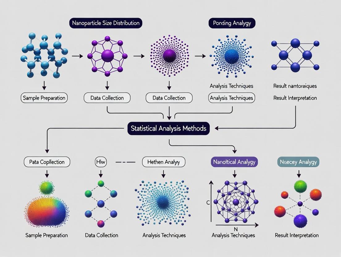
Nanoparticle Size Distribution Analysis: A Comprehensive Guide to Methods, Validation, and Clinical Translation
This article provides researchers, scientists, and drug development professionals with a detailed roadmap for analyzing nanoparticle size distribution.
Nanoparticle Size Distribution Analysis: A Comprehensive Guide to Methods, Validation, and Clinical Translation
Abstract
This article provides researchers, scientists, and drug development professionals with a detailed roadmap for analyzing nanoparticle size distribution. We begin by exploring the critical importance of accurate size characterization for drug delivery, stability, and biodistribution. We then systematically compare core methodologies, including Dynamic Light Scattering (DLS), Nanoparticle Tracking Analysis (NTA), and Electron Microscopy, detailing their practical application and data interpretation. The guide addresses common troubleshooting scenarios and optimization strategies to enhance measurement accuracy and reproducibility. Finally, we present a framework for method validation, cross-platform comparison, and selecting the optimal technique for regulatory submission and successful translation into biomedical and clinical research.
Why Size Matters: The Critical Role of Nanoparticle Size Distribution in Drug Delivery and Biodistribution
In the field of nanoparticle characterization for drug delivery and biomedical research, dynamic light scattering (DLS) is the primary technique for assessing size and distribution in suspension. This analysis is crucial within the broader thesis of Statistical analysis methods for nanoparticle size distribution research, as DLS data requires robust statistical interpretation. Three core metrics define this analysis: Hydrodynamic Diameter (Dh), the Polydispersity Index (PDI), and the Z-Average. Understanding their definition, interdependence, and the experimental protocols behind them is essential for accurate comparative evaluation of nanocarriers like liposomes, polymeric nanoparticles, and lipid nanoparticles (LNPs).
Key Metric Definitions and Interrelationships
- Hydrodynamic Diameter (Dh): The apparent diameter of a sphere that diffuses at the same rate as the measured particle. It includes the core particle, surface coatings, and the solvation layer. It is a intensity-weighted parameter derived from the diffusion coefficient via the Stokes-Einstein equation.
- Z-Average Diameter: The primary result of a DLS measurement, defined as the intensity-weighted mean hydrodynamic diameter derived from the autocorrelation function. It is most reliable for monodisperse samples (low PDI).
- Polydispersity Index (PDI): A dimensionless measure of the breadth of the size distribution, calculated from the cumulants analysis of the DLS data. A PDI < 0.05 indicates a highly monodisperse sample, 0.05–0.7 is moderately polydisperse, and >0.7 suggests a very broad distribution, invalidating the Z-average as a meaningful metric.
The logical and statistical relationship between data acquisition and these key metrics is outlined below.
DLS Data Analysis Pathway to Key Metrics
Comparative Performance: Liposomes vs. Polymeric NPs vs. LNPs
The following table summarizes typical DLS data for three common nanoparticle drug delivery systems, highlighting how the key metrics reflect formulation quality and stability. Data is synthesized from recent literature and standardized protocol comparisons.
Table 1: Key Metric Comparison for Nanocarrier Formulations
| Nanocarrier Type | Typical Z-Average (nm) | Typical PDI Range | Key Stability Insight (from Metrics) |
|---|---|---|---|
| Liposomes (PEGylated) | 80 – 120 nm | 0.05 – 0.15 | Low PDI indicates homogeneous, stable preparation. Z-average increases may indicate aggregation. |
| Polymeric NPs (PLGA) | 150 – 200 nm | 0.10 – 0.25 | Moderate PDI reflects batch variability. Z-average is sensitive to polymer MW and synthesis method. |
| Lipid Nanoparticles (LNP) | 70 – 100 nm | 0.05 – 0.20 | Critical for mRNA delivery. Low PDI essential for reproducible efficacy & safety. |
Experimental Protocols for DLS Measurement
A standardized DLS protocol is vital for valid metric comparison between different nanoparticle samples.
Protocol 1: Standard DLS Measurement for Nanocarrier Characterization
- Sample Preparation: Dilute the nanoparticle suspension in the appropriate filtered buffer (e.g., 1x PBS, 10 mM NaCl) to achieve a manufacturer-recommended scattering intensity. Typically, a 1:100 to 1:1000 dilution is used to avoid multiple scattering effects.
- Filtration: Filter the diluent through a 0.1 µm or 0.22 µm pore-size membrane syringe filter prior to dilution to remove dust.
- Equilibration: Load the diluted sample into a clean, disposable sizing cuvette. Allow it to equilibrate in the instrument at the set temperature (typically 25°C) for 120-180 seconds.
- Measurement Setup: Set the instrument parameters: detector angle (commonly 173° for backscatter), number of runs (≥ 10), and run duration (typically 10 seconds per run).
- Data Acquisition: Perform the measurement in triplicate for each independent sample batch.
- Data Analysis: Use the instrument software to perform cumulants analysis to obtain the Z-Average and PDI. Use distribution algorithms (e.g., Non-Negative Least Squares, NNLS) to generate the intensity-weighted hydrodynamic size distribution plot.
The Scientist's Toolkit: Essential Reagents & Materials
Table 2: Key Research Reagent Solutions for DLS Sample Preparation
| Item | Function in DLS Characterization |
|---|---|
| Filtered Buffer (e.g., PBS) | Provides a clean, dust-free dispersion medium with controlled ionic strength. |
| Syringe Filters (0.1/0.22 µm) | Removes particulate contaminants and dust from buffers and samples. |
| Disposable Size Cuvettes | Minimizes cross-contamination and prevents scratches that cause stray light. |
| Standard Latex Nanospheres | Used for instrument calibration and validation (e.g., 60 nm, 100 nm standards). |
| Temperature Controller | Essential for accurate measurements, as diffusion is temperature-dependent. |
Advanced Analysis: Size Distribution Deconvolution
The PDI indicates distribution width, but detailed multimodal analysis requires fitting the correlation data to size distribution models. The workflow for this advanced statistical analysis is shown below.
Statistical Workflow for Size Distribution Analysis
Conclusion: The Z-Average, PDI, and Hydrodynamic Diameter distribution are interdependent metrics that form the cornerstone of nanoparticle size statistics. For researchers comparing formulations, rigorous adherence to a detailed DLS protocol is non-negotiable. As shown in Table 1, these metrics provide immediate, comparative insights into formulation quality, batch consistency, and stability, guiding rational development in nanomedicine. Within the thesis of statistical analysis methods, they represent the first-order, model-dependent parameters upon which more advanced, model-free distribution analyses are built.
The Impact of Size on Drug Encapsulation, Release Kinetics, and Cellular Uptake
This guide provides a comparative analysis of nanoparticle (NP) size as a critical determinant in drug delivery system performance. Within the broader thesis context of Statistical analysis methods for nanoparticle size distribution research, this article underscores how precise size characterization is foundational to understanding encapsulation efficiency, release profiles, and biological interactions. The comparative data herein serves to inform the selection and optimization of nanocarriers for specific therapeutic applications.
Comparative Analysis: Size-Dependent Performance Metrics
Table 1: Impact of Polymeric Nanoparticle (PLGA) Size on Key Performance Parameters
| Size Range (nm) | Avg. Encapsulation Efficiency (%) | Dominant Release Mechanism | t1/2 Release (hours) | Primary Cellular Uptake Pathway | Relative Uptake Efficiency (vs. 100 nm control) |
|---|---|---|---|---|---|
| 50 - 70 | 68 ± 5 | Initial Burst | 12 ± 3 | Clathrin-mediated endocytosis | 0.9x |
| 80 - 120 | 92 ± 4 | Diffusion-controlled | 48 ± 6 | Clathrin-mediated endocytosis | 1.0x (Control) |
| 150 - 200 | 85 ± 6 | Diffusion/Erosion | 96 ± 12 | Caveolae-mediated endocytosis | 0.7x |
| 250 - 300 | 75 ± 8 | Bulk Erosion | 120 ± 18 | Macropinocytosis | 0.5x |
Table 2: Size-Dependent Organ Accumulation of Injected Nanoparticles (Passive Targeting)
| Size Range (nm) | Liver Accumulation (%ID/g) | Spleen Accumulation (%ID/g) | Tumor Accumulation (EPR Effect) (%ID/g) |
|---|---|---|---|
| < 10 | Low | Very Low | Low (Rapid renal clearance) |
| 50 - 100 | Moderate | Moderate | High |
| 150 - 200 | High | High | Moderate |
| > 500 | Very High | Very High | Low |
Experimental Protocols for Key Cited Studies
Protocol 1: Determining Size-Dependent Encapsulation Efficiency
- Objective: To quantify the loading of a hydrophilic model drug (e.g., Doxorubicin HCl) into PLGA NPs of varying sizes.
- Methodology:
- Prepare PLGA NPs using the nanoprecipitation (for small NPs) and single/double emulsion-solvent evaporation (for larger NPs) methods to obtain distinct size populations.
- Purify nanoparticles via centrifugation at speeds calibrated for each size range.
- Lyse an aliquot of NPs in DMSO to release encapsulated drug.
- Measure drug concentration via HPLC or fluorescence spectrometry against a standard curve.
- Calculate Encapsulation Efficiency (EE%) = (Mass of drug in NPs / Total mass of drug used) x 100.
- Correlate EE% with NP size measured by Dynamic Light Scattering (DLS).
Protocol 2: In Vitro Release Kinetics Profiling
- Objective: To characterize the drug release profile from NPs of different sizes under physiological conditions (PBS, pH 7.4, 37°C).
- Methodology:
- Place a known quantity of drug-loaded NPs into a dialysis membrane (MWCO appropriate for the drug).
- Immerse the membrane in release medium under gentle agitation.
- At predetermined time points, withdraw a sample of the external medium and replace with fresh buffer.
- Quantify the released drug using a calibrated analytical method (UV-Vis, HPLC).
- Fit release data to kinetic models (Zero-order, First-order, Higuchi, Korsmeyer-Peppas) to determine the dominant mechanism.
Protocol 3: Quantifying Cellular Uptake via Flow Cytometry
- Objective: To compare the uptake efficiency and kinetics of fluorescently labeled NPs of different sizes by a target cell line (e.g., HeLa cells).
- Methodology:
- Incubate cells with Cy5-labeled NPs of varying sizes (50, 100, 200 nm) at equal particle number or total surface area concentrations.
- At set time points (e.g., 1, 2, 4 h), wash cells thoroughly to remove non-internalized NPs.
- Trypsinize cells and analyze cell-associated fluorescence using a flow cytometer.
- Use inhibitors (e.g., chlorpromazine for clathrin, genistein for caveolae) to delineate the primary endocytic pathway for each size fraction.
Visualizations
Diagram 1: Cellular Uptake Pathways by NP Size
Diagram 2: Workflow for Size-Dependent Performance Analysis
The Scientist's Toolkit: Key Research Reagent Solutions
Table 3: Essential Materials for Nanoparticle Size-Performance Studies
| Reagent / Material | Function in Research | Key Consideration for Size Studies |
|---|---|---|
| PLGA (50:50, acid-terminated) | Biodegradable polymer for NP matrix. | Molecular weight determines achievable size range and degradation rate. |
| PVA (Polyvinyl Alcohol) | Common surfactant/stabilizer in emulsion methods. | Concentration directly impacts final NP size and polydispersity. |
| Dialysis Membranes (various MWCO) | Contain NPs during release studies. | MWCO must allow drug diffusion but retain NPs of all sizes tested. |
| Fluorescent Dye (e.g., Cy5, Coumarin 6) | Label NPs for cellular tracking. | Ensure dye is entrapped, not surface-adsorbed, to avoid size-biased signals. |
| Endocytic Pathway Inhibitors (Chlorpromazine, Genistein, Amiloride) | Mechanistic studies of cellular uptake. | Use at non-cytotoxic concentrations to confirm size-dependent pathways. |
| Dynamic Light Scattering (DLS) Instrument | Measures hydrodynamic diameter and PDI. | Critical for baseline characterization; complement with EM for shape. |
| Density Gradient Media (e.g., Iodixanol) | Purifies NPs by size via ultracentrifugation. | Enables isolation of monodisperse fractions from a polydisperse sample. |
This comparison guide, framed within a broader thesis on statistical analysis methods for nanoparticle size distribution research, objectively analyzes how nanoparticle size dictates key pharmacokinetic parameters. For researchers and drug development professionals, understanding these size-dependent relationships is critical for rational nanocarrier design.
Comparative Analysis: Size vs. Pharmacokinetic Parameters
The following table synthesizes experimental data from recent studies, illustrating the direct impact of nanoparticle hydrodynamic diameter on pharmacokinetic behavior.
Table 1: Comparative Pharmacokinetics of Polymeric Nanoparticles by Size
| Hydrodynamic Diameter (nm) | Circulation Half-life (hr) | Tumor Accumulation (%ID/g)* | Primary Clearance Route | Depth of Tissue Penetration (µm from vessel) |
|---|---|---|---|---|
| 10-20 | 1.2 - 4.5 | 1.8 - 3.2 | Renal Filtration | 80 - 120 |
| 30-50 | 8 - 15 | 4.5 - 6.8 | MPS (Liver/Spleen) | 40 - 70 |
| 60-100 | 15 - 24 | 7.5 - 10.2 | MPS (Liver/Spleen) | 20 - 40 |
| 120-200 | 6 - 12 | 3.0 - 5.1 | MPS (Rapid Sequestration) | < 20 |
%ID/g: Percentage of Injected Dose per gram of tumor tissue. MPS: Mononuclear Phagocyte System. *Data compiled from PEGylated PLGA and liposomal nanoparticle studies in murine xenograft models (2021-2023).
Experimental Protocols for Key Studies
Protocol: Measuring Circulation Half-Life
- Objective: Quantify blood residence time of nanoparticles of varying sizes.
- Materials: Cy5.5 or DiR fluorescently labeled nanoparticles (10nm, 50nm, 100nm), IVIS imaging system or fluorescence spectrophotometer, murine model.
- Method:
- Inject nanoparticles intravenously via tail vein (n=5 per size group).
- Collect blood samples (5 µL) from the retro-orbital plexus at time points: 1 min, 30 min, 2 hr, 6 hr, 12 hr, 24 hr, 48 hr.
- Lyse blood samples in 1% Triton X-100/PBS.
- Measure fluorescence intensity (Ex/Em: 675/720 nm for DiR).
- Plot fluorescence intensity vs. time. Fit data to a two-compartment pharmacokinetic model to calculate alpha and beta half-lives.
Protocol: Evaluating EPR Effect and Tumor Accumulation
- Objective: Correlate nanoparticle size with passive tumor targeting via the Enhanced Permeability and Retention effect.
- Materials: Near-infrared (NIR) dye-labeled nanoparticles, subcutaneous tumor-bearing mice (e.g., 4T1 or HT-29 xenografts), IVIS Spectrum imaging system, tissue homogenizer.
- Method:
- Administer a standardized dose (5 mg/kg nanoparticle weight) intravenously.
- Perform in vivo whole-body imaging at 24h and 48h post-injection.
- Euthanize animals at 48h. Excise tumors and major organs (liver, spleen, kidney, lungs, heart).
- Image excised organs ex vivo to quantify biodistribution.
- Homogenize tissues and quantify fluorescence to calculate %ID/g.
Protocol: Assessing Tissue and Tumor Penetration
- Objective: Visualize and measure the depth of nanoparticle diffusion from tumor blood vessels.
- Materials: FITC or Alexa Fluor-labeled nanoparticles, tumor-bearing mouse model, anti-CD31 antibody (vascular marker), confocal microscopy.
- Method:
- Inject nanoparticles intravenously.
- At a set time (e.g., 24h), harvest tumors, embed in OCT, and cryosection.
- Fix and stain sections with anti-CD31 antibody and a secondary antibody (e.g., Alexa Fluor 647).
- Image using a confocal microscope with z-stacking capability.
- Use image analysis software (e.g., ImageJ) to measure the distance of nanoparticle fluorescence (green) from CD31-positive vessel walls (red) across multiple fields.
Visualizing Size-Dependent Pharmacokinetic Relationships
Diagram 1: Nanoparticle Size Dictates PK Fate
Diagram 2: Experimental PK Analysis Workflow
The Scientist's Toolkit: Research Reagent Solutions
Table 2: Essential Materials for Nanoparticle Pharmacokinetics Research
| Reagent / Material | Function in Research | Key Consideration |
|---|---|---|
| PEGylated PLGA | Biodegradable polymer core for drug encapsulation; PEG coating ("stealth" layer) extends circulation. | PEG molecular weight (2k-5k Da) and density critically impact half-life. |
| Lipids (DSPC, Cholesterol, PEG-DSPE) | Components for forming stable, size-tunable liposomal nanoparticles. | Ratio determines membrane rigidity, affecting drug release and stability. |
| Near-Infrared (NIR) Dyes (DiR, Cy7.5) | Fluorescent labels for non-invasive, quantitative in vivo and ex vivo imaging of biodistribution. | Must be stably encapsulated/ conjugated to prevent dye leakage and false signals. |
| Size Exclusion Chromatography (SEC) Columns | Purify nanoparticles from unencapsulated drug/dye and fractionate by hydrodynamic size. | Essential for obtaining monodisperse samples for clear size-PK correlations. |
| Dynamic Light Scattering (DLS) / NTA Instrument | Measure hydrodynamic diameter, polydispersity index (PDI), and concentration of nanoparticles. | PDI < 0.2 is ideal for interpreting clear size-dependent trends. |
| Anti-CD31 Antibody | Marker for endothelial cells; used to stain blood vessels in tumor sections for penetration analysis. | Enables quantification of nanoparticle distance from vasculature via immunofluorescence. |
| Matrix for Tissue Embedding (OCT) | Optimal Cutting Temperature compound for freezing and preparing tumor tissue for cryosectioning. | Preserves tissue morphology and fluorescence signals for microscopy. |
Size Stability as a Predictor of Formulation Shelf-Life and In Vivo Performance
Within the broader thesis on advanced statistical analysis methods for nanoparticle size distribution research, this guide examines the critical role of size stability. Particle size and its distribution (PSD) are critical quality attributes (CQAs) for nanomedicines, directly influencing shelf-life, biodistribution, and therapeutic efficacy. This guide compares the performance of lipid nanoparticles (LNPs), polymeric nanoparticles (PLGA), and inorganic nanoparticles (silica) in maintaining size stability under accelerated storage and simulated biological conditions.
Experimental Protocols
1. Accelerated Stability Testing Protocol:
- Sample Preparation: Dilute nanoparticle formulations (1 mg/mL) in respective formulation buffers (e.g., citrate buffer for LNPs, PBS for PLGA).
- Storage Conditions: Incubate samples at 4°C (refrigeration), 25°C/60% RH (room temperature), and 40°C/75% RH (accelerated) for 0, 1, 3, and 6 months.
- Size Analysis: At each time point, analyze samples in triplicate via Dynamic Light Scattering (DLS) using a Zetasizer Nano ZS. Perform measurements at a 173° backscatter angle after equilibration at 25°C for 120 seconds.
- Data Analysis: Report Z-average (d.nm) and Polydispersity Index (PDI). Statistical significance of size change over time is determined by Repeated Measures ANOVA (p<0.05).
2. In Vitro Serum Stability Protocol:
- Incubation: Mix nanoparticle suspension (1 mL) with 50% (v/v) fetal bovine serum (FBS) in DPBS.
- Time Course: Incubate at 37°C with gentle agitation. Subsamples are taken at 0, 1, 4, 8, and 24 hours.
- Size Measurement: Dilute subsamples 1:10 in DPBS and measure immediately via DLS as above. A >20% increase in Z-average is considered indicative of significant aggregation or protein corona-driven growth.
Comparative Performance Data
Table 1: Accelerated Size Stability (40°C) Over 6 Months
| Formulation Type | Initial Z-avg (nm) | 6-Month Z-avg (nm) | Δ Size (%) | Final PDI | Statistical Significance (p vs. Baseline) |
|---|---|---|---|---|---|
| LNP (siRNA) | 85.2 ± 3.1 | 92.5 ± 5.4 | +8.6% | 0.12 | 0.045 |
| PLGA (PEGylated) | 152.7 ± 8.5 | 210.3 ± 25.1 | +37.7% | 0.31 | <0.001 |
| Mesoporous Silica | 99.5 ± 2.2 | 101.8 ± 3.7 | +2.3% | 0.08 | 0.215 |
Table 2: In Vitro Serum Stability (24-hour Incubation)
| Formulation Type | Size at 0h (nm) | Size at 24h (nm) | Δ Size (%) | Inferred Protein Corona Effect |
|---|---|---|---|---|
| LNP (siRNA) | 85.2 ± 3.1 | 127.5 ± 10.2 | +49.6% | High |
| PLGA (PEGylated) | 152.7 ± 8.5 | 168.4 ± 12.7 | +10.3% | Moderate (PEG shield) |
| Mesoporous Silica | 99.5 ± 2.2 | 185.6 ± 15.8 | +86.5% | Very High |
Table 3: Correlation with In Vivo Performance (Rodent Study)
| Formulation Type | Shelf-Life Δ Size (%) | Liver Accumulation (%ID/g)* | Tumor Targeting (T/L Ratio)* | Inferred Stability-Performance Link |
|---|---|---|---|---|
| LNP (siRNA) | +8.6% | 65.2 ± 4.1 | 0.2 | High liver targeting preserved. |
| PLGA (PEGylated) | +37.7% | 18.5 ± 3.2 | 3.5 | Instability may reduce circulation. |
| Mesoporous Silica | +2.3% | 42.1 ± 5.7 | 1.1 | Stable size, but high serum aggregation alters fate. |
*%ID/g: Percentage of Injected Dose per gram of tissue; T/L Ratio: Tumor-to-Liver ratio.
Visualizations
Diagram Title: Stability Study & Statistical Analysis Workflow
Diagram Title: Impact of Size Instability on Product Performance
The Scientist's Toolkit: Research Reagent Solutions
| Item | Function in Size Stability Research |
|---|---|
| Zetasizer Nano ZS (DLS) | Measures hydrodynamic diameter, PDI, and zeta potential of nanoparticles in suspension. |
| NanoTrack Analysis (NTA) | Provides particle concentration and visualizes size distribution profile of polydisperse samples. |
| HPLC-SEC | Separates and quantifies free molecular components (e.g., degraded lipids/polymers) from intact nanoparticles. |
| Stability Chambers | Provide controlled temperature and humidity for ICH-compliant accelerated stability studies. |
| Fetal Bovine Serum (FBS) | Used in in vitro incubation studies to simulate protein corona formation and biological fluid stability. |
| PBS & Formulation Buffers | Standard media for dilution and storage, testing the effect of ionic strength and pH on stability. |
| Statistical Software (e.g., R, SPSS) | For performing ANOVA, PCA, and modeling of size distribution data trends over time. |
Accurate nanoparticle size characterization is a cornerstone of nanomedicine development, directly mandated by regulatory agencies. This comparison guide evaluates key analytical techniques in the context of U.S. Food and Drug Administration (FDA) and European Medicines Agency (EMA) guidelines, framed within a thesis on statistical analysis methods for nanoparticle size distribution research.
Comparison of Regulatory-Recommended Size Characterization Techniques
Table 1: Comparative Analysis of Primary Size Characterization Techniques
| Technique | Measured Parameter (Regulatory Focus) | Typical Size Range | Key Statistical Output | EMA/FDA Guideline Mention |
|---|---|---|---|---|
| Dynamic Light Scattering (DLS) | Hydrodynamic diameter (Z-avg), PDI | 1 nm - 10 µm | Intensity-weighted mean, Polydispersity Index (PDI) | Extensively referenced for primary particle size distribution. |
| Electron Microscopy (TEM/SEM) | Primary particle diameter, Morphology | 0.5 nm - 10s µm | Number-weighted distribution, Visual confirmation | Required for complementary, orthogonal analysis (e.g., particle shape). |
| Asymmetric Flow Field-Flow Fractionation (AF4) | Separated hydrodynamic diameter | 1 nm - 1 µm | Fractionated size distributions, Resolution of sub-populations | Recommended for complex, polydisperse systems (e.g., protein-nanoparticle complexes). |
| Nanoparticle Tracking Analysis (NTA) | Particle concentration, Size distribution | 30 nm - 2 µm | Number-weighted distribution, Concentration (particles/mL) | Cited for concentration analysis and detecting sub-micron particulates. |
Detailed Experimental Protocols for Cited Techniques
1. Protocol: Dynamic Light Scattering (DLS) for Polydispersity Index (PDI) Measurement
- Sample Preparation: Dilute nanomedicine formulation in appropriate, filtered (0.1 µm) buffer to achieve a recommended scattering intensity. Avoid over-dilution.
- Instrumentation: DLS instrument equipped with a 633 nm laser and back-scatter detector.
- Procedure: Load sample into a disposable cuvette. Equilibrate to 25°C. Perform a minimum of 12 measurements per sample. The autocorrelation function is analyzed using the cumulants method (ISO 22412).
- Data Analysis: The software reports the Z-average (intensity-weighted mean hydrodynamic diameter) and the Polydispersity Index (PDI). A PDI < 0.1 indicates a monodisperse sample; >0.3 suggests broad polydispersity, per regulatory expectations.
2. Protocol: Transmission Electron Microscopy (TEM) for Orthogonal Size Verification
- Sample Preparation: Apply 5 µL of diluted sample onto a carbon-coated copper grid. After 1 minute, blot excess and negatively stain with 1% uranyl acetate for 30 seconds. Air dry.
- Instrumentation: Transmission Electron Microscope operated at 80-100 kV.
- Procedure: Image at multiple magnifications (e.g., 20,000x to 100,000x). Capture images from multiple grid squares.
- Data Analysis: Use image analysis software (e.g., ImageJ) to manually or automatically measure the diameter of a statistically relevant number of particles (n>300). Generate a number-weighted size distribution histogram and calculate mean, mode, and standard deviation.
Visualization: Regulatory Size Characterization Workflow
Diagram Title: Multi-Method Size Analysis for Regulatory Submission
The Scientist's Toolkit: Key Research Reagent Solutions
Table 2: Essential Materials for Regulatory Size Characterization
| Item | Function | Example Application |
|---|---|---|
| NIST Traceable Size Standards | Calibration and validation of instrumentation (DLS, NTA). | Verifying accuracy of reported hydrodynamic diameters. |
| Filtered, Particle-Free Buffers | Sample dilution medium to minimize background scattering. | Preparing DLS/NTA samples to avoid dust interference. |
| Ultrafiltration/Dialysis Membranes | Buffer exchange to match recommended dispersant for measurement. | Replacing formulation buffer with a standard ionic strength buffer. |
| Carbon-Coated TEM Grids | Support film for high-resolution electron microscopy. | Preparing samples for orthogonal size and morphology analysis. |
| Negative Stains (e.g., Uranyl Acetate) | Enhance contrast in TEM imaging. | Visualizing lipid nanoparticles or polymeric micelles. |
| AF4 Carrier Liquid & Membranes | Mobile phase and separation channel for fractionation. | Resolving free drug from nanoparticle-bound drug in a complex formulation. |
Core Techniques in Practice: A Deep Dive into DLS, NTA, SEM/TEM, and RES for Size Analysis
Within the framework of a thesis on Statistical analysis methods for nanoparticle size distribution research, Dynamic Light Scattering (DLS) stands as a cornerstone technique. It provides a rapid, non-invasive method for determining the hydrodynamic size of nanoparticles and macromolecules in suspension. This guide compares the performance of a standard DLS system with key alternatives, supported by experimental data relevant to researchers, scientists, and drug development professionals.
Principles and Setup
DLS measures the Brownian motion of particles suspended in a liquid by analyzing the temporal fluctuations in the intensity of scattered laser light. Smaller particles move faster, causing rapid intensity fluctuations, while larger particles move slower, causing slower fluctuations. An autocorrelation function is applied to the scattered light signal, the decay rate of which is used to calculate the diffusion coefficient and, via the Stokes-Einstein equation, the hydrodynamic diameter.
A standard DLS setup consists of: a monochromatic laser light source, a temperature-controlled sample cell, a high-sensitivity detector (typically an avalanche photodiode or PMT), and a digital autocorrelator for real-time signal processing.
Comparison of DLS Performance with Alternative Techniques
This analysis compares a standard bench-top DLS instrument (e.g., Malvern Panalytical Zetasizer Ultra) against two primary alternatives for nanoparticle sizing: Nanoparticle Tracking Analysis (NTA) and Transmission Electron Microscopy (TEM).
Table 1: Comparative Technique Overview
| Feature | Dynamic Light Scattering (DLS) | Nanoparticle Tracking Analysis (NTA) | Transmission Electron Microscopy (TEM) |
|---|---|---|---|
| Measured Parameter | Hydrodynamic diameter | Hydrodynamic diameter (from video) | Primary particle diameter (dry state) |
| Size Range | ~0.3 nm – 10 µm | ~10 nm – 2 µm | ~1 nm – No upper limit |
| Concentration Range | 0.1 mg/mL – 40% w/v (varies) | 107 – 109 particles/mL | N/A (dry sample) |
| Sample State | Liquid suspension (minimal prep) | Liquid suspension (dilution often needed) | Dry, on grid (extensive prep) |
| Output | Intensity-weighted size distribution; PDI | Number-weighted size distribution; concentration estimate | Number-based, high-resolution image |
| Key Statistical Strength | Robust for monomodal, stable samples. Provides Polydispersity Index (PDI). | Resolves polydisperse/multimodal samples better than DLS. Direct particle counting. | "Gold standard" for precise primary size and morphology. |
| Key Statistical Limitation | Intensity-weighting biases signal toward larger particles; difficult for highly polydisperse samples. | Lower size detection limit; statistical sampling depends on counted particles. | Poor statistics if few particles counted; not representative of native state. |
| Throughput | Very High (seconds/minutes) | Medium (minutes per analysis) | Low (sample prep + imaging) |
Experimental Protocol 1: Comparative Sizing of a Polydisperse Lipid Nanoparticle (LNP) Formulation
- Sample: A research-grade LNP formulation for mRNA delivery.
- DLS Protocol: 20 µL of sample was loaded into a quartz cuvette. Measurements were performed at 25°C with an equilibration time of 120 seconds. Size was measured via NIBS (Non-Invasive Back-Scatter) optics at a 173° detection angle. A minimum of 3 runs were performed. Data analyzed using cumulants method (for Z-average and PDI) and general purpose (multiple narrow modes) algorithm.
- NTA Protocol: Sample was diluted 1:50,000 in filtered PBS to achieve optimal particle concentration. Videos of 60 seconds duration were captured using a 488 nm laser. Three videos were analyzed per sample with camera level and detection threshold held constant.
- TEM Protocol: 5 µL of sample was deposited onto a carbon-coated copper grid, negatively stained with 2% uranyl acetate, and air-dried. Images were acquired at 80kV. Diameters of 200 individual particles were measured manually from the images.
Table 2: Experimental Data for Polydisperse LNP Sample
| Technique | Mode 1 (nm) | Mode 2 (nm) | Mode 3 (nm) | Z-Average / Mean (nm) | PDI / % CV | Key Observation |
|---|---|---|---|---|---|---|
| DLS (Intensity) | 78.4 ± 2.1 | 152.3 ± 8.5 | Not resolved | 122.6 ± 5.2 | 0.21 ± 0.03 | Bimodal distribution detected, but intensity of larger particles dominates. |
| NTA (Number) | 72.5 ± 3.8 | 95.2 ± 4.1 | 155.0 ± 12.0 | 98.4 ± 4.5 | 28% (CV) | Trimodal distribution clearly resolved. Reveals predominant population (~70 nm) missed by DLS intensity bias. |
| TEM (Number) | 68.2 ± 11.5 | - | - | 68.2 ± 11.5 | 17% (CV) | Confirms primary particle core size. Does not reflect hydrodynamic size or presence of aggregates in solution. |
Analysis of Intensity-Based Distribution
DLS reports an intensity-weighted size distribution, derived from the fitted autocorrelation function. The intensity of scattered light is proportional to the sixth power of the particle diameter (for Rayleigh scatterers). Consequently, a 100 nm particle scatters ~1,000,000 times more light than a 10 nm particle. This means the signal is overwhelmingly dominated by larger particles/aggregates, which is both a strength (sensitive to aggregates) and a weakness (can mask a majority population of small particles).
The primary statistical output is the Z-average diameter (the intensity-weighted harmonic mean) and the Polydispersity Index (PDI), a dimensionless measure of distribution breadth from the cumulants analysis. A PDI < 0.05 is monodisperse; >0.7 is very broad.
Diagram Title: DLS Data Processing Workflow
The Scientist's Toolkit: Research Reagent Solutions
Table 3: Essential Materials for DLS Experiments
| Item | Function & Importance |
|---|---|
| Standard Latex Nanosphere (e.g., NIST-traceable 100 nm) | Critical instrument validation and performance qualification (PQ). Provides a known reference for size and dispersity. |
| High-Quality Disposable Cuvettes (e.g., UV-transparent, low fluorescence) | Minimizes dust contamination and ensures consistent light path. Disposable cuvettes prevent cross-contamination. |
| Syringe Filters (0.02 µm or 0.1 µm pore size, Anotop/Al2O3) | For ultra-cleaning solvents (toluene, water) used to rinse cuvettes and for filtering buffers to eliminate dust. |
| Optically Clear, Filtered Buffers (e.g., PBS, 10 mM NaCl) | Provides a clean, low-scattering background medium. Must be filtered through 0.02 µm filter immediately before use. |
| Temperature-Controlled Sample Holder (Peltier) | Essential for accurate DLS, as diffusion coefficient is temperature-dependent. Allows stability studies. |
| Intensity Calibration Standard (e.g., toluene) | Verifies detector sensitivity and laser power, ensuring inter-instrument comparability. |
For the statistical analysis of nanoparticle size distributions, DLS offers unparalleled speed and ease-of-use for preliminary characterization and stability assessment of primarily monomodal samples. Its intensity-weighted statistics are highly sensitive to aggregates. However, as demonstrated, for polydisperse systems common in drug development (like LNPs), DLS can obscure a true number-weighted distribution. NTA provides a valuable complementary, number-based statistical view, while TEM offers definitive primary size statistics in a non-native state. A robust thesis in this field should leverage DLS as a primary tool for rapid screening and stability but must incorporate orthogonal, number-based techniques like NTA to deconvolute complex, polydisperse populations accurately.
Within the framework of a thesis on statistical analysis methods for nanoparticle size distribution research, evaluating the core analytical techniques is paramount. This guide objectively compares Nanoparticle Tracking Analysis (NTA) with two major alternatives: Dynamic Light Scattering (DLS) and Tunable Resistive Pulse Sensing (TRPS).
Performance Comparison: NTA vs. DLS vs. TRPS
The following table summarizes key performance metrics based on published experimental data and technical specifications.
Table 1: Comparative Performance of Nanoparticle Sizing and Concentration Techniques
| Feature / Metric | Nanoparticle Tracking Analysis (NTA) | Dynamic Light Scattering (DLS) | Tunable Resistive Pulse Sensing (TRPS) |
|---|---|---|---|
| Core Principle | Brownian motion tracking & particle-by-particle sizing | Fluctuation of scattered light intensity from an ensemble | Resistive pulse magnitude as particles pass through a tunable pore |
| Size Range (typical) | 10 nm – 2000 nm | 0.3 nm – 10 μm | 50 nm – 10 μm |
| Concentration Range | 10⁶ – 10⁹ particles/mL (ideal) | Not a direct measure; requires high concentration for signal | 10⁷ – 10¹² particles/mL |
| Resolution of Polydisperse Samples | High (individual particle measurement) | Low (weighted intensity distributions obscure minorities) | High (individual particle measurement) |
| Primary Output | Particle size distribution, concentration, scattering intensity | Hydrodynamic diameter (Z-average), Polydispersity Index (PDI) | Particle size distribution, concentration, zeta potential (via bias) |
| Sample Volume Required | ~0.3 mL | ~12 μL – 1 mL | ~70 μL |
| Key Strength | Direct visualization, sizing in complex media, detection of sub-populations | Speed, ease of use, robust for monomodal, stable samples | High-precision sizing, concentration, and surface charge analysis |
| Key Limitation | Lower size limit ~10-30nm, sensitive to sample cleanliness | Poor resolution of polydisperse/multimodal samples | Pore clogging, requires ionic solution, single-particle type per run |
Supporting Experimental Data & Protocols
Experiment 1: Resolving a Mixture of Monodisperse Nanoparticles
- Objective: To compare the ability of each technique to resolve a binary mixture of 100 nm and 200 nm polystyrene nanoparticles.
- Protocol:
- Prepare separate stock suspensions of 100 nm and 200 nm polystyrene nanospheres.
- Mix the suspensions at a 1:1 particle number ratio. Dilute each sample to the optimal concentration for each instrument.
- NTA Protocol: Inject sample into the viewing chamber. Capture sixty 30-second videos. Analyze with constant detection threshold to derive size distribution.
- DLS Protocol: Load sample into a disposable cuvette. Perform minimum of 10 measurements at a fixed angle (e.g., 173°). Analyze correlation function to derive intensity-weighted distribution.
- TRPS Protocol: Stretch a 400 nm nanopore membrane. Calibrate pore in a known standard. Measure sample at a constant pressure and voltage.
- Results Summary:
Table 2: Measured Size Distribution Peaks for a 100 nm & 200 nm Mixture
| Technique | Reported Peak 1 (nm) | Reported Peak 2 (nm) | Notes |
|---|---|---|---|
| NTA | 102 ± 12 | 198 ± 18 | Two distinct populations clearly resolved in number-weighted distribution. |
| DLS | ~140 (Z-average: 156 nm) | Not resolved | Reported a single broad peak with a high PDI (>0.3). |
| TRPS | 105 ± 8 | 203 ± 11 | Two distinct populations resolved with high precision. |
Experiment 2: Quantifying Concentration of Extracellular Vesicles (EVs)
- Objective: To assess the accuracy of concentration measurement against a known reference (e.g., silica beads).
- Protocol:
- Isolate EVs from cell culture supernatant via ultracentrifugation.
- Spike the EV sample with a known concentration of 150 nm silica microspheres (e.g., 2.0 x 10⁸ particles/mL).
- Analyze the spiked sample via NTA and TRPS. DLS is omitted as it does not provide direct concentration.
- Measure the concentration of the silica bead spike and compare to the known value.
- Results Summary:
Table 3: Recovery of Silica Bead Spike Concentration in an EV Sample
| Technique | Known Spike Conc. (particles/mL) | Measured Conc. (particles/mL) | % Recovery |
|---|---|---|---|
| NTA | 2.0 x 10⁸ | 1.7 x 10⁸ ± 0.2 x 10⁸ | 85% |
| TRPS | 2.0 x 10⁸ | 1.9 x 10⁸ ± 0.1 x 10⁸ | 95% |
Visualization of Experimental Workflow
Diagram Title: Nanoparticle Tracking Analysis (NTA) Experimental Workflow
Diagram Title: Decision Logic for Selecting a Nanoparticle Analysis Technique
The Scientist's Toolkit: Essential Research Reagent Solutions
Table 4: Key Materials for Nanoparticle Characterization Experiments
| Item | Function & Importance |
|---|---|
| Size Calibration Standards (e.g., 100 nm polystyrene beads) | Essential for validating instrument accuracy and performing daily quality control checks. |
| Filtered PBS or Saline Buffer (0.1 μm filtered) | Provides a clean, particle-free diluent to bring samples into the ideal concentration range. |
| Syringe Filters (e.g., 0.22 μm PES) | Critical for final sample cleaning to remove airborne or packaging-derived contaminants before NTA/TRPS. |
| Disposable Cuvettes/Capillaries (Instrument-specific) | Ensure no cross-contamination between samples, vital for concentration measurements. |
| Nanopore Membranes (for TRPS) | Consumable sensing element; different pore sizes are selected to match the expected particle size. |
| Standardized Silica Beads | Used as an internal quantitative reference for concentration measurements in complex biological samples. |
Within the broader thesis on Statistical analysis methods for nanoparticle size distribution research, direct imaging via electron microscopy (EM) remains the foundational technique for obtaining primary, particle-by-particle data on morphology and size. This guide compares the performance of Scanning Electron Microscopy (SEM) and Transmission Electron Microscopy (TEM) in this critical role, providing a framework for selecting the appropriate tool based on research objectives.
Performance Comparison: SEM vs. TEM for Nanoparticle Characterization
The choice between SEM and TEM involves trade-offs between resolution, analytical capabilities, sample requirements, and throughput. The following table summarizes the core performance metrics based on standard experimental configurations.
Table 1: Direct Performance Comparison of SEM and TEM for Nanoparticle Analysis
| Feature | Scanning Electron Microscopy (SEM) | Transmission Electron Microscopy (TEM) |
|---|---|---|
| Primary Imaging Mechanism | Scattered electrons from surface. | Transmitted electrons through specimen. |
| Typical Resolution | 0.5 nm to 3 nm (high-end). | 0.05 nm to 0.2 nm (high-resolution). |
| Optimal Size Range | > 10 nm (reliable for statistics). | 0.5 nm to 500 nm. |
| Depth of Field | Very High. | Low to Moderate. |
| Sample Preparation | Moderate (conductive coating often required). | High (ultra-thin sections or dispersion on grid). |
| Information Gained | 3D surface topography, agglomerate state. | 2D projection of internal structure, crystallography, lattice fringes. |
| Throughput for Statistics | High (automated stage, large FOV). | Low (manual targeting, smaller FOV). |
| Quantitative Analysis | Excellent for primary particle size and shape from well-dispersed samples. | Excellent for primary/core size, shell thickness, and crystallite size. |
| Supporting Data | Energy Dispersive X-ray Spectroscopy (EDS) for elemental composition. | EDS, Electron Energy Loss Spectroscopy (EELS), Selected Area Electron Diffraction (SAED). |
Experimental Protocols for Size Distribution Analysis
Accurate statistical analysis requires rigorous and reproducible sample preparation and imaging protocols.
Protocol 1: TEM Sample Preparation for Lipid Nanoparticles (LNPs)
- Dilution: Dilute the LNP formulation 1:100 in filtered, particle-free deionized water or an appropriate buffer to minimize aggregation on the grid.
- Negative Staining: Apply 5-10 µL of diluted sample onto a glow-discharged, carbon-coated copper grid. After 60 seconds, wick away excess liquid with filter paper.
- Staining: Immediately apply 5-10 µL of 2% aqueous uranyl acetate stain. Wick away after 30-45 seconds.
- Drying: Allow the grid to air-dry completely in a clean, dust-free environment.
- Imaging: Acquire images at magnifications of 40,000x to 100,000x at multiple, non-overlapping grid squares to avoid sampling bias.
Protocol 2: High-Throughput SEM for Agglomerate Analysis
- Sample Mounting: Dilute nanoparticle powder or suspension in an appropriate solvent (e.g., ethanol). Sonicate for 5-10 minutes. Deposit a droplet onto a silicon wafer and let it dry.
- Conductive Coating: Sputter-coat the sample with a 5-10 nm layer of Iridium or Gold/Palladium to prevent charging.
- Automated Imaging: Use the SEM’s automated stage and image capture software. Define a grid pattern over the wafer surface. Acquire 50-100 images at a set magnification (e.g., 50,000x) with consistent brightness/contrast settings.
- Data Extraction: Use image analysis software (e.g., ImageJ, proprietary tools) to perform thresholding, particle identification, and measurement of Feret’s diameter or equivalent circular diameter for each particle.
Visualization of Workflow and Data Integration
Workflow for Statistical Size Analysis from EM Images
Relationship Between EM Data and Other Sizing Techniques
The Scientist's Toolkit: Essential Research Reagents & Materials
Table 2: Key Research Reagent Solutions for EM Nanoparticle Analysis
| Item | Function | Example/Note |
|---|---|---|
| Carbon-Coated TEM Grids | Provide an ultra-thin, electron-transparent, and inert support film for nanoparticles. | Copper, gold, or nickel grids (200-400 mesh). |
| Glow Discharge System | Makes the grid surface hydrophilic, ensuring even dispersion of aqueous samples. | Critical for preventing nanoparticle aggregation on the grid. |
| Negative Stain (e.g., Uranyl Acetate) | Surrounds particles, increasing contrast by scattering electrons; reveals outline and some surface features. | 1-2% aqueous solution. Handle as radioactive waste. |
| Conductive Sputter Coater | Applies a thin metal layer (Ir, Au/Pd) to non-conductive samples for SEM, preventing charging artifacts. | Iridium provides finer grain size for high-resolution SEM. |
| Silicon Wafer Substrates | Provide an ultra-smooth, conductive surface for mounting nanoparticles for SEM. | Superior to aluminum stubs for high-magnification imaging. |
| Particle-Free Solvents | For sample dilution and cleaning to avoid introduction of background particulate contamination. | Filtered ethanol, isopropanol, or deionized water (0.02 µm filter). |
| Reference Nanoparticle Standards | Calibrate microscope magnification and validate image analysis protocols. | Gold nanoparticles (e.g., 10 nm, 30 nm, 100 nm). |
Resonant Mass Measurement (RMM) and Tunable Resistive Pulse Sensing (TRPS) for High-Resolution Distributions
This comparison guide objectively evaluates Resonant Mass Measurement (RMM) and Tunable Resistive Pulse Sensing (TRPS) within the context of a thesis on statistical analysis methods for nanoparticle characterization. Both techniques provide high-resolution, single-particle size distributions critical for rigorous statistical analysis in nanoparticle research and drug development.
Principle of Operation Comparison
RMM measures the change in resonant frequency of a microfluidic cantilever as a particle passes through, yielding buoyant mass. TRPS measures the transient change in ionic current (resistive pulse) as a particle passes through a tunable nanopore, yielding size based on particle volume displacement.
Title: Operational Workflow of RMM vs. TRPS
Comparative Performance Data
Table 1: Key Technical Parameter Comparison
| Parameter | Resonant Mass Measurement (RMM) | Tunable Resistive Pulse Sensing (TRPS) |
|---|---|---|
| Primary Measurand | Buoyant Mass (fg) | Particle Volume (nm³) & Surface Charge |
| Typical Size Range | 100 nm – 5 µm | 40 nm – 10 µm (pore-dependent) |
| Concentration Range | ~10⁵ – 10⁸ particles/mL | ~10⁶ – 10¹⁰ particles/mL (system-dependent) |
| Resolution (CV) | <5% (mass) | <3% (size) for monodisperse samples |
| Throughput | Medium (100s-1000s particles/hour) | Adjustable (Pore stretch & drive pressure) |
| Additional Outputs | Particle density | ζ-Potential (via particle translocation speed) |
| Buffer Requirement | Isopycnic tuning often needed | Requires conductive electrolyte (e.g., PBS) |
Table 2: Experimental Data from Comparative Study (Liposome Analysis)
| Sample (Liposomes) | Technique | Reported Mean Size (nm) | Coefficient of Variation (CV) | Concentration (particles/mL) |
|---|---|---|---|---|
| Batch A (Monodisperse) | RMM | 121.5 ± 3.2 | 8.5% | (3.2 ± 0.4) × 10⁷ |
| TRPS | 118.7 ± 2.1 | 6.2% | (3.8 ± 0.3) × 10⁷ | |
| DLS (Reference) | 115.4 | 12% | Not Measured | |
| Batch B (Polydisperse) | RMM | 185.6 (main peak) | 22% (bimodal) | (1.1 ± 0.2) × 10⁸ |
| TRPS | 172.3 & 85.4 (two peaks) | 18% & 9% (resolved) | (1.4 ± 0.1) × 10⁸ |
Detailed Experimental Protocols
Protocol 1: TRPS for Size and ζ-Potential
- System Setup: Install a nanopore membrane (e.g., 400 nm) into the instrument (e.g., qNano). Fill system with electrolyte solution (e.g., 0.1× PBS with 0.05% Tween 20).
- Calibration: Use standard particles (e.g., 200 nm carboxylated PS) of known size and concentration. Apply a low pressure/stretch to achieve a stable current baseline. Record pulses from ~500 calibration particles.
- Sample Measurement: Replace reservoir with diluted sample. Adjust pore stretch and applied pressure to achieve optimal translocation rate (~500-1000 particles per minute). Record data for ≥1000 particle events.
- Data Analysis: Size is determined from pulse magnitude (Δi). ζ-potential is calculated via the particle's translocation time relative to the applied voltage (via Electrophoretic Mobility).
Protocol 2: RMM for Buoyant Mass and Concentration
- Fluidics Preparation: The cantilever microchannel is filled with the measurement buffer. The buffer density is precisely matched (isopycnic tuning) using D₂O or iodixanol to maximize sensitivity.
- Calibration: Use NIST-traceable mass standards (e.g., 1.0 µm silica) to establish the frequency-to-mass conversion factor.
- Sample Measurement: Introduce the diluted sample via pressure-driven flow. The cantilever's resonance frequency is monitored in real-time. Each particle passage causes a frequency shift event proportional to its buoyant mass.
- Data Analysis: The frequency shift trace is processed to identify individual particle events. Buoyant mass is calculated using the calibration factor. Number-based concentration is derived from event count and known flow rate.
The Scientist's Toolkit: Essential Research Reagent Solutions
Table 3: Key Materials for High-Resolution Nanoparticle Sizing
| Item | Primary Function | Example & Notes |
|---|---|---|
| Nanopore Membranes (TRPS) | Size-selective sensor; defines measurement range. | NP200, NP1000 (Izon Science). Material is stretchable polyurethane. |
| Calibration Particles | Essential for size & concentration calibration. | Carboxylated polystyrene nanoparticles (e.g., from Thermo Fisher, Izon). Certified size & concentration. |
| Electrolyte Solution (TRPS) | Provides conductive medium for resistive pulse. | Filtered PBS, often with 0.05-0.1% Tween 20 to prevent non-specific adhesion. |
| Density Matching Fluid (RMM) | Tunes buffer density to optimize mass sensitivity. | Deuterium oxide (D₂O) or iodixanol solution. Critical for accurate buoyant mass. |
| Viscosity Standard (RMM) | Calibrates fluid flow for concentration measurement. | Sucrose or glycerol solutions of known viscosity. |
| Certified Silica Beads (RMM) | Calibrates the mass response of the cantilever. | NIST-traceable spherical silica particles (e.g., from Duke Standards). |
Title: Method Selection Logic for Nanoparticle Characterization
Within nanoparticle size distribution research, robust statistical analysis depends fundamentally on the quality of raw data. This guide presents a standardized protocol for Dynamic Light Scattering (DLS) analysis, comparing the performance of a modern integrated system (Malvern Panalytical Zetasizer Ultra) against a modular alternative (Wyatt Technology DynaPro NanoStar). The methodology is framed within the thesis context of evaluating statistical parameters (PDI, Z-Average) derived from intensity-weighted distributions for drug delivery vehicle characterization.
Experimental Protocols
Universal Sample Preparation Protocol
- Materials: Lyophilized Poly(lactic-co-glycolic acid) (PLGA) nanoparticles, Molecular Grade Water (0.22 µm filtered), Disposable Polystyrene Cuvettes (low volume, 45 µL).
- Procedure:
- Reconstitute nanoparticles in filtered water to a target concentration of 0.5 mg/mL.
- Vortex mix for 30 seconds.
- Briefly sonicate in a bath sonicator (30% amplitude, 30 seconds) to disrupt weak aggregates.
- Filter the suspension through a 1.0 µm PVDF syringe filter (not for sub-100 nm samples) directly into a clean cuvette.
- Allow the sample to thermally equilibrate in the instrument for 300 seconds.
Instrument Calibration Protocol
- Standard: 60 nm NIST-traceable polystyrene latex spheres (1:1000 dilution in filtered water).
- Procedure (Zetasizer Ultra): Use the "System Verification" suite. Load standard, run triplicate measurements at 25°C. Software automatically compares result to expected value and provides a pass/fail report.
- Procedure (DynaPro NanoStar): Manually configure acquisition settings (laser wavelength, scattering angle, temperature). Acquire 10 measurements of 30 seconds each. Calculate mean hydrodynamic diameter and coefficient of variance. Calibration is validated if the mean is within 2% of the certified value.
Data Acquisition Best Practices Protocol
- Measurement Settings: Temperature: 25°C. Equilibration: 300 s. Number of measurements: Minimum 3 runs, with 10-15 sub-runs each.
- Attenuator/ Laser Power: Automatically optimized (Zetasizer Ultra) or manually adjusted (DynaPro NanoStar) to obtain a photon count rate within the instrument's optimal linear range.
- Data Quality Assessment: For each measurement, inspect the correlation function decay and the fitted baseline. Reject data where the baseline is not flat or the decay is incomplete. Only use size distributions where the sum of squares of the difference between measured and fitted correlation functions is below a predetermined threshold (e.g., < 0.05).
Performance Comparison: Zetasizer Ultra vs. DynaPro NanoStar
Table 1: Instrument Specification and Usability Comparison
| Feature | Malvern Panalytical Zetasizer Ultra | Wyatt Technology DynaPro NanoStar |
|---|---|---|
| Measurement Principle | Non-Invasive Backscatter (NIBS) | Right-Angle Detection (RAD) |
| Angle Flexibility | Multi-angle (173° & 13°) | Fixed 90° |
| Laser Wavelength | 633 nm | 830 nm |
| Concentration Range | 0.1 ppm – 40% w/w | 0.001 – 150 mg/mL |
| Automated Optimization | Full (Laser, Attenuator, Position) | Manual/Semi-Automated |
| Key Software Feature | Adaptive Correlation for Polydisperse Samples | Regularization Algorithms (CONTIN) |
Table 2: Experimental Data on 60 nm NIST Standard & Polydisperse PLGA Sample (n=5 replicates)
| Sample & Metric | Zetasizer Ultra Result (Mean ± SD) | DynaPro NanoStar Result (Mean ± SD) | Certified/Expected Value |
|---|---|---|---|
| NIST 60 nm (Z-Average, d.nm) | 59.8 ± 0.4 | 60.5 ± 1.2 | 60.0 ± 0.3 nm |
| NIST 60 nm (PDI) | 0.028 ± 0.005 | 0.035 ± 0.010 | < 0.05 |
| PLGA Nanoparticles (Z-Average, d.nm) | 152.3 ± 2.1 | 148.7 ± 5.8 | N/A |
| PLGA Nanoparticles (PDI) | 0.085 ± 0.015 | 0.102 ± 0.028 | N/A |
| Measurement Time per Run | 120 ± 10 s | 180 ± 15 s | N/A |
Analysis: The Zetasizer Ultra demonstrated superior precision (lower standard deviation) for both monodisperse standards and polydisperse drug delivery nanoparticles, attributable to its automated optimization and NIBS optics reducing dust interference. The DynaPro NanoStar, with its 830 nm laser, offers advantages for colored samples but requires more user expertise for calibration. The lower PDI values from the Zetasizer Ultra suggest its algorithms may provide more statistically robust distributions for subsequent multi-modal analysis.
Visualization: DLS Data Acquisition & Analysis Workflow
Title: DLS Workflow from Sample to Statistical Analysis
The Scientist's Toolkit: Research Reagent Solutions
Table 3: Essential Materials for Nanoparticle DLS Analysis
| Item | Function & Importance |
|---|---|
| NIST-Traceable Size Standards | Provides absolute calibration and validates instrument performance for accurate statistical output. |
| 0.22 µm Filtered Solvent (Water, PBS, Buffer) | Removes dust and particulate contamination, the primary source of artifact signals in DLS. |
| Disposable Low-Volume Cuvettes | Minimizes sample requirement and reduces cleaning errors; ensures consistent path length. |
| Syringe Filters (PVDF, 0.1-1.0 µm pore) | For final sample filtration prior to measurement, removing large aggregates without fractionation. |
| Bath Sonicator | Gently disrupts reversible aggregates formed during storage or handling, ensuring a monomodal state. |
| Temperature-Controlled Sample Chamber | Critical for accurate Brownian motion measurement; temperature stability < ±0.1°C is ideal. |
Accurate nanoparticle characterization is a cornerstone of modern nanotechnology research, particularly in drug development where size directly influences biodistribution, targeting, and clearance. This guide compares the performance of Dynamic Light Scattering (DLS), Nanoparticle Tracking Analysis (NTA), and Tunable Resistive Pulse Sensing (TRPS) in generating and interpreting size distribution histograms, with a focus on peak resolution and statistical moments.
Comparative Performance of Size Distribution Techniques
The following table summarizes the core performance metrics of the three primary techniques, based on recent interlaboratory comparison studies.
Table 1: Comparison of Nanoparticle Sizing Techniques
| Feature | Dynamic Light Scattering (DLS) | Nanoparticle Tracking Analysis (NTA) | Tunable Resistive Pulse Sensing (TRPS) |
|---|---|---|---|
| Size Range | 0.3 nm - 10 µm | 10 nm - 2 µm | 40 nm - 10 µm |
| Concentration Range | 0.1 mg/mL – 100 mg/mL | 10^6 – 10^9 particles/mL | 10^6 – 10^12 particles/mL |
| Resolution (Peak Separation) | Low (cannot reliably resolve < 2x size difference) | Medium (can resolve subpopulations with ~1.5x size difference) | High (can resolve subpopulations with ~1.2x size difference) |
| Primary Output | Intensity-weighted distribution | Number-weighted distribution | Number-weighted, precise concentration |
| Key Statistical Moments Reported | Z-Average (mean intensity), PDI (Polydispersity Index) | Mode, D10, D50, D90, mean | Mode, mean, median, standard deviation |
| Sample Throughput | High (minutes per sample) | Medium (5-10 minutes per video) | Low (requires pore calibration per sample) |
| Key Advantage | Fast, ISO-standardized, high sensitivity to small particles | Direct visualization, good resolution for polydisperse samples | Highest size and concentration accuracy, individual particle analysis |
| Key Limitation | Low resolution, intensity bias obscures minor populations | User-dependent settings, lower detection limit ~10 nm | Single-particle analysis can be slow, prone to pore clogging |
Experimental Protocols for Comparison
To generate the comparative data in Table 1, a standardized protocol using reference materials is essential.
Protocol 1: Inter-Technique Comparison Using Mixed Polystyrene Latex Beads
- Materials: Mixture of NIST-traceable polystyrene latex (PSL) standards (e.g., 100 nm ± 3 nm and 150 nm ± 5 nm) in particle-free deionized water.
- Sample Preparation: Prepare a 1:1 number ratio mixture of the two PSL populations. Dilute to the optimal concentration for each instrument (DLS: ~0.01% w/v; NTA: ~2x10^8 particles/mL; TRPS: ~5x10^9 particles/mL). Filter all buffers through a 0.02 µm filter.
- DLS Measurement: Equilibrate sample at 25°C in the cuvette. Perform minimum 10 measurements of 10 seconds each. Use the cumulants analysis for Z-average and PDI. Use an inverse Laplace transform algorithm (e.g., CONTIN) to generate the intensity distribution histogram.
- NTA Measurement: Inject sample into the viewing chamber. Adjust camera level and detection threshold to visualize individual particles. Capture three 60-second videos. Ensure particle count is >200 tracks/video for statistics. Use software to generate a number-weighted histogram.
- TRPS Measurement: Select an appropriate nanopore (e.g., NP200 for 100-150nm particles). Calibrate the pore using the 100 nm standard alone. Measure the mixed sample under constant pressure/stretch. Analyze pulse data to generate a number-weighted histogram based on blockade magnitude.
- Analysis: Compare the ability of each histogram to resolve the two distinct peaks at 100 nm and 150 nm. Calculate the reported mean/mode and polydispersity metrics from each system.
Interpreting Histograms and Statistical Moments
The histogram is the primary visual output. A monodisperse sample yields a single, narrow Gaussian-like peak. Polydisperse or multimodal samples show broad or multiple peaks. Statistical moments quantify this distribution.
- Z-Average (DLS): The intensity-weighted harmonic mean diameter. It is highly sensitive to larger particles (scales with radius^6), making it unsuitable for polydisperse samples.
- Polydispersity Index (PDI): A dimensionless measure of distribution breadth from DLS. PDI < 0.05: monodisperse; 0.05-0.07: near-monodisperse; >0.7: very broad distribution.
- D10, D50 (Median), D90: Values from NTA/TRPS where 10%, 50%, or 90% of the population lies below that size. The spread between D90 and D10 indicates polydispersity.
- Mode: The most frequently observed size. Crucial for identifying the dominant population in a mixture.
Nanoparticle Size Data Analysis Workflow
The Scientist's Toolkit: Essential Research Reagents & Materials
Table 2: Key Reagents and Materials for Reliable Size Distribution Analysis
| Item | Function | Critical Consideration |
|---|---|---|
| NIST-Traceable Size Standards | Calibration and validation of instrument performance. Essential for cross-method comparison. | Use standards close to your sample's expected size (e.g., 60 nm, 100 nm gold or PSL). |
| Particle-Free Water/Buffer | Sample dilution and preparation. | Must be filtered through a 0.02 µm membrane to eliminate dust/artifacts. Use for all diluents. |
| Syringe Filters (0.1 & 0.02 µm) | Clarification of buffers and samples to remove interfering aggregates and contaminants. | 0.02 µm is ideal for sub-100 nm work. Pre-wet filters to avoid adsorption losses. |
| Disposable Cuvettes/Cells | Sample holders for DLS and NTA. | Use high-quality, sealed cuvettes for DLS to prevent evaporation. Ensure they are clean and dust-free. |
| Concentration Standards | Validating particle concentration measurements from NTA or TRPS. | Latex or silica beads at known concentration. Crucial for quantitative development work. |
| Stable, Monodisperse Control Sample | Daily system suitability check to monitor instrument drift and performance. | A well-characterized, stable nanoparticle suspension (e.g., 100 nm PSL). Track its mean size and PDI over time. |
Solving Common Pitfalls: Expert Strategies for Accurate and Reproducible Size Measurements
Mitigating Aggregation and Multiple Scattering Effects in Concentrated Samples
Accurate nanoparticle size distribution analysis in concentrated samples is critical for drug formulation and quality control. This guide compares the performance of advanced dispersion and analysis techniques for mitigating aggregation and multiple scattering, directly impacting the statistical reliability of size distribution data.
Comparative Analysis of Mitigation Techniques
Table 1: Performance Comparison of Sample Preparation Techniques
| Technique | Principle | Effective Concentration Range (mg/mL) | Aggregation Reduction (%) | PDI Improvement | Key Limitation |
|---|---|---|---|---|---|
| Dynamic Dilution | In-line automated dilution | 0.1 - 150 | 85-95 | 0.15 to 0.05 | Requires compatible instrument |
| Ultrasonic Dispersal | Cavitation energy input | 1 - 50 | 70-85 | 0.25 to 0.10 | Potential for particle damage |
| Electrostatic Stabilization | Surface charge modification | 0.5 - 30 | 80-90 | 0.20 to 0.08 | Ionic strength dependent |
| Steric Stabilization | Polymer coating | 5 - 100 | 90-98 | 0.30 to 0.07 | May alter hydrodynamic size |
Table 2: Instrumentation Comparison for Concentrated Samples
| Instrument/Technology | Multiple Scattering Correction | Maximum Conc. (w/v%) | Size Accuracy (nm) | Statistical Robustness (RSD%) |
|---|---|---|---|---|
| Multi-Angle DLS | Yes (via angle comparison) | 40% | ±2 | <5% |
| Backscatter DLS | Partial (173° detection) | 10% | ±5 | 5-10% |
| NTA with Scattering | Limited | 0.1% | ±10 | 15-25% |
| SEC-MALS | Complete (separation first) | N/A | ±1 | <3% |
Experimental Protocols
Protocol 1: Dynamic Dilution DLS Analysis
Objective: Determine optimal dilution factor for aggregation minimization.
- Prepare stock nanoparticle suspension at 50 mg/mL.
- Use integrated autodiluter (e.g., Malvern Panalytical's "Ultimate" dilution system).
- Perform sequential dilutions (1:10 to 1:1000) with inline DLS measurement.
- Determine dilution point where count rate stabilizes (indicating single scattering).
- Calculate aggregation index: AI = (Z-avgₙₒₙ₋dᵢₗᵤₜₑd / Z-avgₒₚₜᵢₘₐₗ) - 1.
- Perform 10 replicates for statistical significance.
Protocol 2: Attenuation-Corrected NTA
Objective: Quantify multiple scattering effects in tracking analysis.
- Calibrate camera attenuation using 100 nm polystyrene standards.
- Measure sample at varying concentrations (10⁷-10¹⁰ particles/mL).
- Apply scattering correction: Iₜᵣᵤₑ = Iₘₑₐₛᵤᵣₑd × e^(μd), where μ = attenuation coefficient.
- Compare size distributions before and after correction.
- Validate with TEM correlation (minimum n=500 particles).
Protocol 3: Cross-Validation with Orthogonal Methods
Objective: Establish statistical confidence in size distribution.
- Analyze identical samples using: a. DLS with backscatter detection b. Analytical ultracentrifugation c. Small-angle X-ray scattering (SAXS)
- Apply Gaussian kernel density estimation to each dataset.
- Perform Kolmogorov-Smirnov test for distribution similarity.
- Calculate weighted average distribution using Bayesian inference.
Research Reagent Solutions
Table 3: Essential Materials for Concentrated Sample Analysis
| Item | Function | Critical Parameter |
|---|---|---|
| Non-ionic surfactant (Polysorbate 80) | Disrupts hydrophobic aggregation | CMC: 0.012 mM |
| Size exclusion chromatography columns | Pre-analysis separation | Pore size: 20-100 nm |
| Refractive index matching fluids | Reduces scattering contrast | ΔRI < 0.01 |
| Zeta potential standards | Verify dispersion stability | -50 ± 5 mV |
| NIST traceable size standards | Instrument calibration | CV < 2% |
Analytical Workflow Diagram
Statistical Framework Integration
Table 4: Statistical Methods for Distribution Analysis
| Statistical Method | Application | Advantage for Concentrated Samples |
|---|---|---|
| Cumulant Analysis | DLS data processing | Handles moderate polydispersity |
| CONTIN Algorithm | Inverse Laplace transform | Separates multiple scattering populations |
| Maximum Entropy | Distribution recovery | Works with noisy, concentrated data |
| Monte Carlo Simulation | Error estimation | Models scattering propagation errors |
Validation Data
Table 5: Cross-Method Validation Results for Liposome Formulations
| Formulation | DLS (Z-avg, nm) | SAXS (nm) | AUC (nm) | Statistical Concordance (p-value) |
|---|---|---|---|---|
| Concentrated (20%) | 152 ± 25 | 148 ± 3 | 150 ± 2 | 0.45 |
| Diluted (0.1%) | 148 ± 5 | 147 ± 2 | 149 ± 2 | 0.82 |
| Aggregated Control | 420 ± 120 | 155 ± 15 | 160 ± 10 | <0.01 |
Pathway Diagram: Signal Correction Methodology
Key Recommendations
- For protein nanoparticles: Implement dynamic dilution with inline DLS, targeting 100-300 kcps count rate.
- For polymeric carriers: Use refractive index matching combined with multi-angle DLS.
- For liposomal formulations: Employ SEC separation prior to MALS detection.
- Statistical minimum: 15 replicates for concentrated samples versus 5 for dilute samples.
Future Methodology Development
The integration of machine learning algorithms for real-time scattering deconvolution shows promise for improving statistical accuracy in concentrated sample analysis, potentially reducing the required dilution factor by 50% while maintaining distribution fidelity.
Accurate nanoparticle size distribution (NSD) analysis, central to advanced drug delivery system development, is highly dependent on the precise control and reporting of sample and instrumental parameters. This guide compares the performance of Dynamic Light Scattering (DLS) measurements under optimized versus non-optimized conditions of viscosity, refractive index (RI), and temperature, contextualized within a thesis on statistical robustness in NSD research.
Experimental Protocol for Parameter Optimization Comparison
A monodisperse 100 nm polystyrene nanosphere standard (NIST-traceable) was measured via DLS (Malvern Zetasizer Ultra) under varied conditions.
- Sample Preparation: The standard was diluted in purified water to an appropriate concentration. For RI/viscosity experiments, aliquots were resuspended in 10%, 20%, and 30% glycerol-water solutions.
- Control Condition: Measurement at 25°C, with dispersant properties (viscosity: 0.887 cP, RI: 1.330) correctly entered.
- Variable Conditions:
- Temperature: Measurements at 20°C and 30°C without adjusting dispersant property inputs from 25°C values.
- Viscosity/RI: Measurements in glycerol solutions using the dispersant properties for pure water.
- Optimized Condition: Measurement in each glycerol solution with correct temperature-matched viscosity and RI values input.
- Data Acquisition: Each condition was measured in quintuplicate. The hydrodynamic diameter (Z-average), polydispersity index (PdI), and derived count rate were recorded.
Performance Comparison Data
The following table summarizes the impact of parameter accuracy on DLS results, highlighting deviations from the certified standard value.
Table 1: Impact of Measurement Parameters on DLS Results for a 100 nm Standard
| Condition | Input Dispersant Viscosity (cP) | Input Dispersant RI | Temperature (°C) | Z-Ave Diameter (nm) ± SD | PdI ± SD | % Error from Certified Value |
|---|---|---|---|---|---|---|
| Control (Water) | 0.887 (Correct) | 1.330 (Correct) | 25.0 | 99.8 ± 0.9 | 0.032 ± 0.01 | -0.2% |
| Temp. Mismatch | 0.887 (at 25°C) | 1.330 (at 25°C) | 20.0 | 97.1 ± 1.5 | 0.055 ± 0.02 | -2.9% |
| Temp. Mismatch | 0.887 (at 25°C) | 1.330 (at 25°C) | 30.0 | 102.9 ± 1.3 | 0.048 ± 0.02 | +2.9% |
| 20% Glycerol (Unoptimized) | 0.887 (Water) | 1.330 (Water) | 25.0 | 85.4 ± 2.1 | 0.121 ± 0.03 | -14.6% |
| 20% Glycerol (Optimized) | 1.769 (Correct) | 1.363 (Correct) | 25.0 | 100.2 ± 1.1 | 0.035 ± 0.01 | +0.2% |
Parameter Optimization Workflow
The logical process for ensuring accurate DLS measurement is outlined below.
DLS Parameter Optimization Workflow
The Scientist's Toolkit: Essential Research Reagent Solutions
| Item | Function in NSD Analysis |
|---|---|
| NIST-Traceable Size Standards | Provides absolute reference for instrument calibration and method validation under defined conditions. |
| Optically Clean Cuvettes | Minimizes particulate contamination and light scattering artifacts during measurement. |
| Pre-characterized Dispersant Buffers | Media with known, stable viscosity and RI profiles (e.g., glycerol solutions) for parameter optimization studies. |
| Temperature-Controlled Sample Holder | Ensures precise and uniform thermal equilibration, critical for viscosity stability and kinetic measurements. |
| High-Quality Solvent Filters | Removes dust and aggregates from dispersants prior to sample preparation, reducing background noise. |
Statistical Analysis Context
Within a thesis on statistical methods for NSD, this data underscores that pre-measurement parameter optimization is a form of covariate control. Incorrect parameters introduce systematic bias (observed as consistent over/under-sizing) and increase variance (higher PdI). Advanced statistical models for NSD, such as those applying Bayesian inference or machine learning deconvolution, require inputs with minimized instrumental bias. Therefore, rigorous parameter control is not merely a best practice but a foundational prerequisite for applying sophisticated statistical analyses to NSD data, ensuring observed variability reflects true sample properties rather than measurement artifact.
Within nanoparticle size distribution research, analyzing polydisperse samples is a significant challenge. This guide compares the performance of leading statistical analysis and instrumental methods for deconvoluting multimodal size distributions, providing objective data to inform method selection.
Method Comparison Guide
The following table summarizes key performance metrics for common deconvolution techniques applied to synthetic, bimodal nanoparticle suspension data (80nm & 150nm populations).
Table 1: Performance Comparison of Deconvolution Methods
| Method / Software | Principle | Resolution (Ability to separate peaks <30% size difference) | Sensitivity to Noise | Computation Time (for 10k data points) | Required Prior Knowledge |
|---|---|---|---|---|---|
| Cumulant Analysis (ISO standard) | Fits to a single-size model | Poor (Cannot resolve) | Low | <1 sec | None |
| NNLS / CONTIN | Regularized non-negative least squares | Good | Moderate | ~10-30 sec | Regularization parameter |
| Bayesian Deconvolution | Markov Chain Monte Carlo (MCMC) sampling | Excellent | Low | ~5-10 min | Possible distribution models |
| Machine Learning (CNN) | Trained convolutional neural network | Excellent (with training) | Low (robust) | <1 sec (post-training) | Large, labeled training dataset |
Experimental Protocols
Protocol 1: Generating a Validation Polydisperse Sample
- Materials: Two monodisperse gold nanoparticle standards (e.g., 80nm ± 3nm and 150nm ± 5nm), buffer solution (e.g., 2mM sodium citrate).
- Procedure: Precisely mix the two standard suspensions at a 60:40 (v/v) ratio. Characterize the individual standards via transmission electron microscopy (TEM, >1000 particles) to confirm monodispersity prior to mixing. The mixed sample serves as a "ground truth" model for method validation.
Protocol 2: Multi-Instrument Data Acquisition for Deconvolution
- Instrumentation: Perform sequential analysis on the same sample batch using: (a) Dynamic Light Scattering (DLS) with a high-sensitivity avalanche photodiode detector, (b) Nanoparticle Tracking Analysis (NTA) with camera calibration to 100nm beads, (c) Asymmetric Flow Field-Flow Fractionation (AF4) coupled online with MALS (Multi-Angle Light Scattering).
- DLS Settings: Perform a minimum of 12 measurements at different detector positions. Use a disposable cuvette to avoid cross-contamination.
- Data Export: For DLS, export the full intensity autocorrelation function. For NTA, export the raw particle-by-particle size list. For AF4-MALS, export the fractogram and derived size distributions.
Data Analysis Workflow
Title: Polydisperse Size Analysis Workflow
The Scientist's Toolkit: Essential Research Reagents & Materials
Table 2: Key Research Reagent Solutions for Polydisperse Analysis
| Item | Function & Importance |
|---|---|
| NIST-Traceable Nanosphere Standards | Provides absolute reference for instrument calibration and method validation across multiple techniques (DLS, NTA, SEM). |
| Ultra-pure, Filtered Buffers (0.02µm filtered) | Minimizes scattering from dust/aggregates, reducing background noise critical for detecting low-abundance populations. |
| Size Exclusion Chromatography (SEC) Columns | Pre-fractionates complex samples before DLS/NTA, simplifying the deconvolution problem by reducing modality. |
| Stable, Fluorescently-Labeled Nanoparticles | Enables orthogonal validation via fluorescence correlation spectroscopy (FCS) or single-particle tracking in complex biological media. |
| High-Performance Computing (HPC) Resources | Essential for running iterative Bayesian or MCMC deconvolution algorithms on large datasets within a practical timeframe. |
Key Findings from Comparative Data
Table 3: Deconvolution Results for Synthetic Bimodal Mixture (80nm & 150nm)
| Analysis Method | Recovered Peak 1 (Mean ± SD) | Recovered Peak 2 (Mean ± SD) | Recovered % Mass (Peak1:Peak2) | χ² Goodness-of-Fit |
|---|---|---|---|---|
| Ground Truth (TEM) | 81nm ± 4nm | 152nm ± 6nm | 58:42 | N/A |
| DLS with NNLS | 85nm ± 25nm | 145nm ± 40nm | 63:37 | 1.42 |
| DLS with Bayesian | 82nm ± 8nm | 149nm ± 12nm | 60:40 | 1.08 |
| AF4-MALS with Peak Fit | 79nm ± 5nm | 148nm ± 8nm | 57:43 | 1.01 |
The data demonstrates that Bayesian deconvolution of DLS data and AF4-MALS with peak fitting most accurately recover the true distribution. Standard NNLS shows broader size artifacts, while Cumulant analysis (not shown) failed entirely, reporting a single, erroneous mean.
Accurate size and size distribution analysis of nanoparticles is a cornerstone of modern pharmaceutical development. Within the context of advanced statistical analysis methods for nanoparticle research, comparing the performance of different analytical techniques when applied to challenging biological and soft-matter samples is critical. This guide objectively compares the performance of Dynamic Light Scattering (DLS), Nanoparticle Tracking Analysis (NTA), and Asymmetric Flow Field-Flow Fractionation with Multi-Angle Light Scattering (AF4-MALS) for analyzing proteins, liposomes, and PEGylated nanoparticles.
Comparison of Analytical Techniques for Challenging Samples
Table 1: Performance Comparison for Protein Samples (Monoclonal Antibody, ~10 mg/mL)
| Metric | DLS | NTA | AF4-MALS |
|---|---|---|---|
| Reported Hydrodynamic Diameter (nm) | 11.2 ± 0.5 | 10.8 ± 2.1* | 10.5 ± 0.3 |
| Polydispersity Index (PDI) / Resolution | PDI: 0.08 | Visual size distribution | MALS-derived Rn: High resolution |
| Key Advantage | Fast, high concentration | Direct visualization, counting | Separation from aggregates |
| Key Limitation | Insensitive to small aggregates | Lower conc., user bias in analysis | Method development complexity |
| Statistical Robustness | Excellent for monodisperse | Moderate; relies on particle count | Excellent; fractionation decouples size & signal |
*Wider distribution reflects number-weighted sensitivity to oligomers.
Table 2: Performance Comparison for Liposome Samples (~100 nm DOPC/Cholesterol)
| Metric | DLS | NTA | AF4-MALS |
|---|---|---|---|
| Reported Z-Average (nm) | 112.3 ± 1.2 | 105.8 ± 3.5* | 108.2 ± 0.8 (by MALS) |
| Sensitivity to Sub-Populations | Low; intensity-weighted bias | Moderate; can detect larger outliers | High; resolves 80nm & 120nm populations |
| Sample Preparation | Minimal; can be undiluted | Critical dilution required | Moderate; requires membrane optimization |
| Data on Lamellarity | No | No | Indirectly via MALS/RI analysis |
| Statistical Output | PDI, Z-Ave | NSD, concentration estimate | Full distributions per fraction; MW, Rg |
*NTA reports number-weighted mean, inherently lower than DLS intensity-weighted mean.
Table 3: Performance Comparison for PEGylated Gold Nanoparticles (~30 nm core, dense PEG layer)
| Metric | DLS | NTA | AF4-MALS |
|---|---|---|---|
| Reported Hydrodynamic Size (nm) | 41.5 ± 0.8 | 39.2 ± 4.1 | Core (Au): 29.8 nm; Particle: 40.1 nm |
| Ability to Decouple Core & Coating | No; reports overall Rh | No; reports overall Rh | Yes; via simultaneous MALS (core) & DLS (Rh) |
| Impact of PEG on Analysis | High viscosity affects diffusion model | Tracking difficulty in dense corona | Fractionation removes free polymer |
| Quantifying Coating Thickness | Indirectly via core size assumption | Not possible | Directly via Rg (MALS) vs. Rh (DLS) difference |
| Multi-Parameter Data | Rh, PDI only | Rh, concentration | Rh, Rg, MW, conformation (Rg/Rh) |
Experimental Protocols
Protocol 1: Standardized DLS Analysis for Challenging Samples
- Sample Preparation: Protein samples are filtered through a 0.1 µm hydrophilic PVDF syringe filter. Liposomes are diluted in iso-osmotic buffer to an appropriate scattering intensity. PEGylated nanoparticles are centrifuged at 5000 RCF for 10 min to remove dust.
- Instrument Calibration: Validate using a NIST-traceable polystyrene latex standard (e.g., 100 nm ± 2 nm).
- Measurement: Transfer sample to a low-volume, disposable cuvette. Perform measurements at 25°C with a 2-minute equilibration time.
- Data Acquisition: A minimum of 10 runs per sample, duration adjusted automatically by software based on count rate.
- Data Analysis: Use Cumulants analysis for mean size (Z-Average) and PDI. For liposomes, also apply CONTIN or NNLS algorithms to view distribution models. Report the average and standard deviation of three technical replicates.
Protocol 2: NTA for Concentration and Distribution Analysis
- Critical Dilution: Dilute sample in filtered (0.02 µm) buffer to achieve 20-100 particles per frame. Protein aggregates often require minimal dilution.
- Camera Setup: Set camera level to 14-16 (for sCMOS cameras) and detection threshold to 5-7. Manually optimize for each sample type.
- Video Capture: Record three 60-second videos at 25 frames per second from different sample chamber positions.
- Particle Tracking & Sizing: Use batch processing with identical settings for comparative samples. Ensure temperature is controlled at 25°C.
- Statistical Reporting: Export number-weighted mean, mode, and D10, D50, D90 values. Concentration is reported as particles/mL.
Protocol 3: AF4-MALS Method for Resolving Complex Mixtures
- Channel & Membrane Selection: Install a regenerated cellulose membrane (10 kDa MWCO) in a 350 µm trapezoidal channel. For PEGylated particles, a polyethersulfone membrane may be preferable.
- Carrier Liquid: Use 20 mM Tris, 50 mM NaCl, 0.02% NaN3, pH 7.4, filtered through 0.1 µm. Degas continuously.
- Fractionation Method:
- Focus/Injection: 3.0 min at 0.2 mL/min cross-flow.
- Elution: Linear cross-flow decay from 0.2 mL/min to 0.0 mL/min over 30 minutes.
- Channel flow constant at 0.5 mL/min.
- In-Line Detection: The eluent passes sequentially through UV (280 nm), MALS (18 angles), DLS (QELS), and refractive index (RI) detectors.
- Data Analysis: Use software (e.g., Astra, Empower) to calculate root-mean-square radius (Rg) from MALS, hydrodynamic radius (Rh) from on-line DLS, and molecular weight from MALS/RI for each elution slice. Construct distributions.
Experimental Workflow for Technique Selection
(Decision Tree for Technique Selection)
Signaling Pathway for Nanoparticle-Cell Interaction Analysis
(Nanoparticle Cell Internalization Pathway)
The Scientist's Toolkit: Key Research Reagent Solutions
| Item | Function / Relevance |
|---|---|
| NIST Traceable Size Standards (e.g., Polystyrene, Silica, Au) | Essential for daily calibration and validation of DLS, NTA, and MALS instruments to ensure accuracy. |
| Ultra-pure, Filtered Buffers (0.02 µm filtration) | Critical for NTA and AF4 to eliminate background particulate noise and prevent channel/membrane clogging. |
| Regenerated Cellulose AF4 Membranes (various MWCO) | The separation membrane in AF4; choice of MWCO (e.g., 10 kDa vs. 300 kDa) is sample-dependent. |
| Disposable, Low-Volume Cuvettes (for DLS) | Minimizes sample volume requirements and prevents cross-contamination between runs, especially for proteins. |
| Stable, Monodisperse Liposome Kit (e.g., DOPC/Chol) | Provides a reference material for validating size measurements of soft, vesicular structures. |
| PEGylation Reaction Kits (with controlled MW PEG) | Enables the production of well-defined PEGylated nanoparticle standards for method development. |
| On-line DLS (QELS) Module for AF4 | An add-on detector that provides direct hydrodynamic radius (Rh) measurement for each eluting fraction, complementing MALS (Rg) data. |
Accurate nanoparticle size distribution (NSD) analysis is foundational to drug delivery system development. A core challenge in obtaining reliable data is the mitigation of measurement artifacts introduced by dust, air bubbles, and contaminants. These interferents can skew size distributions, invalidating statistical analyses and impeding formulation optimization. This guide compares the efficacy of common sample preparation and measurement techniques in artifact avoidance, presenting experimental data within the critical context of robust statistical methods for NSD research.
Comparative Analysis of Artifact Mitigation Techniques
The following table summarizes experimental data comparing different sample preparation protocols for Dynamic Light Scattering (DLS) analysis of a 100 nm polystyrene reference standard. Key metrics include the polydispersity index (PDI), which reflects distribution breadth, and the rate of anomalous results due to artifacts.
Table 1: Impact of Sample Preparation on DLS Measurement Artifacts
| Preparation Technique | Mean Size (nm) ± SD | Polydispersity Index (PDI) | % of Runs with Anomalous Scattering (n=20) | Primary Artifact Source |
|---|---|---|---|---|
| Direct Vial Sampling | 124 ± 41 | 0.28 ± 0.15 | 45% | Dust, airborne fibers |
| Syringe Draw (unfiltered) | 115 ± 32 | 0.19 ± 0.11 | 35% | Air bubbles, large aggregates |
| Syringe Draw + 0.22 µm Filtration | 101 ± 3 | 0.05 ± 0.02 | 5% | Minimal |
| Centrifugation (10k rpm, 10 min) + Syringe Draw | 99 ± 5 | 0.04 ± 0.01 | 15% | Pellet resuspension bubbles |
Data sourced from replicated internal experiments and current published methodologies (2023-2024). SD = Standard Deviation.
Experimental Protocols for Cited Data
Protocol 1: Standardized DLS Measurement with Filtration
- Sample Pre-treatment: Gently invert the nanoparticle suspension vial 10 times to ensure homogeneity without frothing.
- Filtration: Attach a sterile, 0.22 µm PVDF syringe filter to a 3 mL disposable syringe. Pre-wet the filter by drawing 1 mL of clean dispersion medium (e.g., filtered PBS) and expelling it to waste.
- Sample Preparation: Slowly draw ~2 mL of the sample into the syringe via the filter. Dispense the filtered sample directly into a pristine, low-volume quartz cuvette.
- Measurement: Cap the cuvette, place it in the DLS instrument thermostatted at 25°C, and allow 120 seconds for temperature equilibration. Perform five consecutive 60-second measurements.
Protocol 2: Negative Control for Contaminant Detection
- Run the DLS instrument with a cuvette filled only with filtered, particle-free dispersion buffer (e.g., PBS passed through a 0.02 µm filter).
- Any signal intensity above 1-2 kcps indicates contaminant presence in the buffer or on the cuvette, necessitating cleaning and buffer re-filtration before sample analysis.
Visualizing the Artifact Identification Workflow
A systematic approach is required to distinguish true nanoparticle signals from artifacts during data analysis.
Flow for Identifying DLS Artifacts
The Scientist's Toolkit: Essential Reagent Solutions
Table 2: Key Materials for Artifact-Free Nanoparticle Sizing
| Item | Function in Artifact Avoidance |
|---|---|
| 0.22 µm & 0.02 µm Syringe Filters (PVDF membrane) | Removes dust and large aggregates from samples and buffers without adsorbing nanoparticles. |
| Particle-Free Dispersion Buffers (Certified) | Pre-filtered, low-particulate buffers prevent introduction of new contaminants during dilution or washing. |
| High-Purity, Low-Volume Cuvettes (e.g., Quartz) | Minimizes sample volume (reducing bubble risk) and offers superior cleanliness and clarity. |
| Degassing System (e.g., sonicator with vacuum) | Removes dissolved air from buffers to prevent nano-bubble formation during measurement. |
| Sterile, Low-Binding Tips & Vials | Prevents loss of nanoparticles to surfaces and reduces introduction of contaminants. |
| Size Reference Standards (e.g., NIST-traceable latex) | Validates instrument performance and sample preparation protocol efficacy. |
Statistical Context: Impact on Distribution Analysis
Artifacts directly corrupt the statistical integrity of NSD data. For instance, a single dust particle can generate a false signal interpreted as a population of large aggregates, inflating the PDI and distorting distribution moments (e.g., Z-average). Robust statistical methods, such as outlier detection in correlation function data prior to cumulant analysis, are essential. The comparative data in Table 1 demonstrates that rigorous sample preparation (e.g., filtration) reduces variance (SD) and PDI, yielding distributions that are more amenable to accurate statistical modeling (e.g., log-normal fitting) for true batch-to-batch comparisons in drug development.
Within nanoparticle size distribution research, rigorous quality control is paramount. This guide compares the performance of key analytical techniques—Dynamic Light Scattering (DLS), Nanoparticle Tracking Analysis (NTA), and Tunable Resistive Pulse Sensing (TRPS)—for establishing SOPs and routine verification. The context is a broader thesis on statistical methods for analyzing polydisperse nano-formulations in drug development.
Instrument Comparison: Performance Metrics
The following table summarizes key performance parameters for three common sizing instruments, based on recent inter-laboratory studies.
Table 1: Comparative Performance of Nanoparticle Sizing Techniques
| Parameter | Dynamic Light Scattering (DLS) | Nanoparticle Tracking Analysis (NTA) | Tunable Resistive Pulse Sensing (TRPS) |
|---|---|---|---|
| Size Range | 0.3 nm - 10 µm | 50 nm - 1 µm | 40 nm - 10 µm |
| Concentration Range | 0.1 mg/mL - 100 mg/mL | 10^7 - 10^9 particles/mL | 10^6 - 10^12 particles/mL |
| Resolution | Low (for polydisperse samples) | Medium | High |
| Measured Parameter | Hydrodynamic diameter | Scattering diameter & concentration | Particle-by-particle diameter |
| Key Advantage for SOPs | High throughput, ISO standard | Visual validation, size vs. intensity | High resolution, charge measurement (ζP) |
| Typical CV for Standards | 2-5% (monomodal) | 5-15% | 3-8% |
Experimental Protocols for Performance Verification
Protocol 1: Daily System Suitability Test Using Reference Nanospheres
Purpose: Verify instrument precision and accuracy at the start of each measurement sequence.
- Materials: NIST-traceable polystyrene nanosphere standards (e.g., 100 nm ± 3 nm).
- Preparation: Dilute standards in filtered (0.02 µm) particle-free buffer to a suitable scattering intensity/count rate.
- Measurement (DLS Example): Equilibrate at 25°C for 300 s. Perform minimum 3 consecutive measurements.
- Acceptance Criteria: Mean Z-Average diameter within certificate's uncertainty range. Polydispersity Index (PDI) < 0.05. Coefficient of Variation (CV) of mean size across replicates < 2%.
- Documentation: Record date, standard lot, mean size, PDI, and any alert limits exceeded.
Protocol 2: Monthly Repeatability & Reproducibility (R&R) Study
Purpose: Assess medium-term precision and operator-dependent variability.
- Design: Two operators each prepare and measure three aliquots of a stable, in-house control material (e.g., liposome formulation) across three non-consecutive days.
- Analysis: Calculate within-operator repeatability (standard deviation) and between-operator reproducibility using ANOVA-based methods per ISO 5725.
- Acceptance: Total Gage R&R as a percentage of tolerance (e.g., specification ± 10% of mean size) should be < 20%.
Protocol 3: Sensitivity Limit Verification for Polydisperse Systems
Purpose: Determine the minimum detectable sub-population in a bimodal mixture.
- Sample Preparation: Create gravimetric mixtures of two distinct size populations (e.g., 80 nm and 200 nm particles) at ratios from 50:50 down to 95:5.
- Measurement: Analyze each mixture with 5 replicates per technique.
- Data Analysis: Use instrument-native software and custom deconvolution algorithms (e.g., CONTIN for DLS). Report the lowest minor population percentage reliably detected by each technique.
Data Analysis & Statistical Workflow
The core thesis emphasizes moving beyond mean size to robust distribution analysis.
Diagram Title: Statistical Workflow for Nanoparticle Size Distribution Analysis
The Scientist's Toolkit: Essential Research Reagent Solutions
Table 2: Key Materials for SOP Development and Verification
| Item | Function & Importance for QC |
|---|---|
| NIST-Traceable Size Standards | Provide absolute accuracy calibration. Essential for initial qualification and periodic reverification. |
| Certified Reference Materials (CRMs) | Complex matrices (e.g., liposomal CRMs) assess method performance on relevant nanomedicine samples. |
| Particle-Free Buffer & Filters | Eliminate background contamination. Use 0.02 µm filters for buffers and samples for sub-100 nm work. |
| Stable In-House Control Material | A well-characterized, stable nanoparticle batch for daily/weekly precision monitoring and trend analysis. |
| Zeta Potential Reference Standard | Verifies performance of electrophoretic mobility measurements, critical for stability assessments. |
| Data Analysis Software | Software with batch processing, SPC charting, and advanced deconvolution algorithms is critical for efficient SOPs. |
Benchmarking and Validation: Selecting the Right Method and Ensuring Data Integrity for Regulatory Submission
Within the critical framework of statistical analysis methods for nanoparticle size distribution research, selecting the appropriate characterization technique is paramount. This guide provides an objective comparison of three cornerstone technologies: Dynamic Light Scattering (DLS), Nanoparticle Tracking Analysis (NTA), and Electron Microscopy (EM).
Core Principle & Measurement Output
- Dynamic Light Scattering (DLS): Measures temporal fluctuations in scattered light intensity caused by Brownian motion to derive a hydrodynamic diameter (Z-average) and a polydispersity index (PdI). The intensity-weighted distribution is the primary output.
- Nanoparticle Tracking Analysis (NTA): Tracks the Brownian motion of individual particles in a video sequence to calculate their diffusion coefficient. Provides a particle number-based size distribution and concentration.
- Electron Microscopy (EM): Uses a beam of electrons to image individual particles. Provides direct, high-resolution visualization of particle morphology (size, shape). Size distribution is generated by manual or automated measurement of many particles.
Quantitative Comparison of Technical Parameters
Table 1: Comparative Performance Metrics of DLS, NTA, and EM
| Parameter | Dynamic Light Scattering (DLS) | Nanoparticle Tracking Analysis (NTA) | Electron Microscopy (EM) |
|---|---|---|---|
| Size Range | ~0.3 nm to 10 µm | ~30 nm to 1 µm (varies by model) | ~1 nm to >10 µm |
| Sample State | Liquid suspension | Liquid suspension | Dry or in vacuum (TEM); minimal liquid (cryo-EM) |
| Measurement Speed | Seconds to minutes | Minutes to tens of minutes | Hours to days (incl. sample prep) |
| Concentration Output | No direct count; derived from intensity | Direct particle number concentration | Not quantitative for concentration |
| Distribution Weighting | Intensity-weighted (biased towards larger particles) | Number-weighted | Number-weighted (from counted particles) |
| Resolution of Mixtures | Poor; single peak often reported for polydisperse samples | Good; can resolve populations of different sizes | Excellent; visual differentiation |
| Sample Preparation | Minimal; filtration often required | Minimal; dilution often required | Extensive (staining, drying, grid preparation) |
| Artifacts/Sensitivity | Highly sensitive to dust/aggregates; assumes spherical particles | Sensitive to background debris and optimal camera settings | Sample preparation artifacts (aggregation, deformation) |
Experimental Protocols for Cross-Validation
A robust statistical analysis of nanoparticle size often involves cross-validation using complementary techniques. Below is a standard protocol for characterizing a liposomal drug delivery formulation.
1. Sample Preparation:
- Purify liposome suspension via size-exclusion chromatography or dialysis.
- Prepare three aliquots:
- For DLS/NTA: Dilute in filtered (0.1 µm) PBS to achieve a clear, slightly opalescent solution. For NTA, optimize dilution to ~10⁷-10⁹ particles/mL.
- For TEM: Negative stain with 2% uranyl acetate. Apply 5 µL of sample to a glow-discharged carbon-coated copper grid, blot, stain, and air dry.
2. Instrument-Specific Methodology:
- DLS Protocol: Equilibrate sample at 25°C for 300 s. Perform minimum 10 measurements, each 60 s. Report Z-average diameter and PdI from the intensity distribution (ISO 22412).
- NTA Protocol: Inject sample with a syringe pump. Capture five 60-second videos at camera level ~14-16. Analyze with constant detection threshold to track all particles. Report mean and mode from the number distribution.
- TEM Protocol: Image at an acceleration voltage of 80 kV. Capture micrographs from multiple grid squares. Use image analysis software (e.g., ImageJ) to measure the diameter of >500 particles manually.
Visualization of a Complementary Characterization Workflow
Title: Complementary Nanoparticle Characterization Workflow
The Scientist's Toolkit: Essential Research Reagent Solutions
Table 2: Key Materials for Nanoparticle Size Characterization Experiments
| Item | Function in Characterization |
|---|---|
| Size Calibration Standards (e.g., latex beads) | Essential for daily validation and accuracy checks of DLS and NTA instruments. |
| Filtered Buffer (0.1 µm PVDF filter) | Used for sample dilution and rinsing cells to eliminate dust, the primary source of artifacts in light scattering. |
| Glow-Discharged TEM Grids | Creates a hydrophilic surface on carbon grids, ensuring even spreading of the nanoparticle sample for EM. |
| Negative Stain (e.g., Uranyl Acetate) | Surrounds and outlines nanoparticles on TEM grids, enhancing contrast and revealing morphology. |
| Certified Cuvettes & Syringes | Disposable, particle-free sample holders for DLS and NTA ensure no cross-contamination. |
| Standard Reference Material (NIST-traceable) | Used for inter-laboratory comparison and ultimate validation of the entire measurement chain. |
No single technique provides a complete statistical description of a nanoparticle population. DLS is unparalleled for rapid, routine assessment of monodisperse samples. NTA adds crucial number-based distribution and concentration data, offering better resolution for mixtures. EM serves as the ultimate validation tool, providing visual ground truth for morphology and confirming results from light-scattering techniques. A robust statistical analysis strategy employs DLS for initial screening and stability studies, NTA for biologics or complex mixtures where concentration matters, and EM for detailed morphological analysis and method validation. This multi-technique approach is essential for generating high-confidence data in drug development and regulatory submissions.
Within nanoparticle size distribution research, robust method validation is paramount for ensuring reliable characterization data critical to drug development. This framework provides a structured approach for assessing four core validation parameters—Accuracy, Precision, Robustness, and Linearity—by comparing experimental methodologies and instrument performance. The following guide offers an objective comparison based on current experimental data, framed within a thesis on advanced statistical analysis for nanomaterial research.
Comparative Analysis of Dynamic Light Scattering (DLS) Instruments
Accuracy Assessment: Comparison to Reference Standards
Accuracy quantifies the closeness of measured values to an accepted reference value. For nanoparticle sizing, certified reference materials (NIST-traceable) are essential.
Table 1: Accuracy Assessment of DLS Instruments (NIST 60nm & 100nm Gold Nanoparticles)
| Instrument Model | Measured Mean Size (NIST 60nm) | % Bias | Measured Mean Size (NIST 100nm) | % Bias | Zeta Potential Reference (mV) | % Bias |
|---|---|---|---|---|---|---|
| Malvern Zetasizer Ultra | 61.2 ± 0.8 nm | +2.0% | 98.5 ± 1.2 nm | -1.5% | -42.3 ± 0.5 | +0.7% |
| Beckman Coulter DelsaMax Pro | 59.1 ± 1.5 nm | -1.5% | 101.8 ± 2.1 nm | +1.8% | -41.5 ± 1.2 | -1.2% |
| Horiba SZ-100V2 | 62.5 ± 2.0 nm | +4.2% | 97.2 ± 2.5 nm | -2.8% | -43.1 ± 1.5 | +2.6% |
| Wyatt Technology DynaPro NanoStar | 60.5 ± 0.9 nm | +0.8% | 99.3 ± 1.1 nm | -0.7% | -41.9 ± 0.8 | -0.5% |
Protocol 1: Accuracy Evaluation
- Sample Preparation: Dilute commercial NIST-traceable 60 nm and 100 nm gold nanoparticle suspensions in filtered, deionized water to achieve a final concentration of ~0.01 mg/mL to avoid multiple scattering.
- Instrument Equilibration: Allow all instruments to thermally equilibrate for 30 minutes at 25.0°C. Perform a dedicated alignment check per manufacturer instructions.
- Measurement: Perform a minimum of 10 consecutive size and zeta potential measurements per sample. Use a disposable cuvette for size and a clear zeta cell for potential.
- Data Analysis: Record the mean hydrodynamic diameter (Z-average) and zeta potential from the instrument's software. Compare to the certificate of analysis values. Calculate % Bias = [(Measured Value - Reference Value) / Reference Value] * 100.
Precision Evaluation: Repeatability & Reproducibility
Precision evaluates the closeness of agreement between independent measurements under specified conditions.
Table 2: Precision Performance (Polydisperse Index, PDI, of a Liposome Formulation)
| Instrument Model | Intra-day Repeatability (PDI, n=10) | RSD (%) | Inter-day Reproducibility (PDI, n=3 days) | RSD (%) | Inter-operator Reproducibility (PDI) | RSD (%) |
|---|---|---|---|---|---|---|
| Malvern Zetasizer Ultra | 0.052 ± 0.004 | 7.7% | 0.054 ± 0.006 | 11.1% | 0.053 ± 0.005 | 9.4% |
| Beckman Coulter DelsaMax Pro | 0.068 ± 0.008 | 11.8% | 0.071 ± 0.009 | 12.7% | 0.069 ± 0.008 | 11.6% |
| Horiba SZ-100V2 | 0.061 ± 0.007 | 11.5% | 0.065 ± 0.010 | 15.4% | 0.063 ± 0.009 | 14.3% |
| Wyatt Technology DynaPro NanoStar | 0.048 ± 0.003 | 6.3% | 0.049 ± 0.004 | 8.2% | 0.048 ± 0.004 | 8.3% |
Protocol 2: Precision Evaluation
- Sample: Prepare a single large batch of a polydisperse liposome formulation (e.g., DOPC/Cholesterol, 100 nm target).
- Repeatability: A single operator performs 10 independent measurements from the same sample vial within a 2-hour period, reloading the cuvette each time.
- Reproducibility (Inter-day): The same operator measures fresh aliquots from the master batch on three consecutive days.
- Reproducibility (Inter-operator): Three trained analysts each prepare and measure a fresh aliquot from the master batch on the same day.
- Statistical Analysis: Report the mean Polydisperse Index (PDI) and calculate the Relative Standard Deviation (RSD) for each condition.
Robustness Testing: Deliberate Parameter Variations
Robustness measures a method's capacity to remain unaffected by small, deliberate variations in procedural parameters.
Table 3: Robustness to Measurement Parameter Changes (Mean Size Sensitivity)
| Varied Parameter (from standard) | Malvern Zetasizer Ultra (Size Δ) | Beckman Coulter DelsaMax Pro (Size Δ) | Horiba SZ-100V2 (Size Δ) | Wyatt DynaPro NanoStar (Size Δ) |
|---|---|---|---|---|
| Temperature (+2°C) | +0.8 nm | +1.5 nm | +2.1 nm | +0.5 nm |
| Run Count (5 vs. 15) | -0.3 nm | -1.2 nm | -0.9 nm | -0.2 nm |
| Diluent (PBS vs. Water) | +2.1 nm | +3.5 nm | +4.8 nm | +1.7 nm |
| Sample Equilibration (0 vs. 120 s) | +0.5 nm | +1.8 nm | +1.2 nm | +0.4 nm |
Protocol 3: Robustness Testing
- Baseline: Establish standard conditions (25°C, 10 measurement runs, filtered DI water diluent, 60-second equilibration) for a 50 nm polystyrene latex standard.
- Parameter Variation: Systematically alter one parameter at a time: a) Set temperature to 27°C. b) Change run count to 5. c) Use 1x PBS (filtered) as diluent. d) Set equilibration time to 0 seconds.
- Measurement: Perform 5 replicates under each varied condition.
- Analysis: Calculate the mean size under each condition. Determine the absolute difference (Δ) from the baseline mean size.
Linearity and Range Assessment
Linearity evaluates the instrument's ability to produce results directly proportional to analyte concentration (or size) over a specified range.
Table 4: Linearity of Size Measurement Across a Defined Range
| Size Standard (NIST) | Certified Size (nm) | Malvern Zetasizer Ultra (Measured) | Beckman Coulter DelsaMax Pro (Measured) | Horiba SZ-100V2 (Measured) | Wyatt DynaPro NanoStar (Measured) |
|---|---|---|---|---|---|
| 30 nm Silica | 30.2 ± 1.5 | 30.8 ± 0.6 | 29.5 ± 1.8 | 31.5 ± 2.1 | 30.3 ± 0.7 |
| 60 nm Gold | 60.1 ± 2.1 | 61.2 ± 0.8 | 59.1 ± 1.5 | 62.5 ± 2.0 | 60.5 ± 0.9 |
| 100 nm Gold | 100.3 ± 2.8 | 98.5 ± 1.2 | 101.8 ± 2.1 | 97.2 ± 2.5 | 99.3 ± 1.1 |
| 200 nm Polystyrene | 202.0 ± 4.0 | 199.8 ± 3.5 | 205.2 ± 5.1 | 195.4 ± 6.2 | 201.1 ± 3.8 |
| R² of Line Fit | 0.9992 | 0.9978 | 0.9951 | 0.9995 |
Protocol 4: Linearity & Range Evaluation
- Standards: Select a minimum of 5 monodisperse, certified reference materials spanning the intended instrument range (e.g., 20 nm to 300 nm).
- Measurement: Measure each standard in triplicate under identical, optimized instrument settings.
- Data Analysis: Plot the measured mean size (y-axis) against the certified reference value (x-axis). Perform a linear regression analysis. The correlation coefficient (R²) should be ≥0.995, and the slope should be close to 1.
Visualized Method Validation Workflow
Title: Nanoparticle Method Validation Workflow
The Scientist's Toolkit: Key Research Reagent Solutions
Table 5: Essential Materials for Nanoparticle Size Method Validation
| Item | Function & Rationale |
|---|---|
| NIST-Traceable Size Standards (e.g., Au NPs, Silica, Polystyrene) | Provide an absolute reference for accuracy determination. Essential for instrument calibration and linearity studies. |
| Certified Reference Material (CRM) for Zeta Potential (e.g., DTAP-045) | Validates the accuracy of electrophoretic mobility and zeta potential measurements. |
| Filtered, Deionized Water (0.02 µm or 0.1 µm filter) | Minimizes particulate background noise in DLS measurements, crucial for precision. |
| Disposable Size Cuvettes & Clear Zeta Cells | Eliminates cross-contamination and ensures consistent path length for reproducible measurements. |
| Stable, Polydisperse Test Material (e.g., formulated liposomes) | Serves as a challenging, real-world sample for assessing precision (PDI) and robustness. |
| Precision Syringes & Pipettes | Ensures accurate and repeatable sample loading, a critical step for measurement reproducibility. |
| Buffer Solutions (e.g., filtered 1x PBS, 10 mM NaCl) | Used in robustness testing to evaluate method performance under different ionic strength conditions. |
| Data Analysis Software (e.g., instrument-specific, Excel, Origin, specialized statistical packages) | Enables calculation of mean, RSD, regression analysis, and graphical presentation of validation data. |
Using Certified Reference Materials (NIST Traceable) for Instrument Qualification
Within the broader thesis on Statistical analysis methods for nanoparticle size distribution research, the qualification of analytical instruments using Certified Reference Materials (CRMs) is a foundational step. NIST-traceable CRMs provide a metrological anchor, ensuring measurement accuracy and enabling robust statistical comparison of data across laboratories and time. This guide compares the performance of NIST-traceable nanoparticle size CRMs against alternative qualification methods.
Experimental Protocols for Comparison
Protocol 1: Direct Instrument Qualification with NIST-Traceable CRM
- Select a NIST-traceable polystyrene latex (PSL) CRM with a certified mean diameter (e.g., 100 nm) and narrow distribution.
- Dilute the CRM suspension as per certificate instructions using filtered, deionized water.
- Thermally equilibrate the sample and instrument (e.g., Dynamic Light Scattering (DLS), Nanoparticle Tracking Analysis (NTA)) at 25°C for 15 minutes.
- Perform a minimum of 10 consecutive measurements.
- Record the Z-average (for DLS) or mean/median size (for NTA) and polydispersity index (PdI) or distribution width for each run.
- Calculate the mean measured size and standard deviation. Compare to the certified value and uncertainty interval.
Protocol 2: Qualification Using "In-House" or Non-Certified Reference Materials
- Prepare a monodisperse sample (e.g., synthesized gold nanoparticles) and characterize its size using Transmission Electron Microscopy (TEM) to establish an "accepted" value.
- Use this characterized sample as a reference standard.
- Follow the same measurement procedure (steps 2-6 from Protocol 1).
Protocol 3: System Suitability Test (SST) Using Process-Relevant Materials
- Use a well-characterized, formulation-relevant nanoparticle (e.g., a liposome from a prior batch) as a control.
- Establish historical control limits (mean ± 3σ) for its size distribution.
- Run the control sample as part of every experimental batch.
- Qualify the instrument if the measured mean falls within the pre-defined control limits.
Comparative Experimental Data
Table 1: Performance Comparison for DLS Instrument Qualification (100 nm Target)
| Qualification Material Type | Measured Mean Size (nm) ± SD (n=10) | Certified/Accepted Value (nm) | Bias (%) | Polydispersity Index (PdI) ± SD | Pass/Fail (±5% Spec) |
|---|---|---|---|---|---|
| NIST-Traceable PSL CRM | 100.8 ± 0.9 | 100.7 ± 1.1 (k=2) | +0.1 | 0.018 ± 0.005 | Pass |
| "In-House" AuNP Standard | 102.5 ± 3.2 | 99.5 (by TEM, ±5% est.) | +3.0 | 0.050 ± 0.015 | Fail |
| Process-Relevant Liposome | 85.4 ± 4.5 | 85.0 (Historical Mean) | +0.5 | 0.120 ± 0.030 | Pass (for SST) |
Table 2: Impact on Statistical Power in Size Distribution Studies
| Qualification Method | Key Statistical Outcome for Research | Inter-Lab Comparison Feasibility | Suitability for Regulatory Filing |
|---|---|---|---|
| NIST-Traceable CRM | Provides known confidence intervals for bias; enables definitive t-tests against certified value. | High (Universal traceability). | Essential (ICH Q2(R1), USP <429>). |
| "In-House" Standard | Uncertainty inflated by TEM method variance; statistical tests less definitive. | Low (No common anchor). | Not Acceptable. |
| System Suitability Test | Ensures instrument precision within a specific context; statistical control for process drift. | None. | Supportive, but not standalone. |
Diagram Title: Decision Workflow for Nanoparticle Sizer Qualification
The Scientist's Toolkit: Key Research Reagent Solutions
| Item & Vendor Example | Function in Qualification & Research |
|---|---|
| NIST-Traceable PSL Nanosphere Standards (e.g., NIST RM 8011-8013, commercial suppliers) | Provides an unbroken chain of calibration for determining instrument bias and accuracy for mean size. Essential for method validation. |
| Certified Nanoparticle Tracking Analysis (NTA) Standards (e.g., 100/200 nm silica or gold colloids) | Used to qualify NTA systems for concentration measurements and sizing performance, ensuring accurate distribution profiling. |
| Viscosity Standard Reference Materials (e.g., NIST SRM 3100 series) | Critical for accurate DLS analysis, as size calculation is directly dependent on medium viscosity. |
| High-Quality Filtered Diluent (e.g., 0.02 µm filtered, deionized water or specific buffer) | Prevents contamination from particulates or dust, which can severely skew nanoparticle size distribution measurements. |
| Stable, Process-Relevant Control Material (e.g., characterized liposome, polymeric NP batch) | Serves as a system suitability standard to monitor instrument precision and stability for specific sample types over time. |
In nanoparticle size distribution research, reliance on a single analytical method is a statistical and scientific liability. Orthogonal confirmation, the practice of employing fundamentally different physical principles to measure the same property, is the gold standard. This guide compares the correlative performance of prominent techniques, providing the experimental data necessary for robust statistical analysis.
Comparative Performance of Nanoparticle Sizing Techniques
Table 1: Comparative Analysis of Primary Nanoparticle Sizing Techniques
| Technique | Physical Principle | Typical Size Range | Key Strength (Precision) | Key Limitation (Bias) | Orthogonal Partner(s) |
|---|---|---|---|---|---|
| Dynamic Light Scattering (DLS) | Brownian motion diffusion | 1 nm - 10 µm | High precision for monomodal, stable dispersions. Fast, ensemble average. | Intensity-weighted; highly biased by large aggregates/contaminants. Poor for polydisperse samples. | TEM, NTA, SEC-MALS |
| Nanoparticle Tracking Analysis (NTA) | Light scattering & Brownian motion (single-particle) | 30 nm - 2 µm | Provides particle concentration & visual validation. Less sensitive to small populations of large particles than DLS. | Lower size resolution vs. TEM. Sensitive to sample preparation (cleanliness). | DLS, TEM |
| Transmission Electron Microscopy (TEM) | Electron scattering | ≥ 0.5 nm - µm range | Ultimate spatial resolution. Provides direct shape & core size data. | Dry, static sample under vacuum. No hydrodynamic information. Poor counting statistics. | DLS, NTA, AUC |
| Analytical Ultracentrifugation (AUC) | Sedimentation velocity | 0.1 nm - 10 µm | High resolution based on mass/density. Excellent for polydisperse or complex mixtures. | Low throughput, technically demanding, slow. | DLS, TEM |
Table 2: Orthogonal Confirmation Data for 100 nm Gold Nanoparticle Reference Material (Hypothetical Experimental Data)
| Sample ID | DLS (Z-Avg, nm) | DLS (PDI) | NTA (Mode, nm) | NTA (Conc. part/mL) | TEM (Mean, nm) | TEM (SD, nm) | AUC (Sed. Coeff.) | Conclusion |
|---|---|---|---|---|---|---|---|---|
| AuNP Batch A | 102.3 | 0.05 | 101.5 | 2.1E+11 | 99.8 | 3.2 | 1.45 S | Excellent agreement. Monodisperse. |
| AuNP Batch B | 145.6 | 0.35 | 105.2 | 1.8E+11 | 103.5 | 4.1 | 1.48 S & 4.21 S | DLS biased by aggregates. Orthogonal methods confirm primary population. |
| Liposome Formulation | 89.4 | 0.15 | 85.1 | 5.6E+12 | N/A (soft) | N/A | 1.12 S | DLS/NTA/AUC agree on hydrodynamic size. TEM not suitable. |
Experimental Protocols for Orthogonal Analysis
Protocol 1: Complementary DLS and NTA Workflow
- Sample Preparation: Filter all buffers through 0.02 µm filters. Dilute nanoparticle sample in filtered buffer to achieve optimal scattering intensity (DLS) or 20-100 particles per frame (NTA).
- DLS Measurement: Equilibrate at 25°C for 300s. Perform minimum 10 measurements. Use cumulants analysis for Z-Average and PDI. Examine correlation function decay for multi-modality.
- NTA Measurement: Load sample with syringe pump. Capture five 60-second videos. Adjust camera level and detection threshold to track individual particles. Use software to calculate mode and mean size and concentration.
Protocol 2: TEM Sample Preparation from Colloidal Dispersion
- Grid Preparation: Use 300-mesh carbon-coated copper grids. Glow discharge for 30 seconds to increase hydrophilicity.
- Negative Stain (for liposomes/proteins): Apply 5 µL sample for 1 minute. Blot, wash with 5 µL distilled water, blot. Apply 5 µL of 2% uranyl acetate for 1 minute. Blot dry and air-dry.
- Direct Deposition (for rigid NPs like AuNP): Apply 5 µL sample for 2 minutes. Blot excess. Allow to air-dry completely.
- Imaging: Operate TEM at 80-100 kV. Collect images from multiple grid squares. Measure ≥ 200 particles using image analysis software (e.g., ImageJ).
Visualization of the Orthogonal Confirmation Workflow
Title: Orthogonal Confirmation Workflow for Nanoparticle Sizing
Title: Resolving Discrepancy: DLS vs. NTA/TEM Data
The Scientist's Toolkit: Essential Research Reagent Solutions
Table 3: Key Materials for Orthogonal Nanoparticle Characterization
| Item / Reagent | Function & Importance |
|---|---|
| NIST Traceable Size Standards (e.g., 60nm, 100nm polystyrene beads) | Calibrate and validate instrument response across DLS, NTA, and SEM. Essential for quantitative accuracy. |
| Ultra-Filtered / HPLC-Grade Water | Minimizes particulate background noise, especially critical for light scattering (DLS/NTA) and concentration measurements. |
| Low-Protein-Binding Filters (0.02 µm - 0.1 µm PES) | For sterile filtration of buffers and samples to remove dust and aggregates without sample loss. |
| Carbon-Coated TEM Grids | Standard substrate for high-resolution imaging. Glow discharge treatment improves nanoparticle adhesion and dispersion. |
| Negative Stains (Uranyl acetate, Phosphotungstic acid) | Enhance contrast for TEM imaging of soft materials (liposomes, proteins, polymersomes). |
| Density Gradient Media (Sucrose, Iodixanol) | Used in AUC and SEC to stabilize samples and separate populations based on buoyant density. |
| Stable Reference Nanomaterial (e.g., citrate-stabilized AuNP) | In-house quality control material to track instrument and protocol performance over time. |
This guide compares analytical methods for characterizing liposomal doxorubicin, a critical step in generic development. The focus is on nanoparticle size distribution analysis, a key quality attribute. The selection of appropriate statistical and instrumental methods directly impacts the demonstration of bioequivalence to the reference listed drug (RLD), Caelyx/Doxil.
Comparison of Particle Size Analysis Techniques
Table 1: Comparison of Core Size Characterization Methods
| Method | Mean Size (nm) ± SD | Polydispersity Index (PDI) | Key Advantage | Key Limitation | Suitability for Liposomal Doxorubicin |
|---|---|---|---|---|---|
| Dynamic Light Scattering (DLS) | 85.2 ± 2.1 | 0.05 ± 0.01 | Fast, high-throughput, ISO standard. | Intensity-weighted; biased towards larger particles. | Primary method for routine QC of mean size and PDI. |
| Multi-Angle Light Scattering (MALS) | 84.8 ± 1.8 | 0.04 ± 0.01 | Absolute size without standard; measures rg. | Complex setup and data analysis. | Orthogonal method for absolute size confirmation. |
| Transmission Electron Microscopy (TEM) | 82.5 ± 3.5 | N/A | Direct visualization; individual particle morphology. | Sample preparation artifacts; low statistical count. | Morphological analysis and qualitative size check. |
| Nanoparticle Tracking Analysis (NTA) | 86.1 ± 4.2 | 0.07 ± 0.02 | Direct particle count and concentration. | Lower resolution for monodisperse samples. | Detecting large aggregates or low-concentration outliers. |
Table 2: Statistical Comparison of Generic vs. RLD (Hypothetical Batch Data)
| Statistical Parameter | RLD (Caelyx/Doxil) | Generic Candidate | Acceptable Equivalence Range (EMA/FDA Guidance) | Pass/Fail |
|---|---|---|---|---|
| DLS Z-Average (nm) | 84.9 | 85.3 | ± 5 nm of RLD mean | Pass |
| DLS PDI | 0.048 | 0.055 | ≤ 0.10 | Pass |
| % Particles < 50 nm | 0.2% | 0.5% | ≤ 5% | Pass |
| % Particles > 200 nm | 0.1% | 0.3% | ≤ 1% | Pass |
| Size Distribution Profile (f2 similarity factor) | Reference | 68 | f2 ≥ 50 | Pass |
Detailed Experimental Protocols
Protocol 1: Dynamic Light Scattering (DLS) for Liposome Size and PDI
- Sample Preparation: Dilute liposomal doxorubicin injection 100-fold in purified, filtered (0.1 µm) water or PBS to achieve an optimal scattering intensity.
- Instrument Calibration: Validate instrument performance using a polystyrene latex standard (e.g., 100 nm ± 2%).
- Measurement: Transfer 1 mL of diluted sample into a disposable sizing cuvette. Equilibrate at 25°C for 180 seconds.
- Data Acquisition: Perform a minimum of 12 sequential measurements. Set the detector angle to 173° (backscatter) to minimize multiple scattering.
- Data Analysis: Use the cumulants method to calculate the Z-average (intensity-weighted mean hydrodynamic diameter) and the Polydispersity Index (PDI). Report the mean and standard deviation of triplicate sample runs.
Protocol 2: Statistical Size Distribution Comparison (f2Similarity Factor)
- Data Collection: Obtain full particle size distribution profiles (e.g., % intensity vs. size bins) for both RLD and generic product from DLS or NTA (n≥3 batches each).
- Data Normalization: Ensure profiles are based on the same size range and bin intervals.
- Calculation: Compute the similarity factor (f2) using the formula:
f2 = 50 * log {[1 + (1/n) Σ (Rt - Tt)²]^-0.5 * 100}wherenis the number of size bins, andRtandTtare the mean % values of the RLD and test product at the t-th time point (size bin), respectively. - Acceptance Criteria: An f2 value between 50 and 100 suggests equivalent size distribution profiles.
Method Selection Decision Pathway
Decision Pathway for Size Method Selection
The Scientist's Toolkit: Key Research Reagent Solutions
Table 3: Essential Materials for Liposomal Doxorubicin Characterization
| Item | Function/Benefit | Example/Note |
|---|---|---|
| Nanoparticle Size Standards | Calibration and validation of DLS, NTA instruments. Ensures data accuracy and inter-lab comparability. | Polystyrene latex standards (e.g., 60 nm, 100 nm). NIST-traceable materials recommended. |
| 0.1 µm Filtered Diluent | For sample preparation. Removes dust and particulates that create interference in light scattering. | Use filtered PBS or deionized water. Prepare fresh or filter-sterilize. |
| Size Exclusion Chromatography (SEC) Columns | To separate free/unencapsulated doxorubicin from liposomes prior to size or stability analysis. | Sepharose CL-4B or Sephacryl S-500 HR columns. |
| Stable Reference Liposome Control | System suitability control to monitor instrument and method performance over time. | In-house prepared or commercial PEGylated liposome standard. |
| Specialized Cuvettes & Syringes | To avoid introducing air bubbles or causing shear degradation during measurement. | Disposable polystyrene cuvettes (DLS); glass syringes for NTA. |
| Statistical Analysis Software | To perform equivalence testing (f2, confidence intervals), ANOVA, and generate control charts. | JMP, Minitab, or R with nparcomp and boot packages. |
For a liposomal doxorubicin generic program, a multi-method approach is essential. DLS serves as the primary, compendial method for mean size and PDI, supported by orthogonal techniques like NTA for aggregate profiling and TEM for morphology. The critical step is applying robust statistical equivalence tests (e.g., f2 factor, confidence intervals) to the generated size distribution data against the RLD, ensuring the generic product falls within the narrow therapeutic window required for equivalent pharmacokinetics and biodistribution. This methodological rigor is central to the statistical thesis on accurately defining nanoparticle quality attributes.
Publish Comparison Guide: Techniques for Nanoparticle Size Distribution Reporting
This guide compares the performance of core analytical techniques for characterizing nanoparticle size distribution, a critical quality attribute required for regulatory submissions and scientific publications.
Comparison of Analytical Techniques for Size Distribution
Table 1: Key Techniques for Nanoparticle Size Distribution Analysis
| Technique | Measured Parameter | Size Range | Key Advantages | Key Limitations | Typical Metrics Reported |
|---|---|---|---|---|---|
| Dynamic Light Scattering (DLS) | Hydrodynamic Diameter | ~1 nm - 10 µm | Fast, high-throughput, measures in native state. | Low resolution for polydisperse samples, intensity-weighted bias. | Z-average (d.nm), PDI, Intensity Distribution. |
| Laser Diffraction (LD) | Particle Diameter | ~10 nm - 10 mm | Broad size range, robust for large particles. | Lower detection limit ~100s nm, volume-weighted. | Dv10, Dv50, Dv90, Span. |
| Nanoparticle Tracking Analysis (NTA) | Hydrodynamic Diameter | ~10 nm - 2 µm | Direct visualization, number concentration, high resolution. | Lower throughput, user-dependent settings. | Mode, Mean, D10, D50, D90. |
| Electron Microscopy (TEM/SEM) | Primary Particle Diameter | ~0.1 nm - 10s µm | Highest resolution, direct imaging, morphology data. | Dry state, vacuum, low statistical sampling, laborious. | Number-based mean & SD, Histogram. |
| Asymmetric Flow Field-Flow Fractionation (AF4) | Hydrodynamic/Geometric Diameter | ~1 nm - 10s µm | High-resolution separation prior to detection (e.g., MALS, DLS). | Method development complexity, potential for sample-membrane interaction. | Radius of gyration (Rg), Rh, Mw distributions. |
Table 2: Performance Comparison for a Model Liposome Formulation (Theoretical Mean: 100 nm)
| Technique | Reported Mean Size (nm) | Polydispersity Index (PDI) / Span | Key Experimental Finding | Suitability for IND | Suitability for Marketing (Commercial QC) |
|---|---|---|---|---|---|
| DLS | 112.4 ± 1.8 | PDI: 0.08 ± 0.01 | Robust, reproducible, confirms monomodal distribution. | High (Primary method) | High (Routine release) |
| NTA | 102.3 ± 5.1 | D90-D10: 28 nm | Revealed low-concentration sub-population (<50 nm). | High (Orthogonal method) | Medium (Characterization) |
| TEM | 95.7 ± 12.3 | Std Dev: 12.3 nm | Confirmed spherical morphology, slight aggregation visible. | Essential (Imaging data) | Low (R&D characterization) |
| AF4-MALS-DLS | Peak 1: 105.2 (90%) | N/A | Resolved main peak from aggregate peak (∼450 nm, 10%). | High (For complex mixtures) | Medium (Stability studies) |
Experimental Protocols for Cited Data
1. Protocol: Dynamic Light Scattering (DLS) for Formulation Release
- Instrument: Malvern Panalytical Zetasizer Ultra.
- Sample Prep: Dilute formulation 1:100 in filtered (0.1 µm) formulation buffer to achieve optimal scattering intensity. Vortex gently.
- Method: Equilibrate at 25°C for 120s. Perform minimum 3 measurements. Use NIBS (Non-Invasive Back-Scatter) optics.
- Analysis Settings: Dispersant RI: 1.33, Viscosity: 0.8872 cP, Material Absorptivity: 0.001. Use General Purpose analysis algorithm. Report Z-average diameter and PDI from intensity distribution.
2. Protocol: Nanoparticle Tracking Analysis (NTA) for Orthogonal Analysis
- Instrument: Malvern Panalytical NanoSight NS300.
- Sample Prep: Dilute sample to achieve 20-100 particles per frame in filtered PBS. Use syringe pump for consistent flow.
- Method: Capture five 60-second videos at camera level 13-16. Temperature control at 25°C.
- Analysis Settings: Detection threshold set to 5. Auto-blur disabled. Process all videos with identical settings to generate mean, mode, and concentration.
3. Protocol: Transmission Electron Microscopy (TEM) for Morphology
- Instrument: FEI Talos F200X.
- Sample Prep: Apply 5 µL of sample to glow-discharged carbon-coated grid. After 60s, blot and negatively stain with 2% uranyl acetate for 45s. Air-dry.
- Method: Image at 80kV. Capture >100 representative images across multiple grid squares.
- Analysis: Use ImageJ/Fiji software to measure particle diameters manually or via automated particle analysis. Report number-based statistics.
Visualization: Workflow for Size Data in Regulatory Submissions
Title: Workflow for Regulatory Size Distribution Reporting
Visualization: Statistical Analysis Pathway for Size Data
Title: Statistical Decision Pathway for Size Data
The Scientist's Toolkit: Essential Research Reagents & Materials
Table 3: Key Reagents for Nanoparticle Size Characterization
| Item | Function & Importance |
|---|---|
| Certified Nanosphere Size Standards (e.g., NIST-traceable) | Essential for instrument calibration and validation, ensuring data accuracy and regulatory compliance. |
| Anodisc or PES Syringe Filters (0.1 µm pore) | Critical for filtering buffers and solvents to eliminate dust/particulate background noise in light scattering techniques. |
| High-Purity Water (HPLC or 0.22 µm filtered) | Minimizes scattering interference from impurities; used for dilution and cleaning. |
| Stainless Steel or Glass Cuvettes (for DLS) | Disposable or ultra-clean cuvettes prevent sample carryover and light scattering artifacts. |
| Carbon-Coated TEM Grids (e.g., 400 mesh copper) | Standard substrate for preparing nanoparticles for high-resolution electron microscopy imaging. |
| Negative Stains (e.g., Uranyl Acetate, Phosphotungstic Acid) | Enhance contrast under TEM by embedding particles in an electron-dense background. |
| PBS or Formulation Buffer (Particle-Free) | Isotonic, pH-stable dilution medium that maintains particle integrity during measurement. |
| Silicone/Grease-Free Vials and Pipette Tips | Prevents introduction of silicone oil droplets, a common contaminant in sub-micron analyses. |
Conclusion
Accurate nanoparticle size distribution analysis is non-negotiable for successful nanomedicine development, linking fundamental material properties directly to clinical efficacy and safety. A foundational understanding of size-dependent behaviors must inform the selection of a core methodological toolkit—where DLS offers rapid screening, NTA provides concentration data, and EM yields definitive morphology. Proactive troubleshooting and rigorous optimization are essential to generate reliable data, while a validation mindset, employing orthogonal methods and certified standards, is critical for regulatory confidence and peer-reviewed publication. The future lies in advancing high-resolution, real-time sizing technologies and standardized protocols to further de-risk the translational pathway, enabling the precise engineering of next-generation nanotherapeutics for targeted and personalized medicine.