Ensuring Nanomedicine Stability: A Comprehensive Guide to Nanoparticle Sample Storage for Research and Development
This definitive guide details the critical best practices for storing characterized nanoparticle samples, from foundational principles to advanced validation strategies.
Ensuring Nanomedicine Stability: A Comprehensive Guide to Nanoparticle Sample Storage for Research and Development
Abstract
This definitive guide details the critical best practices for storing characterized nanoparticle samples, from foundational principles to advanced validation strategies. Designed for researchers, scientists, and drug development professionals, it addresses the core challenges of preserving nanoparticle integrity, physicochemical properties, and biological functionality over time. Covering exploratory factors, methodological protocols, troubleshooting strategies, and comparative validation techniques, the article provides a systematic framework to prevent costly sample degradation, ensure reproducible data, and accelerate the translation of nanotherapeutics from lab to clinic.
Why Nanoparticle Stability Matters: Core Principles and Degradation Risks in Storage
Technical Support Center: Troubleshooting Nanoparticle Stability
Troubleshooting Guides
Issue 1: Observed Increase in Nanoparticle Size Over Time (Weeks/Months)
- Possible Cause: Aggregation (flocculation) or Ostwald Ripening.
- Diagnostic Steps:
- Perform Dynamic Light Scattering (DLS) to measure hydrodynamic diameter and polydispersity index (PDI). An increase in both suggests aggregation.
- Analyze the same sample via Transmission Electron Microscopy (TEM). TEM can distinguish between aggregates (particles clustered together) and larger individual particles (suggestive of ripening).
- Check zeta potential. A decrease in absolute zeta potential value (e.g., from |-30 mV| to |-10 mV|) indicates reduced electrostatic stabilization, leading to aggregation.
- Solution Path:
- For Aggregation: Increase electrostatic or steric repulsion. Consider adjusting pH away from the isoelectric point, adding/optimizing a steric stabilizer (e.g., PEG), or transferring to a different buffer ionic strength.
- For Ostwald Ripening: Store samples at a constant, low temperature. Ensure the nanoparticle material has very low solubility in the dispersion medium. Consider adding a stabilizing agent that binds to the nanoparticle surface to reduce solubility.
Issue 2: Loss of Nanoparticle Functional Activity or Optical Properties
- Possible Cause: Chemical degradation (e.g., oxidation, hydrolysis, ligand desorption).
- Diagnostic Steps:
- Use UV-Vis spectroscopy to track shifts or damping of plasmonic peaks (for metals) or absorption/emission profiles (for quantum dots).
- Employ techniques like X-ray Photoelectron Spectroscopy (XPS) or Fourier-Transform Infrared Spectroscopy (FTIR) to analyze surface chemistry changes.
- Monitor the pH of the dispersion over time; a shift may indicate hydrolysis or reactive species generation.
- Solution Path: Store samples under inert atmosphere (argon/nitrogen), in the dark, and at low temperatures. Add appropriate antioxidants or chelating agents. Ensure container compatibility (use glass or specific polymer vials).
Issue 3: Formation of a Precipitate or Gel-Like Layer
- Possible Cause: Severe, irreversible aggregation leading to sedimentation or gelation.
- Diagnostic Steps: Visual inspection, followed by DLS of the supernatant vs. the pellet/residue.
- Solution Path: This often indicates a failed stabilization protocol. Re-formulate the dispersion medium from scratch, paying attention to ligand density, pH, and salt concentration. Sonication may temporarily re-disperse, but is not a long-term fix.
Frequently Asked Questions (FAQs)
Q1: What is the most critical parameter to monitor for nanoparticle stability? A: The zeta potential is a key indicator of electrostatic stability. For aqueous dispersions, a large absolute zeta potential (typically > |±30| mV) suggests good stability against aggregation. However, a full stability assessment requires monitoring size (by DLS), morphology (by TEM), and chemical state over time.
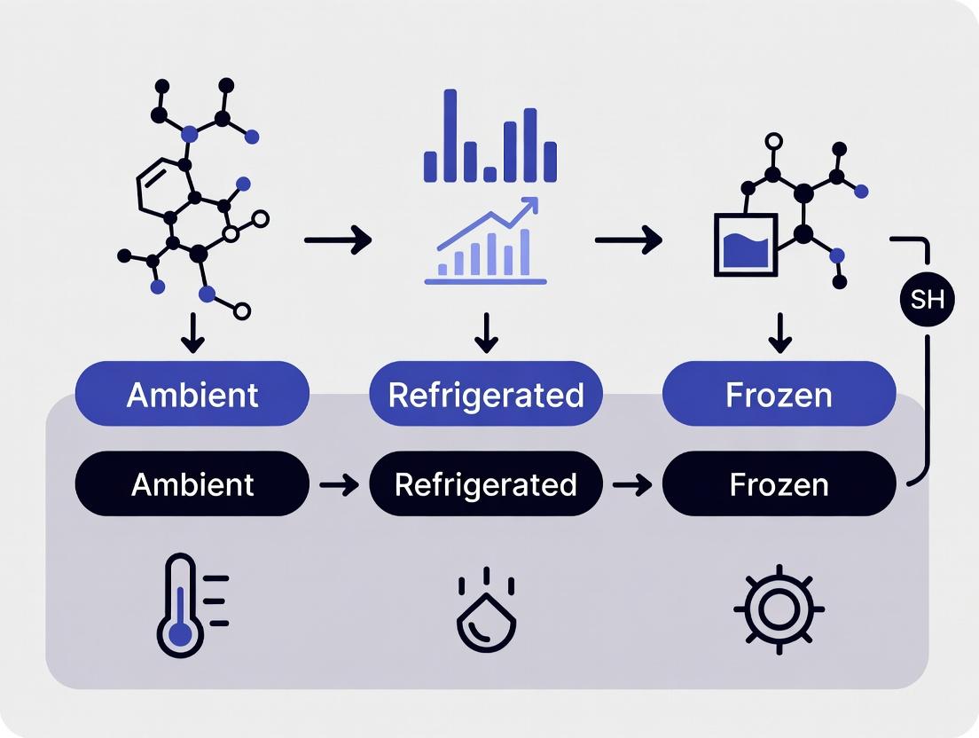
Q2: Should I store my nanoparticle samples in the refrigerator (4°C) or freezer (-20°C)? A: It depends on the formulation. 4°C is generally safer for most aqueous dispersions to avoid freeze-thaw stresses that can cause aggregation. -20°C or -80°C may be used for long-term archival if cryoprotectants (e.g., sucrose, glycerol) are added to prevent ice crystal damage. Always test freeze-thaw cycles on an aliquot first.
Q3: How often should I characterize my stored nanoparticle samples? A: Establish a stability testing protocol. Perform key analyses (size, zeta, UV-Vis) at defined time points: initially (t=0), then at 1 week, 1 month, 3 months, 6 months, and 1 year. This generates a stability profile.
Q4: What is the best container material for long-term nanoparticle storage? A: Borosilicate glass vials are inert and preferred for most samples. For particles sensitive to surface adsorption, use low-protein-binding polypropylene tubes. Always avoid containers made with plasticizers that can leach out.
Q5: How does Ostwald Ripening differ from aggregation? A: In aggregation, individual particles clump together but retain their original size. In Ostwald Ripening, larger particles grow at the expense of smaller ones due to the dissolution and re-deposition of material. This leads to a shift in the core size distribution, not just clustering.
Table 1: Impact of Storage Conditions on Gold Nanoparticle (10 nm, Citrate-Stabilized) Stability Over 6 Months
| Storage Condition | Temp (°C) | Atmosphere | [NaCl] | Size Increase (DLS, nm) | Zeta Potential Change (mV) | Observable Precipitation? |
|---|---|---|---|---|---|---|
| Aqueous Solution | 4 | Air | 0 mM | +2.1 | -38 to -35 | No |
| Aqueous Solution | 25 | Air | 0 mM | +5.5 | -38 to -28 | No |
| Aqueous Solution | 4 | Air | 50 mM | +45.0 (Aggregated) | -38 to -12 | Yes |
| Lyophilized w/ Sucrose | -20 | Air | N/A | +1.8 (after reconstitution) | -38 to -36 | No |
Table 2: Common Stabilizers and Their Mechanisms
| Stabilizer Class | Example | Primary Function | Best For |
|---|---|---|---|
| Electrostatic | Citrate, CTAB | Provides surface charge for repulsion | Au, Ag NPs in low-ionic strength buffers |
| Steric | PEG, PVP | Creates physical barrier via polymer brush | Broad, improves biocompatibility |
| Electrosteric | Polyelectrolytes, charged PEG | Combines charge and physical barrier | High ionic strength environments (e.g., PBS) |
| Ligand/Shell | Oleic acid, Silica shell | Passivates surface, reduces chemical reactivity | Quantum dots, iron oxide NPs |
Experimental Protocols
Protocol 1: Accelerated Stability Testing via DLS and Zeta Potential Objective: To predict long-term stability by monitoring changes in hydrodynamic diameter and surface charge under stress. Materials: DLS/Zeta Potential Analyzer, temperature-controlled sample holder, filtered (0.22 µm) dispersion medium. Procedure:
- Filter all buffers and media before use.
- Measure the hydrodynamic diameter (Z-average), PDI, and zeta potential of a freshly prepared or dialyzed sample (t=0). Perform in triplicate.
- Aliquot the sample into stable, inert vials.
- Subject aliquots to different stress conditions: elevated temperature (e.g., 40°C, 60°C), light exposure, or added electrolyte.
- At defined intervals (e.g., 1, 3, 7 days), remove an aliquot, equilibrate to room temp, and repeat measurements in step 2.
- Plot size and zeta potential versus time. A sharp change indicates instability.
Protocol 2: Distinguishing Aggregation from Ostwald Ripening via TEM Objective: To visually identify the mechanism of particle growth. Materials: TEM grid, TEM instrument. Procedure:
- Prepare TEM grids from the nanoparticle dispersion at initial state and after observed size increase via DLS.
- Image multiple grid squares at appropriate magnifications to obtain a representative population.
- Analysis: If particles appear in close-packed clusters, the mechanism is aggregation. If particles remain well-separated but the population shows a clear increase in individual particle core diameter and a loss of the smallest particles, the mechanism is Ostwald ripening.
Visualizations
(Diagram Title: Nanoparticle Stability Enemies and Key Monitoring Parameters)
(Diagram Title: Nanoparticle Stability Issue Diagnostic Workflow)
The Scientist's Toolkit: Research Reagent Solutions
| Item | Function/Application |
|---|---|
| Amicon Ultra Centrifugal Filters | For buffer exchange, concentration, and removal of unbound ligands/salts to "clean" samples before storage. |
| Dialysis Cassettes (MWCO appropriate) | Gentle alternative for buffer exchange over longer periods, minimizing shear forces that can cause aggregation. |
| Trehalose or Sucrose | Cryoprotectant and lyoprotectant. Helps maintain nanoparticle dispersion during freeze-drying (lyophilization) and prevents aggregation upon reconstitution. |
| Argon/Nitrogen Gas Canister | For creating an inert atmosphere in storage vials to prevent oxidation of sensitive nanoparticles (e.g., quantum dots, iron oxide). |
| Amber Glass Vials | Protects light-sensitive nanoparticles (e.g., certain dyes, perovskites) from photodegradation during storage. |
| PEG-Thiol (e.g., mPEG-SH) | Common reagent for introducing a steric stabilization layer on gold and other metal nanoparticles, improving stability in high-salt and biological media. |
| Sodium Citrate | Classic electrostatic stabilizer and reducing agent for gold nanoparticles. Used in synthesis and as a stabilizing additive. |
| 0.22 µm PES Syringe Filters | Essential for filtering all dispersion media to remove particulate contaminants that can act as nucleation sites for aggregation. |
| Zirconium Oxide Cuvettes | For DLS measurements of samples in aggressive organic solvents, where standard disposable plastic cuvettes may dissolve. |
Troubleshooting Guides & FAQs
FAQ: Common Storage-Related Issues
Q1: After 4 weeks of storage at 4°C, my nanoparticle size (by DLS) has increased significantly. What are the likely causes and how can I troubleshoot this?
A: An increase in hydrodynamic diameter is a classic sign of aggregation or instability. Likely causes and solutions are:
- Cause 1: Inadequate Stabilization. Electrostatic or steric stabilizers may be degrading or adsorbing to container walls.
- Troubleshoot: Re-measure zeta potential. A shift towards neutral values (e.g., from ±30 mV to ±10 mV) confirms electrostatic destabilization. Consider adding fresh stabilizer (e.g., 0.1% w/v polysorbate 80) before storage.
- Cause 2: Cold Denaturation or "Ostwald Ripening." For some polymer or lipid nanoparticles, 4°C can cause crystallization or molecular rearrangement, leading to growth.
- Troubleshoot: Test stability at room temperature (25°C) or a controlled 20°C for 1 week as a comparative experiment. Use isothermal titration calorimetry (ITC) to study binding constant changes.
- Cause 3: Container Interaction.
- Troubleshoot: Switch from polypropylene to low-binding, siliconized vials. Pre-rinse storage vials with a 1% serum albumin solution or the dispersion medium to create a passivating layer.
Q2: The Polydispersity Index (PDI) of my sample is increasing over time, even though the mean size is stable. What does this indicate?
A: A rising PDI indicates a broadening of the size distribution, often a precursor to visible aggregation or sedimentation. It suggests heterogeneous degradation or interactions.
- Actionable Protocol: Perform Asymmetric Flow Field-Flow Fractionation (AF4) coupled with multi-angle light scattering (MALS). This will separate populations by size and reveal if the increase is due to a small fraction of large aggregates or a general shift. Centrifugation (e.g., 10,000 x g for 10 min) followed by DLS analysis of the supernatant can also isolate and assess the non-aggregated fraction.
Q3: My nanoparticles' zeta potential is becoming less negative/more positive during storage. Why does this happen and how can I prevent it?
A: Zeta potential drift signals changes in surface chemistry. Common reasons:
- Ion Adsorption/Desorption: Ions from the dispersion medium (e.g., phosphates, citrates) or leaching from container walls can adsorb, shielding surface charge.
- Chemical Degradation: Surface groups (e.g., carboxylates) may esterify or hydrolyze.
- Prevention Protocol: Use purified, deionized water with a consistent, low ionic strength buffer (e.g., 1 mM HEPES, pH 7.4). Chelate metal ions using 1 mM EDTA. Store under inert atmosphere (N2 blanket) to prevent oxidative degradation of surface ligands.
Q4: How can I verify if the surface chemistry (e.g., PEG density, targeting ligand) has changed during storage?
A: Direct surface analysis is required. Implement these complementary techniques:
- Protocol 1: Fluorescence Spectroscopy. For fluorescently-tagged ligands, use fluorescence correlation spectroscopy (FCS) to measure changes in diffusion time, correlating to ligand density.
- Protocol 2: NMR Spectroscopy. Perform ¹H NMR in D2O. The intensity ratio of peaks specific to PEG (δ ~3.6 ppm) or other ligands vs. core nanoparticle signals provides a quantitative measure of surface coating integrity.
- Protocol 3: X-ray Photoelectron Spectroscopy (XPS). This surface-sensitive technique (<10 nm depth) can detect changes in elemental composition (e.g., C, O, N, S) and chemical states of surface atoms before and after storage.
Data Presentation: Typical Degradation Ranges
Table 1: Quantifiable Changes in Key Properties Indicative of Instability
| Property | Stable Sample Range | "Warning" Change During Storage | "Critical Failure" Change | Likely Mechanism | ||
|---|---|---|---|---|---|---|
| Size (Dh) | Baseline ± 10% | Increase of 10-25% | Increase > 25% or multimodal distribution | Aggregation, Ostwald ripening | ||
| PDI | < 0.1 (monodisperse) 0.1-0.2 (moderate) | Increase by > 0.05 units | PDI > 0.25 for formerly monodisperse samples | Heterogeneous aggregation/degradation | ||
| Zeta Potential | > | ±30 | mV (high stability) | Shift of > 10 mV towards neutral | Crossing ±10 mV threshold | Surface group degradation, ion adsorption |
| Surface Ligand Density | Baseline ± 5% (by NMR/XPS) | Loss of 5-20% | Loss > 20% | Desorption, chemical degradation, microbial action |
Experimental Protocols for Stability Assessment
Protocol: Comprehensive Pre- and Post-Storage Characterization Workflow
- Sample Preparation: Aliquot the master nanoparticle batch into identical, low-binding vials. Fill vials completely (minimize headspace) or standardize headspace-to-volume ratio.
- Baseline Characterization (Day 0):
- Size & PDI: Measure by Dynamic Light Scattering (DLS) at 3 angles (e.g., 90°, 15°, 173° backscatter) at 25°C. Perform minimum 3 runs.
- Zeta Potential: Measure by Phase Analysis Light Scattering (M3-PALS) in folded capillary cells. Use appropriate dispersion medium for the electrode.
- Surface Chemistry: Record ¹H NMR spectrum in D2O. Calculate ligand density via peak integration.
- Storage Conditions: Store aliquots under varied, documented conditions: 4°C, 25°C, 40°C (for accelerated studies), and under light protection vs. ambient light.
- Post-Storage Analysis (e.g., Day 7, 30, 90):
- Visual Inspection: Note any sedimentation, color change, or opalescence.
- Gentle Re-dispersion: Invert vial 20 times. Do not vortex or sonicate unless this is part of the intended use protocol.
- Repeat Baseline Characterization identically to Day 0.
- Advanced Analysis: Subject one aliquot per condition to AF4-MALS-UV-DLS for population deconvolution.
Visualization: Stability Assessment Workflow
Title: Nanoparticle Stability Assessment Workflow
The Scientist's Toolkit: Research Reagent Solutions
Table 2: Essential Materials for Nanoparticle Storage Studies
| Item | Function & Importance |
|---|---|
| Low-Binding, Siliconized Microtubes/Vials | Minimizes nanoparticle adhesion to container walls, preserving concentration and surface chemistry. |
| Inert Headspace Gas (N2 or Argon) | Prevents oxidative degradation of sensitive surface ligands (e.g., thiols, amines) by displacing oxygen. |
| Cryoprotectants (e.g., Trehalose, Sucrose, 5% w/v) | Forms a glassy matrix during freeze-drying, preventing aggregation and maintaining size upon reconstitution. |
| Sterile, 0.22 µm Filters | Used to filter-sterilize dispersion media to prevent microbial growth during long-term storage. |
| Chelating Agents (e.g., 0.1-1 mM EDTA) | Binds trace metal ions in buffer that can catalyze oxidative reactions or bridge nanoparticles. |
| High-Purity, Low-Ionic Strength Buffers (e.g., HEPES, Tris) | Maintains stable pH without introducing high salt concentrations that can screen electrostatic stabilization. |
| Reference Nanosphere Standards (e.g., NIST-traceable) | Essential for daily calibration of DLS and Zeta Potential instruments to ensure measurement accuracy. |
| Desiccant (for lyophilized samples) | Ensures moisture-free environment for long-term storage of freeze-dried nanoparticles. |
Article Title: The Impact of Storage on Biological Functionality: Ligand Orientation, Targeting Efficiency, and Drug Release Profiles.
Context: This support center provides troubleshooting guidance framed within the thesis: Best practices for storing characterized nanoparticle samples to preserve their engineered biological functions.
Troubleshooting Guides & FAQs
FAQ Section 1: Ligand Orientation and Surface Conformation
Q1: After 4 weeks of storage at 4°C, our PEGylated nanoparticles show a 40% reduction in cell targeting. What could be the issue? A: This is a classic sign of ligand reorientation or desorption. Storage conditions can cause surface ligands (e.g., antibodies, peptides) to undergo conformational changes or detach, masking active targeting sites.
- Troubleshooting Steps:
- Verify: Perform a quantitative ligand binding assay (e.g., ELISA-style plate capture) on fresh vs. stored samples.
- Check Storage Buffer: Ensure it contains stabilizing agents (e.g., 0.1% BSA, 1-5% sucrose) to minimize surface tension changes.
- Protocol - Ligand Binding Quantification:
- Coat a plate with your target receptor.
- Incubate with serial dilutions of fresh and stored nanoparticles.
- Detect bound nanoparticles via a tag on the nanoparticle core (e.g., fluorescent dye, elemental tag for ICP-MS).
- Compare the binding curves to calculate active ligand density.
Q2: Our DLS data shows unchanged hydrodynamic size, but FTIR suggests altered surface chemistry. Is the sample still usable for in vivo studies? A: Not recommended without further validation. DLS is insensitive to minor conformational changes. Altered FTIR peaks indicate chemical degradation or rearrangement of surface groups, which directly impacts biorecognition.
- Action Protocol:
- Perform an Activity Assay: Use a simple, rapid functional assay (e.g., inhibition of cell adhesion, receptor binding in vitro) before proceeding to complex animal studies.
- Switch to More Stable Conjugation: If recurring, consider using covalent, chemoselective conjugation chemistries (e.g., click chemistry) over physical adsorption.
FAQ Section 2: Targeting Efficiency
Q3: How does freeze-thaw cycling affect the active targeting of lipid nanoparticles (LNPs)? A: Freeze-thaw cycles induce ice crystal formation, causing particle aggregation and shear forces that can strip or denature surface ligands.
- Preventive Protocol: Cryopreservation Best Practice
- Use a cryoprotectant (e.g., 10% trehalose or 5% DMSO).
- Flash-freeze in liquid nitrogen or a dry ice/ethanol bath.
- Store at -80°C, not -20°C.
- Thaw rapidly in a 37°C water bath with gentle agitation.
- Perform a post-thaw size measurement (DLS) and a single-point targeting validation using a cell-based flow cytometry assay.
Q4: Should we store targeting ligand-functionalized samples in solution or lyophilized? A: It depends on the ligand stability. See Table 1.
Table 1: Storage Format Impact on Targeting Ligand Integrity
| Storage Format | Recommended For | Risk to Targeting Efficiency | Key Stabilizer |
|---|---|---|---|
| 4°C Solution | Short-term (< 1 week), antibodies, proteins | Microbial growth, ligand hydrolysis | 0.02% sodium azide, 1% BSA |
| -80°C Solution | Medium-term (months), most ligands | Freeze-thaw damage, aggregation | 5-10% cryoprotectant (sucrose/trehalose) |
| Lyophilized | Long-term (years), peptides, aptamers | Improper reconstitution, moisture | Matrix former (e.g., trehalose, mannitol) |
FAQ Section 3: Drug Release Profiles
Q5: The drug release profile of our PLGA nanoparticles accelerated significantly after 3 months of storage. Why? A: Hydrolytic degradation of the polymer matrix (PLGA, PLA) continues during storage, altering porosity and erosion rates.
- Diagnostic Protocol: Monitoring Matrix Integrity
- GPC/SEC: Measure the molecular weight distribution of polymer extracted from stored vs. fresh nanoparticles. A left-shift indicates chain scission.
- DSC: Analyze the glass transition temperature (Tg). A lowered Tg suggests increased chain mobility and faster degradation.
- Correlate: Plot % drug released at 24h against polymer Mw from stored batches to establish a predictive stability model.
Q6: For stimuli-responsive (e.g., pH-sensitive) nanoparticles, how do we verify the "trigger" remains functional post-storage? A: The responsive moiety (e.g., hydrazone bond, ionizable lipid) can degrade.
- Validation Workflow Protocol:
- Control Release Medium: Perform the standard drug release assay in both trigger (e.g., pH 5.0 buffer) and non-trigger (e.g., pH 7.4 buffer) conditions.
- Calculate Trigger Efficiency:
% Triggered Release = (% Release at trigger condition) - (% Release at non-trigger condition). - Compare: A decrease in "Triggered Release" value for stored samples indicates loss of stimuli-responsive function. See Diagram 1.
Diagram 1: Workflow for Validating Stimuli-Response Post-Storage
The Scientist's Toolkit: Research Reagent Solutions
Table 2: Essential Materials for Storage Stability Studies
| Item | Function & Rationale |
|---|---|
| Trehalose (Di-saccharide) | Cryo- & lyo-protectant. Forms stable glassy matrix, replaces water H-bonds with biomolecules (ligands). |
| BSA (Bovine Serum Albumin) | Surface passivator in storage buffer. Prevents non-specific adsorption of nanoparticles and ligands to vial walls. |
| SPDP Crosslinker (NHS-PEG-Maleimide) | For creating stable, thioether-linked ligand conjugates. More stable than amine-reactive (NHS-ester) alone in aqueous storage. |
| Size Exclusion Chromatography (SEC) Columns | Critical for cleaning samples pre-storage (removing unencapsulated drug/free ligand) and analyzing aggregates post-storage. |
| Inert Vial (e.g., Glass with Teflon-lined cap) | Prevents leaching of plasticizers (from plastic vials) that can adsorb to nanoparticles and alter surface properties. |
| Oxygen Scavenger Sachets | For storage of lyophilized or inert-atmosphere-packed samples. Prevents oxidative degradation of lipids, polymers, and sensitive ligands. |
| Fluorescently-Labeled Target Receptor | Essential reagent for direct, quantitative measurement of targeting ligand availability via flow cytometry or fluorescence anisotropy. |
Diagram 2: How Storage Stressors Degrade Nanoparticle Function
Technical Support Center: Troubleshooting Guides & FAQs
Q1: Our characterized silver nanoparticle suspension shows unexpected aggregation and a color change from yellow to gray after one month of storage at 4°C. What could be the cause? A: This is a common issue linked to temperature-induced Ostwald ripening and container interaction. Storage at 4°C can slow but not prevent thermodynamic processes. Aggregation is accelerated if nanoparticles are stored in a standard polypropylene tube. Ions (e.g., chloride) can leach from certain plastics, destabilizing the electrostatic stabilization of AgNPs. Best Practice: Transfer suspension to a certified low-binding, non-leaching container (e.g., COC polymer or borosilicate glass with PTFE-lined cap) and store at a controlled -20°C to minimize kinetic energy and ion migration. Always note the ionic strength and pH of the suspension buffer in metadata.
Q2: We observed a significant decrease in the fluorescence quantum yield of our characterized CdSe/ZnS quantum dots when samples are repeatedly taken from the main stock. What factors should we investigate? A: This indicates photodegradation and oxygen sensitivity. Each time the container is opened, the sample is exposed to:
- Ambient Light: Causes photo-oxidation of the surface ligands and core.
- Oxygen: Leads to the formation of surface defect sites, quenching fluorescence. Protocol for Remediation:
- Aliquot the master stock into single-use, amber-colored borosilicate glass vials.
- Purge the headspace with inert gas (Argon or Nitrogen) before sealing.
- Store aliquots at 4°C in the dark (use a light-blocking box).
- Thaw/use each aliquot only once.
Q3: For lipid nanoparticle (LNP) formulations containing siRNA, we see a loss of encapsulation efficiency and biological activity after 6 weeks, even at -80°C. Could the container material be a factor? A: Yes. At ultra-low temperatures (-80°C), certain plastics like polycarbonate or standard polypropylene become brittle and can develop micro-fissures. This breaches the sterile barrier and can allow ice crystal nucleation at the polymer surface, physically disrupting LNP integrity upon freeze-thaw cycles. Solution: Use cryogenic vials specifically designed for -80°C to -196°C, made from materials like polyolefin (e.g., Corning Cryostor). Always employ a controlled, slow-rate freezing protocol (e.g., -1°C/min) before transfer to -80°C.
Q4: How do we choose between glass and plastic for long-term storage of characterized gold nanoparticle conjugates (antibody-functionalized)? A: The choice hinges on the conjugation chemistry and concentration.
| Container Material | Advantages | Disadvantages | Recommended Use Case |
|---|---|---|---|
| Borosilicate Glass (Amber) | Chemically inert, superior barrier to O₂, excellent for light-sensitive samples. | Protein/nucleic acid can adsorb to surface; risk of breakage; can be costly. | Long-term (>6 month) storage of high-value, light-sensitive conjugates. Use with passivating agents (e.g., BSA, Trehalose). |
| Low-Binding Polypropylene | Low biomolecule adsorption, shatterproof, cost-effective. | Permeable to oxygen and water vapor over time; potential for additive leaching. | Short-to-medium term storage, working aliquots, high-throughput applications. |
| Cyclic Olefin Copolymer (COC) | Excellent clarity, high chemical resistance, very low leaching and adsorption. | Higher cost than standard plastics; limited supplier options. | Critical applications requiring minimal sample-container interaction and visual inspection. |
Protocol for Container Compatibility Testing:
- Split the characterized nanoparticle sample into three candidate containers.
- Store under identical, defined conditions (e.g., 4°C, dark).
- At t=0, 1 week, 1 month, analyze key parameters: Hydrodynamic Diameter (DLS), Zeta Potential, UV-Vis Plasmon Peak (for AuNPs), and functional activity (e.g., ELISA binding).
- Compare data against the t=0 baseline to identify the most inert container.
Q5: What is the recommended protocol for creating a "stable" baseline when characterizing nanoparticles for storage studies? A: A rigorous pre-storage characterization protocol is essential for meaningful data.
Detailed Experimental Protocol: Pre-Storage Characterization Objective: To establish a comprehensive baseline profile of the nanoparticle sample prior to stability studies. Materials: Nanoparticle suspension, Zetasizer or equivalent, UV-Vis-NIR spectrophotometer, pH meter, sterile filtered buffer. Procedure:
- Equilibration: Allow the sample to equilibrate to room temperature (22-25°C) for 30 minutes in the dark.
- Homogenization: Gently vortex the sample for 10 seconds. Do not sonicate unless it is a standard part of the formulation protocol.
- Physical Characterization:
- Size & PDI: Perform Dynamic Light Scattering (DLS) measurement in triplicate. Report the Z-average diameter and Polydispersity Index (PDI).
- Surface Charge: Measure Zeta Potential via Electrophoretic Light Scattering in the appropriate dispersion medium (e.g., 1mM KCl). Perform minimum 5 runs.
- Optical Characterization:
- Record the full UV-Vis-NIR spectrum (e.g., 300-1100 nm for AuNPs, 350-800 for QDs).
- Note the Absorption Maxima (λmax) and the Absorbance at λmax.
- Chemical/Environmental Recording:
- Measure and record the exact pH of the suspension.
- Record the Buffer/Medium Composition and any stabilizing agents (e.g., 0.1% BSA, 1mM citrate).
- Note the Primary Container Material and Fill Volume.
- Documentation: Save all raw data files and label with a unique sample ID linked to a master log.
The Scientist's Toolkit: Research Reagent & Material Solutions
| Item | Function & Rationale |
|---|---|
| Amber Borosilicate Glass Vials | Provides a chemically inert, light-blocking (UV/visible) environment, minimizing photodegradation and leaching. Critical for light-sensitive samples (e.g., QDs, photosensitizers). |
| Argon/Nitrogen Gas Canister | Used to purge headspace in storage vials, displacing oxygen to prevent oxidative degradation of nanoparticle cores, surfaces, or encapsulated cargo. |
| Cryoprotectants (e.g., Trehalose, Sucrose) | Non-reducing disaccharides that form a glassy matrix during freezing, immobilizing nanoparticles and preventing ice crystal-induced aggregation or fusion (e.g., for LNPs, liposomes). |
| Low-Binding, Non-Leaching Tubes (COC, Certified PP) | Minimizes the loss of precious sample via surface adsorption and prevents the introduction of contaminants (plasticizers, mold release agents) that can destabilize colloids. |
| Portable, Calibrated pH Meter | Essential for verifying suspension pH before and after storage. Small pH shifts can dramatically alter zeta potential and colloidal stability. |
| Stability Chambers (Precision Temp/Humidity) | Allows for controlled, ICH-compliant stability studies at set temperatures (e.g., 4°C, 25°C/60% RH) to model real-world or accelerated storage conditions. |
| Sterile, Low-Particulate Buffer Kits | Ensures dispersion media does not introduce contaminants (bacterial, ionic, particulate) that can seed aggregation or provide reactive species. |
Visualizations
Technical Support Center: Troubleshooting Pre-Characterization & Storage
FAQs & Troubleshooting Guides
Q1: We characterized our nanoparticles (NPs) in suspension before storage, but after 4 weeks at 4°C, the particle size by DLS has increased dramatically. What went wrong? A: This indicates aggregation or instability. The likely issue is an incomplete pre-storage characterization baseline. You may have measured size but overlooked critical parameters.
- Troubleshooting Steps:
- Re-measure Zeta Potential: A magnitude below |±20| mV suggests insufficient electrostatic stabilization. Your storage buffer may lack necessary ionic strength or pH control.
- Check for Contamination: Replicate your initial DLS measurement with the original sample aliquot (if saved). A discrepancy suggests bacterial or fungal growth. Implement sterile filtration (0.22 µm) before storage.
- Re-analyze Polydispersity Index (PdI): An initial PdI > 0.2 was a warning sign of an inherently polydisperse sample, prone to further aggregation. Pre-storage purification (e.g., centrifugation, filtration, SEC) is required.
Q2: Our functionalized NPs show a 40% loss of targeting ligand binding after 6 months at -80°C. Why would freezing cause this? A: This is a common issue with incomplete pre-storage analysis. Freezing can cause "cold denaturation" of surface biomolecules or lead to ligand shedding via ice crystal formation.
- Troubleshooting Steps:
- Verify Pre-Storage Binding Assay: Ensure your initial binding efficiency measurement (e.g., SPR, ELISA) was quantitative, not just qualitative.
- Implement Cryoprotection: Add sucrose or trehalose (5-10% w/v) to the formulation. Pre-storage characterization must then be repeated with the cryoprotectant present to confirm it doesn't itself cause aggregation.
- Switch Storage Method: Consider lyophilization. However, pre-storage characterization must be expanded to include tests for stability after reconstitution.
Q3: How many characterization techniques are absolutely necessary for a valid pre-storage baseline? A: There is no single number, but a minimum panel covering physical, chemical, and functional integrity is non-negotiable. See the table below.
Table 1: Minimum Viable Pre-Storage Characterization Panel for Nanoparticle Samples
| Parameter | Primary Technique(s) | Target Acceptance Criteria | Rationale for Stability Baseline | ||
|---|---|---|---|---|---|
| Size & Distribution | Dynamic Light Scattering (DLS) | PDI < 0.2 | Monodispersity predicts resistance to aggregation. | ||
| Surface Charge | Zeta Potential Measurement | ±30 | mV | Indicates colloidal stability; predicts interaction with storage vessels. | |
| Concentration | UV-Vis Spectroscopy, ICP-MS | Accurate mg/mL or particle #/mL | Essential for dose consistency in future experiments. | ||
| Morphology | Transmission Electron Microscopy (TEM) | Uniform shape, no pre-existing aggregates | Visual validation of DLS data and structural integrity. | ||
| Chemical Identity | FTIR, XPS | Conjugation efficiency > 90% | Confirms ligand attachment before storage degradation. | ||
| Functional Activity | Cell Binding/Internalization Assay | >85% relative activity | Establishes benchmark for post-storage potency loss. |
Experimental Protocol: Comprehensive Pre-Storage Characterization Workflow
Title: Protocol for Establishing a Nanoparticle Stability Baseline.
Materials:
- Purified nanoparticle sample.
- Storage buffers (target buffer and cryoprotectant-containing buffer if needed).
- DLS/Zeta Potential instrument (e.g., Malvern Zetasizer).
- UV-Vis Spectrophotometer.
- Access to TEM/SEM services.
Method:
- Homogenization: Vortex the final synthesized/batch sample for 2 minutes. Do not sonicate unless previously validated.
- Size & Zeta Potential (Triplicate Measurement):
- Load 1 mL of sample into a clean DLS cuvette or zeta cell.
- Equilibrate at 25°C for 2 minutes.
- Run 3-13 measurements per replicate as per instrument software.
- Record the Z-Average size (d.nm), PdI, and Zeta Potential (mV). Acceptance: PdI < 0.25, Zeta Potential consistent with formulation theory.
- Concentration Determination:
- For Plasmonic NPs (Au, Ag): Measure absorbance at λmax (e.g., ~520 nm for 20 nm AuNPs). Calculate concentration using the Beer-Lambert law with the known extinction coefficient.
- For Others: Use a quantified assay (e.g., BCA for protein NPs, ICP-MS for elemental cores).
- Morphological Validation (TEM):
- Deposit 10 µL of sample on a carbon-coated grid for 1 minute. Wick away excess.
- Negative stain with 2% uranyl acetate if required.
- Image at minimum 50,000x magnification. Capture images from multiple grid squares.
- Functional Assay (Ligand-Specific):
- Perform your standard binding/in vitro activity assay (e.g., flow cytometry with target cells, ELISA).
- Compare activity to a positive control (e.g., free ligand) and negative control (unfunctionalized NP). Report as % relative activity.
- Documentation & Aliquotting:
- Record all data in Table 1 format. This is your Stability Baseline.
- Aseptically aliquot the characterized sample into pre-labeled, inert vials (e.g., polypropylene) for storage under defined conditions.
Diagram Title: Pre-Storage Characterization Decision Workflow
The Scientist's Toolkit: Essential Reagents & Materials
Table 2: Key Research Reagent Solutions for Pre-Storage Characterization
| Item | Function | Critical Note |
|---|---|---|
| Zeta Potential Titrant Kits (e.g., HCl/NaOH) | To determine the isolectric point (pI) and stability across pH. | Perform a pH stability sweep (pH 3-10) to identify optimal storage pH. |
| Sterile Syringe Filters (0.22 µm, PES membrane) | For aseptic filtration prior to storage to prevent microbial growth. | Pre-wet filter with buffer to minimize NP adsorption losses. |
| Disposable Zeta Cells & DLS Cuvettes | For accurate, cross-contamination-free measurements. | Always use high-quality, clean, and dust-free consumables. |
| Cryoprotectants (Trehalose, Sucrose) | To protect NPs from ice crystal damage during freeze-thaw cycles. | Must be screened for compatibility; can increase viscosity affecting DLS. |
| Negative Stains for TEM (2% Uranyl Acetate) | To enhance contrast for imaging soft-material NPs. | Handle as hazardous waste. Alternative: phosphotungstic acid. |
| Reference Nanospheres (e.g., NIST-traceable PS beads) | For instrument calibration and validation of DLS/TEM measurements. | Run a reference sample at the start of each characterization session. |
Step-by-Step Protocols: Optimizing Storage Conditions for Different Nanoparticle Classes
Technical Support Center & Troubleshooting Guide
FAQs & Troubleshooting
Q1: Our nanoparticles aggregate upon reconstitution after lyophilization. What is the primary cause and how can we prevent it?
- A: Aggregation is most commonly caused by inadequate cryoprotection during freezing or collapse during primary drying. To prevent it: 1) Increase the concentration of your primary cryoprotectant (e.g., sucrose, trehalose) to a 5:1 to 10:1 (w/w) sugar-to-nanoparticle ratio. 2) Ensure the formulation pH is away from the nanoparticle's isoelectric point to maximize electrostatic stabilization. 3) Optimize the freezing rate; a faster freeze (using a shell freezer or liquid nitrogen) creates smaller ice crystals, reducing physical stress. 4) Verify that the product temperature during primary drying remains at least 2-3°C below the collapse temperature (T꜀) of your formulation.
Q2: How do we determine the critical collapse temperature (T꜀) for our nanoparticle formulation?
- A: The T꜀ is best determined experimentally using Freeze-Drying Microscopy (FDM). This technique visually observes the point at which the dried product structure collapses under vacuum at varying temperatures. If FDM is unavailable, a conservative proxy is the glass transition temperature of the maximally freeze-concentrated solute (Tg') of your amorphous cryoprotectant, measured by Differential Scanning Calorimetry (DSC). For sucrose-based formulations, Tg' is typically around -32°C to -34°C.
Q3: Our cycle time is excessively long (>72 hours). What parameters can we safely adjust to shorten it without compromising product quality?
- A: Focus on the primary drying stage. You can: 1) Increase Shelf Temperature: Ramp the temperature as close as possible to the T꜀ of your formulation (e.g., -25°C to -30°C for common sugars) while monitoring product temperature via probes. 2) Optimize Chamber Pressure: If using a manifold, ensure it is at the optimal range for ice sublimation (typically 0.1-0.3 mBar). For cake resistance-limited cycles, a slightly higher pressure (0.2-0.4 mBar) can improve heat transfer. 3) Reduce Cake Thickness: If possible, reduce the fill depth in your vials (<1 cm is ideal). A thicker cake significantly increases resistance and drying time.
Q4: What is the recommended method for reconstituting lyophilized nanoparticles to ensure complete recovery of initial properties?
- A: For characterized nanoparticle samples, controlled reconstitution is vital. 1) Use the original dispersion medium (e.g., purified water, specific buffer) pre-equilibrated to the storage temperature. 2) Add the medium gently along the vial wall to avoid foaming or direct high-pressure stream onto the cake. 3) Allow the cake to hydrate fully by letting it sit undisturbed for 1-5 minutes before gentle manual swirling (not vortexing) until fully dissolved. 4) Characterize key parameters (size, PDI, concentration) post-reconstitution and compare to pre-lyophilization data.
Q5: How should we select between sucrose and trehalose as a cryoprotectant?
- A: Both are excellent. The choice can be based on your stability data and downstream use. See the comparison table below.
Table 1: Cryoprotectant Comparison for Nanoparticle Lyophilization
| Parameter | Sucrose | Trehalose | Mannitol |
|---|---|---|---|
| Primary Mechanism | Amorphous stabilizer (Vitrification) | Amorphous stabilizer (Vitrification) | Crystalline bulking agent |
| Typical Conc. Range | 2-10% (w/v) | 2-10% (w/v) | 2-5% (w/v) |
| Tg' (approx.) | -32°C to -34°C | -30°C | -32°C (as amorphous) |
| Advantages | High stabilizing efficiency, common | Higher chemical stability, better for long-term storage | Provides elegant cake structure, good for combination |
| Disadvantages | Can hydrolyze at low pH | More expensive | Can crystallize, offering little cryoprotection alone |
| Best For | Most nanoparticle formulations, neutral pH | Long-term storage, sensitive nanoparticles | As a bulking agent combined with amorphous protectors |
Experimental Protocols
Protocol 1: Formulation Screening for Cryoprotection Objective: To identify the optimal cryoprotectant type and concentration to prevent nanoparticle aggregation.
- Prepare nanoparticle sample (e.g., 1 mL at 1 mg/mL).
- Dialyze against a series of cryoprotectant solutions (e.g., 1%, 5%, 10% w/v of sucrose, trehalose, and a sucrose:mannitol 4:1 mixture).
- Aliquot 0.5 mL of each formulated sample into 3 mL lyophilization vials.
- Freeze samples using a standardized method (e.g., -80°C freezer for 4 hours or shell freezer).
- Lyophilize using a conservative cycle (Primary drying: -40°C, 0.1 mBar, 48 hrs; Secondary drying: +25°C, 0.01 mBar, 12 hrs).
- Reconstitute with the original volume of sterile water.
- Characterize particle size (DLS), PDI, and zeta potential. Compare to pre-lyophilization controls.
- Data Presentation: Tabulate post-reconstitution size and PDI for each condition. The formulation yielding values closest to the control is optimal.
Protocol 2: Cycle Optimization via Conservative Ramp Objective: To establish a safe, efficient primary drying shelf temperature.
- Load vials with optimized formulation from Protocol 1, equipped with temperature probes in the center of the product in several vials.
- Start the lyophilizer. Freeze the product to -45°C and hold for 2 hours.
- Set the chamber pressure to 0.1 mBar. Begin primary drying with the shelf temperature at -40°C.
- Gradually increase the shelf temperature by +5°C increments every 3-5 hours, closely monitoring the product temperature.
- Continue until the product temperature approaches (within 2-3°C) of the known Tg' or T꜀.
- Hold the shelf temperature at this maximum safe level until the product temperature converges with the shelf temperature (indicating sublimation endpoint).
- Proceed with secondary drying (e.g., +25°C, 0.01 mBar, 8-10 hrs).
- Data Presentation: Create a table logging shelf temperature, product temperature, and chamber pressure at each time increment to define the optimized ramp profile.
Visualizations
Title: Lyophilization Workflow for Nanoparticles
Title: Troubleshooting Aggregation in Lyophilized Nanoparticles
The Scientist's Toolkit: Key Reagent Solutions
| Item | Function & Rationale |
|---|---|
| Sucrose (Molecular Biology Grade) | Primary amorphous cryoprotectant. Forms a glassy matrix that immobilizes nanoparticles, preventing aggregation and stabilizing structure during freezing and drying. |
| Trehalose Dihydrate (Pharma Grade) | Alternative disaccharide cryoprotectant with higher chemical stability and resistance to hydrolysis, often preferred for long-term storage stability studies. |
| Mannitol (USP Grade) | Crystalline bulking agent. Provides structural integrity to the lyophilized cake, preventing blow-out, but offers minimal cryoprotection on its own. |
| Poloxamer 188 (Pluronic F-68) | Non-ionic surfactant. Used as a secondary stabilizer in formulations to prevent surface adsorption and ice crystal-induced aggregation during freezing. |
| Histidine or Citrate Buffer | Buffering agents. Maintain pH away from the nanoparticle's isoelectric point during freezing (pH shift can occur), ensuring electrostatic repulsion is maintained. |
| Type I Borosilicate Lyophilization Vials | Chemically inert, thermal shock-resistant containers designed for use under high vacuum and low-temperature conditions. |
| Butyl Rubber Lyophilization Stoppers | Provide an elastomeric closure that allows for water vapor escape during drying and creates a hermetic seal upon full stoppering under vacuum. |
Troubleshooting Guides & FAQs
Q1: My stored gold nanoparticle (AuNP) solution shows visible aggregation after one week. What are the primary causes and solutions?
A: Primary causes are incorrect buffer ionic strength or pH. Citrate-capped AuNPs are stable in low-ionic-strength buffers (~1-10 mM) at pH near or slightly above the nanoparticle's isoelectric point. Aggregation indicates the ionic strength is too high, neutralizing surface charge. Solution: Dialyze the aggregated sample against a fresh, low-salt buffer (e.g., 2 mM sodium citrate, pH 7.0). Centrifuge gently to remove large aggregates and re-characterize by DLS and UV-Vis.
Q2: How do I prevent bacterial/fungal growth in my long-term (months) nanoparticle suspension without affecting surface chemistry?
A: Use sterile filtration (0.22 µm) and aseptic technique. For preservatives, sodium azide (NaN₃) is common but can be reactive with certain surface ligands. For biological ligands, consider 0.01% ProClin 300.
- Protocol: To a 10 mL nanoparticle suspension in PBS, add 10 µL of a 1% (v/v) ProClin 300 stock solution in ethanol. Mix gently by inversion. Store at 4°C.
Q3: What is the recommended storage concentration for lipid nanoparticles (LNPs) to prevent fusion or degradation?
A: High concentrations can promote fusion. For long-term stability, store LNPs at a concentration of 0.1-1.0 mg/mL lipid in a sucrose-rich, cryoprotective buffer (see Table 1). Avoid freeze-thaw cycles. Aliquot samples for single-use.
Q4: My fluorescently tagged nanoparticles show quenching after storage. Is this reversible?
A: Quenching may be due to fluorophore degradation or nanoparticle aggregation bringing fluorophores too close. Check for aggregation via DLS. If aggregated, quenching may be partially reversible by re-dispersing particles via gentle sonication. If no aggregation, the fluorophore may be chemically degraded (irreversible) due to light exposure or reactive buffer components. Solution: Always store in the dark (amber vials) at 4°C in an inert, oxygen-scavenging buffer.
Recommended Storage Buffers & Preservatives
Table 1: Recommended Buffer and Preservative Conditions for Common Nanoparticle Types
| Nanoparticle Type | Recommended Buffer | Ionic Strength | Preservative & Concentration | Storage Temp | Max Recommended Conc. |
|---|---|---|---|---|---|
| Citrate-capped Au/Ag NPs | 2 mM Sodium Citrate, pH 7.0-8.0 | Low (<5 mM) | 0.02% NaN₃ | 4°C | 10 nM (particle number) |
| PEGylated Inorganic NPs | 10 mM HEPES, pH 7.4 | Low-Moderate | 0.01% ProClin 300 | 4°C | 1 mg/mL |
| Lipid Nanoparticles (LNPs) | 10 mM Tris, 10% (w/v) Sucrose, pH 7.4 | Low | Sterile filtration only | 4°C or -80°C (frozen) | 1 mg/mL lipid |
| Polymeric NPs (PLGA) | 1x PBS, pH 7.4 | Physiological | 0.05% (v/v) Tween 20 & 0.02% NaN₃ | 4°C | 5 mg/mL |
| Quantum Dots (PEG) | 50 mM Borate, pH 9.0 | Moderate | 0.02% NaN₃, 1 mM β-ME* | 4°C (dark) | 5 µM |
*β-ME (β-mercaptoethanol) prevents oxidation of surface thiols.
Detailed Experimental Protocol: Evaluating Storage Stability
Title: Protocol for Assessing Nanoparticle Storage Stability via DLS and UV-Vis Spectroscopy.
Objective: To quantitatively monitor changes in hydrodynamic size and plasmonic properties of nanoparticles over time under different storage conditions.
Materials: Nanoparticle sample, dialysis tubing (appropriate MWCO), storage buffers (see Table 1), amber vials, 0.22 µm syringe filters.
Methodology:
- Buffer Exchange: Dialyze 5 mL of the characterized nanoparticle sample against 1 L of the desired storage buffer for 24 hours at 4°C with two buffer changes.
- Aliquot & Preserve: Filter the dialyzed suspension through a 0.22 µm filter. Add preservative if required (see Table 1). Aliquot into sterile amber vials (e.g., 500 µL each).
- Storage: Store aliquots under test conditions (e.g., 4°C in dark, 25°C in dark, -20°C).
- Time-point Analysis: At predetermined intervals (Day 0, 7, 30, 90), retrieve one aliquot per condition.
- DLS: Equilibrate sample to room temp for 15 min. Gently invert 5x. Measure hydrodynamic diameter and PDI in triplicate.
- UV-Vis Spectroscopy: Scan absorbance from 200-800 nm. Record the wavelength and intensity of the surface plasmon resonance (SPR) peak for metal NPs, or the first exciton peak for QDs.
- Data Interpretation: A >10% increase in mean diameter or a significant broadening of the SPR peak indicates instability/aggregation.
Visualization: Nanoparticle Storage Stability Assessment Workflow
Diagram Title: NP Storage Stability Testing Workflow
The Scientist's Toolkit: Key Research Reagent Solutions
Table 2: Essential Materials for Nanoparticle Liquid-State Storage Experiments
| Item | Function & Rationale |
|---|---|
| Ultrapure Water (≥18 MΩ·cm) | Prevents unintended ionic contamination during buffer preparation, crucial for charge-stabilized NPs. |
| HEPES Buffer | A zwitterionic, biological buffer that maintains pH (7.2-8.2) with minimal metal ion complexing, ideal for surface functionalization studies. |
| Sucrose (Molecular Biology Grade) | Acts as a cryoprotectant and density stabilizer; prevents fusion and aggregation of lipid-based NPs during storage. |
| Sodium Azide (NaN₃), 1% stock | Broad-spectrum antimicrobial preservative for inorganic NP suspensions. Caution: Highly toxic and reactive with certain organic ligands. |
| ProClin 300 | A low-toxicity, broad-spectrum preservative effective at low concentrations (0.005-0.02%), suitable for NPs with biological surface moieties. |
| Amber HPLC Vials (2 mL, Screw Top) | Provides light-sensitive protection for fluorescent or photoactive nanoparticles; prevents evaporation. |
| Regenerated Cellulose Dialysis Membranes | Allows for gentle, efficient buffer exchange without significant nanoparticle loss due to adsorption (compared to some other polymers). |
| 0.22 µm PES Syringe Filters | For sterile filtration of nanoparticle suspensions prior to storage to eliminate microbial contaminants. |
| Dynamic Light Scattering (DLS) Cell | A clean, disposable cuvette for accurate, contaminant-free size distribution measurements during stability monitoring. |
Troubleshooting Guides & FAQs
Q1: Our nanoparticle samples stored at -80°C have shown aggregation upon thawing. What are the likely causes and solutions? A: Aggregation post-thaw is commonly due to ice crystal formation or cryoprotectant absence. Ensure samples are flash-frozen in liquid nitrogen before -80°C transfer. Use a suitable cryoprotectant (e.g., 1-5% trehalose or 5% DMSO) in your formulation buffer. Avoid repeated freeze-thaw cycles by aliquoting.
Q2: We observe inconsistent results from assays using antibodies stored at 4°C. How should protein-based reagents be stored? A: For short-term (<1 month), store in a stabilized buffer with 0.02% sodium azide at 4°C. For long-term, aliquot, add glycerol (50% v/v), and store at -20°C. Avoid frost-free freezers. Always spin down briefly before use to collect condensation.
Q3: Our LN2 storage dewar has a rapid nitrogen loss rate. What should we check? A: Perform a visual inspection of the inner chamber for cracks. Check the lid seal and neck plug for integrity. Ensure the vacuum level is within the manufacturer's specification. If loss persists, the vacuum jacket may be compromised, requiring professional service.
Q4: Samples in a -20°C freezer were compromised during a power outage. What is the best protocol for backup storage? A: Critical characterized nanoparticle samples should always have a backup aliquot stored in a separate, ideally -80°C or LN2, facility. Implement 24/7 temperature monitoring with remote alerts. Consider a UPS for freezers. For -20°C storage, a full freezer can stay cold for ~24-48 hours if unopened.
Q5: How do we choose between -80°C and liquid nitrogen vapor phase for long-term storage of lipid nanoparticle (LNP) formulations? A: For LNPs designed for nucleic acid delivery, -80°C is often sufficient for 1-2 years if properly formulated with cryoprotectants. For master cell banks used to produce viral vectors for nanoparticles, or for irreplaceable samples, liquid nitrogen vapor phase (-150°C to -196°C) is the gold standard for indefinite stability.
Data Presentation: Storage Condition Specifications
Table 1: Temperature Regime Comparison for Nanoparticle Storage
| Parameter | 4°C (Refrigeration) | -20°C (Freezing) | -80°C (Ultra-low) | Liquid N2 (Vapor Phase) |
|---|---|---|---|---|
| Typical Use Case | Short-term, stable formulations | Reagents, some antibodies | Long-term nanoparticle stocks, proteins | Primary cell stocks, viral vectors, master samples |
| Max Stability Period | Days to weeks | Months to 1-2 years | 2-5 years (varies) | Indefinite (theoretical) |
| Key Risk Factors | Microbial growth, chemical degradation | Ice crystal formation, frost-free cycles | Power failure, seal integrity on tubes | Canister failure, sample cross-contamination |
| Recommended Vial | Sterile microtube | Screw-cap, O-ring seal | Cryogenic vial, internal thread | Certified cryogenic vial (e.g., Nalgene) |
| Cool-down Protocol | Direct placement | Gradual or flash-freeze | Flash-freeze in LN2 or dry ice/isopropanol slurry before transfer | Controlled-rate freezer or LN2 immersion |
| Thawing Protocol | On bench | 4°C or on ice | Rapid in 37°C water bath (for LNPs) | Rapid in 37°C water bath, with secondary container |
Table 2: Cryoprotectant Guidelines for Nanoparticle Formulations
| Nanoparticle Type | Recommended Cryoprotectant | Typical Concentration | Critical Pre-storage Step |
|---|---|---|---|
| Polymeric NPs (PLGA) | Sucrose | 5-10% (w/v) | Sterile filtration (0.22 µm) of cryoprotectant solution |
| Lipid NPs (LNPs) | Trehalose | 5% (w/v) | Flash-freezing after adding cryoprotectant |
| Liposomes | Sucrose/Trehalose | 10% (w/v) | Size characterization post-thaw to check fusion |
| Inorganic (Gold NPs) | PBS (often none) | N/A | Characterization of surface plasmon resonance post-thaw |
Experimental Protocols
Protocol 1: Flash-Freezing Nanoparticle Aliquots for -80°C Storage
- Materials: Characterized nanoparticle suspension, cryoprotectant solution, appropriate cryovials, isopropanol, dry ice, or liquid nitrogen in a shallow dewar.
- Method:
- Aliquot the nanoparticle formulation into cryovials (recommended volume ≤ 1 mL).
- Add an equal volume of 2X cryoprotectant solution (e.g., 10% trehalose) dropwise while gently vortexing, or formulate directly with cryoprotectant.
- Immediately place vials in a pre-cooled isopropanol/dry ice freezing chamber or suspend them in the vapor phase of liquid nitrogen for 10-15 minutes.
- Quickly transfer the frozen vials to a pre-cooled rack in the -80°C freezer. Record the location.
- Validation: Thaw one aliquot after 24 hours. Perform DLS for size (PDI) and zeta potential measurement. Compare to pre-freeze data. A >20% change in hydrodynamic diameter indicates formulation instability.
Protocol 2: Transferring Master Stocks to Liquid Nitrogen Vapor Phase
- Materials: Flash-frozen cryovials, permanent markers, liquid nitrogen dewar, cryogenic gloves, face shield, inventory system.
- Method:
- Ensure vials are securely sealed and properly labeled with cryo-resistant labels.
- Pre-cool a goblet or cane in the vapor phase for 5 minutes.
- Using tongs and PPE, quickly transfer frozen vials from -80°C or dry ice to the pre-cooled cane.
- Immediately lower the cane into the designated vapor phase location (-150°C to -196°C). Never store in liquid phase for tubes, as it risks explosion upon retrieval.
- Update the dewar map and electronic inventory with vial IDs, location, and date.
- Safety: Always wear a face shield and cryogenic gloves. Work in a well-ventilated area to avoid oxygen depletion.
The Scientist's Toolkit: Research Reagent Solutions
Table 3: Essential Materials for Nanoparticle Storage & Stability Testing
| Item | Function | Example/Catalog Consideration |
|---|---|---|
| Cryogenic Vials | Safe containment at ultra-low temps; prevent cracking/leaks | Nalgene 5000-0020 (2.0 mL), with silicone O-ring |
| Cryoprotectants | Inhibit ice crystal formation, stabilize particle structure | Trehalose (low chemical reactivity), Sucrose, DMSO (for cellular components) |
| Controlled-Rate Freezer | Programmable cooling to optimize vitrification, reduce stress | Planer Kryo 560-16 (for critical cell-nanoparticle constructs) |
| Sterile Filters | Ensure cryoprotectant solutions are particle-free | PES membrane, 0.22 µm pore size, low protein binding |
| Temperature Data Loggers | Continuous monitoring with alarm capabilities | ELPRO LIBERO PDF (for GMP environments) |
| Dry Ice Shipper | Safe transport of frozen samples at -78°C | EPS foam containers (e.g., Striker) meeting IATA regulations |
| Dynamic Light Scattering (DLS) Instrument | Post-thaw stability assessment (size, PDI) | Malvern Zetasizer Nano ZS |
| Cryogenic Gloves & Face Shield | Mandatory PPE for handling LN2 | Mitten-style gloves with gauntlet and full-face shield |
Visualizations
Title: Nanoparticle Storage Decision Pathway
Title: Sample Preparation for Long-Term Storage Workflow
Troubleshooting Guides & FAQs
This technical support center addresses common issues encountered during the storage of characterized nanoparticle samples, a critical component of a thesis on best practices for long-term sample integrity.
Lipid Nanoparticles (LNPs)
Q1: After 4 weeks of storage at 4°C, my mRNA-LNP formulation shows a significant drop in transfection efficiency. What could be the cause? A: This is typically caused by hydrolysis of the ionizable lipid or degradation of the encapsulated mRNA. Ensure storage under an inert atmosphere (e.g., argon) and in a non-aqueous buffer (e.g., sucrose or trehalose in TRIS, pH ~7.4). Avoid repeated freeze-thaw cycles; aliquot samples and store at -80°C for long-term stability.
Q2: My LNPs are aggregating upon thawing from -80°C storage. How can I prevent this? A: Aggregation upon thawing often results from insufficient cryoprotectant concentration or a slow freeze/thaw rate. Increase cryoprotectant (e.g., sucrose) to 10% w/v. Implement a controlled, slow thawing process by placing the vial on ice for 60-90 minutes.
Polymeric Nanoparticles (e.g., PLGA)
Q3: My characterized PLGA nanoparticle size increases significantly after 1 month of storage at 4°C. Why? A: PLGA nanoparticles are susceptible to polymer hydrolysis and swelling. Storage in aqueous media leads to water uptake and eventual particle fusion. Lyophilization is the recommended storage method. Use a 5% sucrose/1% hydroxypropyl methylcellulose (HPMC) matrix as a cryo/lyoprotectant before freezing and lyophilization.
Q4: The drug release profile of my stored PLGA NPs has accelerated. How do I stabilize it? A: Accelerated release indicates advanced polymer degradation. This is often temperature and pH-driven. For long-term storage, lyophilize the nanoparticles and store the powder at -20°C under desiccant. Reconstitute with chilled buffer only when needed.
Metallic Nanoparticles (e.g., Gold, Silver NPs)
Q5: My citrate-capped gold nanoparticle solution is forming a precipitate at 4°C. A: Citrate capping is dynamic and can desorb, leading to loss of electrostatic stabilization and aggregation. Store at room temperature (20-25°C) in the dark, as cooling can destabilize the capping layer. Consider adding a low concentration of a stabilizing agent (e.g., 0.1% polyethylene glycol (PEG)) or transfer to a borate buffer (pH 9) for improved shelf-life.
Q6: The surface plasmon resonance (SPR) peak of my silver NPs broadens over time. What does this indicate? A: Peak broadening is a direct indicator of aggregation and/or shape deformation. Ensure complete removal of reaction byproducts via dialysis. Store in amber vials to prevent light-induced oxidation and aggregation. Passivate the surface with a stable ligand like polyvinylpyrrolidone (PVP) or a PEG-thiol.
Liposomes
Q7: My drug-loaded liposomes show drug leakage and increased size after repeated analysis. A: This is caused by membrane perturbation during handling and temperature fluctuations. Store liposomes in the gel phase (e.g., at 4°C for DSPC-based liposomes) to reduce membrane fluidity and permeability. Always handle above the phase transition temperature (Tm) for sizing measurements. Use hydrogenated phospholipids for greater oxidative stability.
Q8: How can I prevent oxidation of phospholipids in liposome formulations? A: Oxidation degrades lipid tails, causing leakiness. Add 0.1% mol/mol of the antioxidant α-tocopherol to the lipid film. Purge headspace with argon or nitrogen before sealing vials. Store in opaque containers at -80°C for long-term storage, and avoid exposure to metals that catalyze oxidation.
Table 1: Recommended Storage Conditions for Nanoparticle Formulations
| Nanoparticle Type | Recommended Temp. | Recommended Medium | Cryoprotectant/Lyoprotectant | Expected Stability | Key Stability Indicator |
|---|---|---|---|---|---|
| mRNA-LNPs | -80 °C | 10% Sucrose, TRIS pH 7.4 | Sucrose/Trehalose (10% w/v) | 6-12 months | PDI (<0.2), Encapsulation % (>90%) |
| PLGA NPs | -20 °C (lyophilized) | Lyophilized Powder | Sucrose/HPMC (5%/1%) | >12 months | Size change (<10%), Residual Moisture (<3%) |
| Citrate-AuNPs | Room Temp (20-25°C) | 0.1 mM Citrate Buffer | N/A (PEG optional) | 3-6 months | SPR Peak FWHM, A520/A600 Ratio |
| Cholesterol Liposomes | 4 °C | HEPES-Buffered Saline, pH 7.4 | Trehalose (5-10% w/v) | 3-6 months | Size (DLS), Lamellarity (SAXS), % Drug Retained |
Table 2: Common Degradation Pathways and Mitigation Strategies
| Failure Mode | Most Susceptible NP Type | Root Cause | Preventive Action |
|---|---|---|---|
| Hydrolysis | LNPs, PLGA NPs | Water, pH, Temp | Lyophilization, Inert Atmosphere, Low-Temp Storage |
| Oxidation | Liposomes, Metallic NPs | Oxygen, Light, Metals | Antioxidants (α-Tocopherol), Argon Purging, Amber Vials |
| Aggregation/Fusion | All Types | Electrolytes, Ice Crystals, Cap Instability | Cryoprotectants, Proper Ionic Strength, Steric Stabilizers |
| Surface Desorption | Metallic NPs, LNPs | Dilution, Temperature, Competitive Ligands | Excess Ligand in Buffer, Optimal Storage Temp |
Experimental Protocols
Protocol 1: Lyophilization of Polymeric Nanoparticles for Long-Term Storage
Objective: To preserve PLGA nanoparticle size, PDI, and drug release profile.
- Post-Synthesis: Purify NPs via centrifugation (20,000 g, 20 min) and resuspend in deionized water.
- Cryoprotectant Addition: Mix the NP suspension with an equal volume of 10% w/v sucrose and 2% w/v HPMC solution. Final concentrations: 5% sucrose, 1% HPMC.
- Freezing: Aliquot 1 mL into sterile lyophilization vials. Snap-freeze in a dry ice/ethanol bath for 30 minutes.
- Primary Drying: Load vials onto a pre-cooled (-50°C) lyophilizer shelf. Apply vacuum (<100 mTorr) for 24 hours while gradually raising shelf temperature to -20°C.
- Secondary Drying: Increase shelf temperature to 25°C over 12 hours, maintaining vacuum, to remove bound water.
- Storage: Crimp vials under inert gas (argon) and store at -20°C with desiccant.
Protocol 2: Assessing Gold Nanoparticle Stability via UV-Vis Spectroscopy
Objective: To monitor aggregation of AuNPs in storage.
- Sample Preparation: Gently invert stored AuNP vials 10x. Dilute sample 1:10 in the same buffer used for storage.
- Measurement: Acquire UV-Vis spectrum from 400-700 nm using a 1 cm path length quartz cuvette.
- Data Analysis: Record the wavelength (λmax) and Full Width at Half Maximum (FWHM) of the Surface Plasmon Resonance (SPR) peak.
- Stability Criteria: A stable sample shows λmax shift < 5 nm and FWHM increase < 10% from the initial reading. Calculate the A520/A600 ratio; a decreasing ratio indicates aggregation.
Visualizations
Title: LNP Long-Term Storage & Thawing Workflow
Title: Primary Degradation Pathways for Major NP Types
The Scientist's Toolkit: Research Reagent Solutions
Table 3: Essential Materials for Nanoparticle Storage & Stability Testing
| Reagent/Material | Function | Application Notes |
|---|---|---|
| Trehalose (Dihydrate) | Cryo-/Lyoprotectant | Forms stable glassy matrix; protects against ice crystal damage during freeze-thaw/lyophilization. Preferred for sensitive biologics in LNPs. |
| Sucrose | Cryoprotectant & Bulking Agent | Cost-effective protectant for LNPs and polymeric NPs. Used in 5-10% w/v concentrations in lyophilization cake formation. |
| α-Tocopherol (Vitamin E) | Lipid Antioxidant | Added at 0.1% mol/mol to lipid mixtures to prevent peroxidation of unsaturated lipids in liposomes and LNPs. |
| Polyvinylpyrrolidone (PVP, MW 40k) | Steric Stabilizer | Passivates metallic NP surfaces (Au, Ag) to prevent aggregation via steric hindrance. Used in 0.1-1% w/v solutions. |
| Hydroxypropyl Methylcellulose (HPMC) | Lyoprotectant & Stabilizer | Prevents nanoparticle fusion and wall adhesion during lyophilization and reconstitution. Often used with sucrose. |
| HEPES Buffer | pH Stabilization | Superior buffering capacity at physiological pH (7.0-8.0) at 4°C compared to phosphate buffers, ideal for liposome/LNP storage. |
| Argon Gas (Ultra High Purity) | Inert Atmosphere | Used to purge vial headspace before sealing to eliminate oxygen, slowing hydrolysis and oxidation reactions. |
| Sterile Cryogenic Vials | Storage Vessel | 2 mL screw-cap vials with silicone O-rings are essential for leak-proof storage at ultra-low temperatures (-80°C). |
Within the framework of best practices for storing characterized nanoparticle samples, the selection and management of container-closure systems is a critical determinant of sample integrity. For sensitive nanomaterials, improper storage can lead to aggregation, degradation, surface chemistry alteration, and contamination. This technical support center addresses common operational challenges with vials and cryovials, emphasizing headspace management to preserve nanoparticle stability for research and drug development.
Troubleshooting Guides & FAQs
Q1: After thawing my nanoparticle suspension from a cryovial, I observe visible aggregates. What went wrong? A: This is a common issue often related to cryoprotectant absence or thermal history. Nanoparticles are susceptible to ice crystal formation and osmotic stress during freeze-thaw cycles.
- Protocol for Cryopreservation: For nanoparticles intended for cryostorage, prepare a suspension with a cryoprotectant (e.g., 5-10% w/v trehalose or 1-5% DMSO). Aliquot the suspension into cryovials, ensuring minimal headspace. Use a controlled-rate freezer, cooling at -1°C/min to -80°C before transferring to liquid nitrogen vapor phase. For thawing, place the cryovial in a 25-37°C water bath with gentle agitation until just melted, then dilute or use immediately.
Q2: My nanoparticle sample in a glass vial has lost significant volume over time. What could cause this, and how can I prevent it? A: Volume loss is typically due to evaporation, exacerbated by inadequate sealing or excessive headspace.
- Evaporation Mitigation Protocol: For long-term storage (especially at 4°C or ambient), use vials with PTFE-lined silicone caps. Ensure caps are torqued to manufacturer specifications. Minimize headspace volume to less than 20% of the total container volume. Consider using parafilm or cap locks for secondary sealing. Store samples upright to minimize closure wetting.
Q3: Why is headspace management specifically critical for nanoparticle suspensions? A: Excessive headspace introduces three major risks: 1) Oxidation: For sensitive lipid or polymer nanoparticles. 2) Evaporation: Concentrates particles, inducing aggregation. 3) Pressure Fluctuations: During freeze-thaw or temperature cycling, large headspace can increase mechanical stress on particles and the container seal.
- Headspace Optimization Protocol: For liquid storage, fill vials to at least 80% of capacity. For freeze-dried samples, fill to maximize volume but ensure sufficient space for stopper insertion without cake disturbance. For cryopreservation, leave a small headspace (~10% of volume) to allow for liquid expansion during freezing.
Q4: I suspect leaching of chemicals from a vial stopper is interfering with my nanoparticle surface plasmon resonance measurements. How can I verify and avoid this? A: Leaching of additives (e.g., vulcanizing agents, plasticizers) is a known compatibility issue.
- Compatibility Testing Protocol: Conduct a control experiment. Place your storage buffer (without nanoparticles) in the suspect vial and a certified "low-extractable" or "USP Class I" glass vial with a fluoropolymer-lined stopper. Incubate under your storage conditions (e.g., -80°C, 4°C) for the intended duration. Analyze both buffers using UV-Vis spectroscopy (scanning 220-400 nm) and Dynamic Light Scattering (to detect particulate leachates). Compare spectra and particle counts.
Q5: Should I use internal thread vials or external thread vials for my aqueous nanoparticle formulations? A: Internal thread (screw-thread) vials with bonded septa generally provide a more consistent seal with lower risk of coring during needle insertion, preferred for sterile, long-term liquid storage. External thread vials are suitable for dry samples or short-term use. See comparison table below.
Data Presentation
Table 1: Container Selection Guide for Nanoparticle Storage
| Container Type | Best Use Case | Key Advantage | Critical Consideration | Recommended Headspace |
|---|---|---|---|---|
| 2 mL Cryovial (Polypropylene) | Cryostorage (-80°C to -196°C) of liquid suspensions | Withstands extreme temperatures, O-ring seal | Leaching risk with organic solvents; use solvent-resistant grades | ~10% for expansion |
| Glass Serum Vial (Type I Borosilicate) | Long-term liquid or lyophilized storage (4°C to -80°C) | Excellent chemical inertness, minimal leaching | Seal integrity depends on stopper/clinch; potential for glass delamination | Liquid: <20%; Lyophilized: Per stopper seating |
| Screw-Thread Vial with Liner | Routine lab storage, transport, frequent access | Resealing capability, variety of liner materials | Liner compatibility must be verified; torque-sensitive | Liquid: <20% |
| Narrow-Mouth Glass Bottle | Bulk stock solutions of nanoparticle precursors | Easy filling/decanting | Poor seal for volatile solvents; high evaporation risk | Not recommended for long-term NP storage |
Table 2: Common Failure Modes & Corrective Actions
| Observed Problem | Potential Root Cause | Corrective Action |
|---|---|---|
| Particle aggregation post-thaw | Rapid freezing, lack of cryoprotectant | Implement controlled-rate freezing; add cryoprotectant. |
| Sample concentration increase | Evaporation due to large headspace/poor seal | Reduce headspace; use validated closure; store upright. |
| Unusual UV-Vis background | Chemical leaching from container/closure | Switch to certified low-extractable vials & fluoropolymer stoppers. |
| Cryovial crack at -196°C | Use of non-cryogenic vial, thermal stress | Use only vials rated for liquid nitrogen storage. |
| Stopper "pop-up" in lyophilized vial | Insufficient headspace for stopper insertion | Optimize fill volume during lyophilization setup. |
Experimental Protocols
Protocol 1: Headspace Gas Management for Oxidation-Sensitive Nanoparticles
Objective: To store lipid-polymer hybrid nanoparticles under an inert atmosphere. Materials: Nanoparticle suspension, glass serum vials (3 mL), butyl rubber stoppers, aluminum crimp seals, crimper, decrimper, argon gas line with needle.
- Dispense nanoparticle suspension to fill ~85% of vial volume.
- Insert a venting needle through the stopper. Place a second needle attached to the argon line, ensuring the tip is at the bottom of the vial.
- Flush the headspace with a gentle stream of argon for 60-90 seconds, displacing ambient air.
- Remove the argon needle, followed immediately by the venting needle.
- Crimp the stopper in place securely with an aluminum seal.
- Verify seal integrity by inverting the vial.
Protocol 2: Integrity Check for Vial-Closure Systems
Objective: To empirically test the seal quality of a vial/closure system for a given storage condition. Materials: Test vials/closures, analytical balance, dye solution (e.g., 0.1% Coomassie Blue).
- Weigh 5 empty, dry vials with their closures (Weight W1).
- Fill each with a known volume of dye solution (e.g., 50% capacity). Seal as per standard procedure.
- Weigh the sealed, filled vials (Weight W2). Initial fill weight = W2 - W1.
- Subject vials to the intended stress condition (e.g., -80°C for 72 hours, then 25°C for 24 hours; repeat for 3 cycles).
- After cycling, dry the exterior of vials thoroughly and reweigh (Weight W3).
- Calculate weight loss %: [(W2 - W3) / (W2 - W1)] * 100.
- Inspect for dye leakage. A >0.5% weight loss or visible leakage indicates an inadequate seal.
Mandatory Visualizations
(Title: Nanoparticle Storage Decision Workflow)
(Title: Headspace Impact on Nanoparticle Stability)
The Scientist's Toolkit: Research Reagent Solutions
| Item | Function in Nanoparticle Storage |
|---|---|
| Type I Borosilicate Glass Vials | Provides chemically inert primary container with minimal ion leaching, critical for pH-sensitive nanoparticles. |
| Fluoropolymer-faced Rubber Stoppers | Creates a sterile seal with extremely low extractable levels, preventing organic contamination. |
| Trehalose (Dihydrate), Molecular Biology Grade | Acts as a non-reducing cryoprotectant and lyoprotectant, forming a glassy matrix to stabilize nanoparticles. |
| Argon Gas (High Purity) | Used to purge headspace, displacing oxygen to prevent oxidation of lipid or metallic nanoparticles. |
| Parafilm M Laboratory Film | Provides a secondary, water-resistant seal for vial threads, reducing evaporation and contamination risk. |
| Controlled-Rate Freezer | Enables slow, reproducible freezing (e.g., -1°C/min) to minimize ice crystal damage to nanoparticle suspensions. |
| Certified Cryogenic Vials | Polypropylene vials designed with specific resin and O-ring to withstand thermal stress at -196°C without cracking. |
| Crimp Sealer & Decapper | Ensures a uniform, airtight seal on serum vials for lyophilized or inert-atmosphere stored samples. |
Technical Support Center
Troubleshooting Guides & FAQs
Q1: Our nanoparticle suspension shows visible aggregation within two weeks of storage at 4°C. What are the primary stability factors to check? A: Immediate factors to investigate are: 1) Buffer Ionic Strength: High salt can screen surface charges, leading to aggregation. Check if storage buffer matches characterization buffer. 2) Temperature Fluctuations: Repeated freeze-thaw cycles or refrigerator door openings can cause instability. Use a data logger to monitor. 3) Container Surface Adsorption: Nanoparticles may be lost on vial walls. Consider adding a carrier protein like BSA (0.1%) or changing from polypropylene to siliconized vials. 4) Biological Contamination: Check for microbial growth under a microscope. Implement sterile filtration (0.22 µm) during preparation.
Q2: How do we differentiate between chemical degradation and physical aggregation in our stability data? A: Implement orthogonal characterization at each time point. Use this diagnostic table:
| Phenomenon | Primary Assay | Supporting Assay | Expected Shift from Baseline |
|---|---|---|---|
| Chemical Degradation | HPLC/UV-Vis Spectra | Mass Spec | New peaks in chromatogram; Change in λ max or absorbance. |
| Physical Aggregation | Dynamic Light Scattering (DLS) | Visual Inspection | >20% increase in PDI; >30% increase in Z-Average (hydrodynamic diameter). |
| Surface Chemistry Change | Zeta Potential Measurement | FTIR | Shift in zeta potential > ±10 mV from baseline. |
| Sedimentation | Turbidity Measurement | Centrifugation | Increase in pellet volume; Decrease in supernatant absorbance. |
Q3: What is the minimum data set for a compliant stability monitoring log entry? A: Each log entry must include these core fields: Sample ID (Unique), Date/Time of Check, Storage Condition (e.g., -80°C, Rack 3B), Analyst Initials, and Key Metrics. Metrics should include at least two quantitative readings from the table above, plus a qualitative assessment (e.g., "clear, colorless suspension").
Q4: Our DLS readings for the same sample vary between stability time points. Is this instrument error or sample instability? A: First, rule out instrument error using a standard reference nanoparticle (e.g., 100 nm polystyrene beads). Follow this protocol:
- Equilibrate sample and standard to room temperature (30 min).
- Vortex sample gently for 5 seconds.
- Perform triplicate measurements of the standard. The Z-Average should be within ±2% of the certified value.
- Perform triplicate measurements of your sample. If the standard passes but your sample readings are inconsistent, it suggests sample heterogeneity or ongoing slow aggregation. Increase sample equilibration time or consider using a stabilizing agent.
Essential Experimental Protocols
Protocol 1: Monthly Stability Check for Lipid Nanoparticle (LNP) Formulations
- Retrieval: Remove one aliquot from the designated storage condition (-80°C, 4°C, etc.). Record the date and time.
- Thawing/Gentle Mixing: For frozen samples, thaw on ice for 60 minutes. Invert the vial 10 times gently—do not vortex.
- Visual Inspection: Note color, opacity, and visible particles against a white and black background.
- DLS & Zeta Potential Measurement: Dilute 10 µL of sample into 1 mL of 1x PBS (filtered, 0.22 µm). Load into a clean, disposable cuvette. Run measurement in triplicate.
- Data Recording: Enter all raw data (Z-Avg, PDI, Zeta Potential) and observations directly into the Stability Monitoring Log (see table below).
- Sample Disposition: The aliquot used for testing must be discarded and not returned to storage.
Protocol 2: Sample Inventory Audit Trail Procedure (Quarterly)
- Physical Count: Access the main storage unit (e.g., -80°C freezer). Locate each sample listed in the digital inventory log.
- Condition Verification: Check for vial integrity, label legibility, and volume.
- Location Reconciliation: Confirm the sample's physical location matches the "Storage Coordinates" (e.g., Freezer A, Shelf 2, Box 5) in the log.
- Discrepancy Resolution: Any missing, misplaced, or degraded samples must be flagged in the log with a note. Investigate immediately.
- Backup Verification: Confirm the most recent version of the digital inventory has been backed up to the secure server.
Data Presentation: Stability Monitoring Log Template
| Sample ID | Date (mm/dd/yyyy) | Storage Condition | Time Point | Z-Avg (nm) | PDI | Zeta Potential (mV) | pH | Visual Inspection | Analyst | Action |
|---|---|---|---|---|---|---|---|---|---|---|
| LNP-101-A1 | 10/26/2023 | 4°C, dark | T=0 | 105.3 | 0.08 | -2.1 | 7.2 | Clear, faint opalescence | JDOE | Baseline |
| LNP-101-A1 | 11/26/2023 | 4°C, dark | 1 Month | 108.7 | 0.11 | -1.8 | 7.1 | Clear, faint opalescence | ASMITH | Continue study |
| LNP-101-A1 | 12/26/2023 | 4°C, dark | 2 Months | 152.4 | 0.25 | -0.5 | 7.0 | Slightly hazy | JDOE | Flag for analysis; Prepare new batch |
The Scientist's Toolkit: Research Reagent Solutions
| Item | Function & Rationale |
|---|---|
| Siliconized Microcentrifuge Tubes | Reduces nanoparticle adsorption to plastic walls, improving sample recovery for low-concentration formulations. |
| Certified Nanosphere Size Standards | Essential for daily validation of DLS and NTA instruments, ensuring accuracy of hydrodynamic diameter measurements. |
| Filtered, Low-Binding PBS (0.22 µm) | Provides a consistent, particle-free medium for sample dilution prior to characterization, preventing artefactual scattering. |
| Paraffin Film | Creates an airtight seal around vial caps for long-term storage, preventing evaporation and potential oxidation. |
| Electronic Data Logger | Continuously monitors and records storage unit temperature, providing an audit trail for regulatory compliance. |
| Inert Cryogenic Labels | Resists fading, freezing, and solvent exposure, ensuring sample identity is maintained throughout the study. |
Workflow & Relationship Diagrams
Title: Nanoparticle Stability Study and Inventory Management Workflow
Title: Root Cause Analysis of Nanoparticle Sample Instability
Solving Common Storage Problems: From Aggregation to Loss of Activity
Troubleshooting Guides & FAQs
FAQ 1: My characterized nanoparticle suspension has developed visible clumps after one month of storage at 4°C. What is my first diagnostic step? Answer: First, assess the aggregation state. Perform Dynamic Light Scattering (DLS) to measure the hydrodynamic diameter and polydispersity index (PDI). Compare these values to the original characterization data. A significant increase in size (>20% of original) and PDI (>0.2) confirms aggregation. Visually inspect for sedimentation or turbidity change.
FAQ 2: I have attempted brief vortexing, but my nanoparticles remain aggregated. What should I try next? Answer: Vortexing often provides insufficient shear force. Proceed to controlled bath sonication. Use a tabletop ultrasonic bath filled with deionized water. Immerse your sealed sample vial and sonicate for 30-second to 2-minute bursts, allowing for cooling between bursts to prevent thermal degradation. Re-measure via DLS after each cycle to monitor dispersion.
FAQ 3: Sonication partially reduced aggregate size, but the PDI remains high. What strategy should I employ? Answer: This indicates a mixture of single particles and persistent aggregates. Implement size-based filtration. Select a syringe filter with a pore size 2-3 times the primary particle diameter (e.g., use a 200 nm filter for 70 nm particles). This physically removes large aggregates but may reduce yield. Always pre-wet the filter with the sample's dispersion medium (e.g., PBS, water) to minimize adsorption losses.
FAQ 4: After filtration, my sample is monodisperse but I need to prevent re-aggregation during long-term storage for drug development studies. What is the best approach? Answer: The most robust strategy is the addition of surface modifiers. These stabilize nanoparticles by increasing steric or electrostatic repulsion. The choice depends on your particle surface chemistry and the storage buffer. Common stabilizers include surfactants (e.g., Polysorbate 20), polymers (e.g., PEG), or proteins (e.g., BSA). Re-characterize the stabilized sample over time to validate the formulation.
FAQ 5: How do I choose between sonication, filtration, and surface modification? Answer: The decision is based on the aggregation mechanism and your sample's purpose. Use the following diagnostic workflow:
Diagram Title: Decision Workflow for Nanoparticle De-Aggregation
Table 1: Efficacy of Common De-Aggregation Methods
| Method | Typical Time | Success Rate* | Key Limitation | Best for Aggregation Type |
|---|---|---|---|---|
| Vortexing | 1-5 min | Low (10-30%) | Low shear force | Weak, reversible agglomerates |
| Bath Sonication | 5-15 min | Moderate (40-70%) | Sample heating | Electrostatic aggregation |
| Probe Sonication | 1-5 min | High (60-90%) | Potential contamination/ degradation | Strong aggregates in small volumes |
| Filtration | <5 min | High (>95% for removed aggregates) | Particle loss, not for >25% v/v solids | Isolating primary particles from mix |
| Surface Modifier Addition | Varies (incubation) | Very High (>80% long-term) | May alter surface properties | Preventing future aggregation |
*Estimated success rate defined as achieving PDI < 0.25 for model gold/copolymer nanoparticles in aqueous buffer.
Table 2: Recommended Surface Modifiers for Long-Term Storage Stability
| Modifier Class | Example | Typical Concentration | Mechanism | Compatibility Notes |
|---|---|---|---|---|
| Non-ionic Surfactant | Polysorbate 20 (Tween 20) | 0.01 - 0.1% w/v | Steric hindrance | Broad; avoid with certain therapeutics |
| Polymer | Polyethylene Glycol (PEG, 5kDa) | 0.1 - 1 mg/mL | Steric stabilization | May require covalent grafting for permanence |
| Protein | Bovine Serum Albumin (BSA) | 0.1 - 1% w/v | Electrosteric layer | Can interfere with targeting ligands |
| Ionic Surfactant | Sodium Dodecyl Sulfate (SDS) | 0.001 - 0.01% w/v | Electrostatic repulsion | Can be cytotoxic, disrupt lipid bilayers |
| Small Molecule | Citric Acid | 1-10 mM | Electrostatic, chelation | Effective for metal oxide NPs, pH-dependent |
Experimental Protocols
Protocol 1: Diagnostic DLS Measurement for Aggregation
- Equipment: DLS instrument, disposable cuvettes or microcuvettes.
- Sample Prep: Gently invert the stored sample vial 5 times. Do not vortex.
- Loading: Pipette 1 mL (or minimum volume per instrument) into a clean cuvette. Avoid bubbles.
- Measurement: Set instrument temperature to 25°C. Allow 2-minute equilibration. Perform minimum 3 runs of 60 seconds each.
- Data Analysis: Record Z-average hydrodynamic diameter (d.nm) and Polydispersity Index (PDI). Compare to historical data. A PDI increase >0.1 suggests new polydispersity/aggregation.
Protocol 2: Bath Sonication for Aggregate Dispersion
- Equipment: Ultrasonic bath, sample vial, deionized water.
- Setup: Fill bath with deionized water to 2-3 cm above the sample level. Set bath temperature to 25°C if possible.
- Sonication: Securely cap the sample vial. Immerse it in the center of the bath. Sonicate for 30 seconds.
- Cooling: Remove vial and let it rest at room temperature for 60 seconds to cool.
- Cycling: Repeat steps 3-4 for 3-6 cycles.
- Check: Perform DLS (Protocol 1) after cycle 3 and cycle 6. Stop when size/PDI plateaus to avoid over-processing.
Protocol 3: Stabilization with PEG Surface Modification
- Reagents: Nanoparticle suspension, mPEG-Thiol (e.g., 5kDa), 1X PBS buffer.
- PEG Solution: Prepare a fresh 10 mg/mL solution of mPEG-Thiol in PBS.
- Reaction: Add PEG solution to nanoparticles under gentle vortexing to achieve a final PEG concentration of 0.1 mg/mL.
- Incubation: Let the reaction proceed for 12-16 hours at 4°C with mild stirring or shaking.
- Purification: Remove excess PEG via centrifugal filtration (using an appropriate MWCO membrane) or dialysis against PBS for 24 hours.
- Characterization: Perform DLS and zeta potential measurement to confirm stabilization and surface charge change.
The Scientist's Toolkit: Research Reagent Solutions
| Item | Function in De-Aggregation/Storage | Key Consideration |
|---|---|---|
| Dynamic Light Scattering (DLS) Instrument | Primary diagnostic tool for measuring hydrodynamic size and PDI to quantify aggregation. | Always measure at consistent temperature. Filter buffers (0.22 µm) before use. |
| Tabletop Ultrasonic Bath | Provides gentle, uniform cavitation energy to break apart weak aggregates via sonication. | Use water as coupling medium; degas water for more consistent power delivery. |
| Syringe Filters (PES membrane) | For size-exclusion filtration to remove large, irreversible aggregates. | Choose pore size carefully. Pre-wet with dispersion medium to minimize sample loss. |
| mPEG-Thiol (5000 Da) | Gold-standard surface modifier for gold and other metallic NPs. Forms a stable steric barrier. | Use fresh solutions for optimal grafting density. Requires purification step post-reaction. |
| Polysorbate 20 (Tween 20) | Non-ionic surfactant for steric stabilization. Simple additive for many nano-formulations. | Can interfere with some colorimetric assays. Optimize concentration to avoid micelle formation. |
| Zeta Potential Analyzer | Measures surface charge. Critical for predicting colloidal stability (target |ζ-potential| > ±30 mV). | Ensure appropriate ionic strength in buffer for meaningful measurement. |
| Sterile, Sealable Vials | For long-term storage. Prevents contamination and evaporation. | Use vials with low protein/binding surfaces (e.g., polypropylene). |
Technical Support Center: Troubleshooting Guides & FAQs
Q1: My nanoparticle suspension in aqueous buffer shows increased polydispersity index (PDI) after 4 weeks of storage at 4°C. What is the likely cause and how can I mitigate it? A: The likely cause is hydrolytic degradation of the nanoparticle surface coating or core (if polymeric/lipid). Hydrolysis breaks chemical bonds, causing aggregation or precipitation. To mitigate:
- Adjust pH: Store at a pH optimal for your nanoparticle's stability (often near neutral, but material-dependent).
- Use lyophilization: For long-term storage, lyophilize samples with appropriate cryoprotectants (e.g., 5% trehalose).
- Change buffer: Avoid buffers like citrate that can catalyze hydrolysis. Consider non-reactive buffers like HEPES for some systems.
Q2: I observe a color change in my metallic nanoparticle colloid (e.g., gold nanospheres) over time, suggesting oxidation. What immediate steps should I take? A: Color change indicates aggregation or surface oxidation. Immediate steps:
- Purge with inert gas: Sparge the sample vial with argon or nitrogen for 10 minutes before sealing.
- Add antioxidants: Introduce a biocompatible antioxidant like sodium ascorbate (0.1-1 mM) or glutathione.
- Transfer to dark, cold storage: Store in an amber vial at 4°C to reduce thermal and photolytic oxidation.
Q3: What concentration of oxygen scavenger is effective for nanoparticle storage, and can it interfere with characterization? A: Common oxygen scavengers like glucose oxidase (GOx) systems are effective at low concentrations. Potential interference must be considered.
| Scavenger System | Typical Working Concentration | Potential Interference with Characterization |
|---|---|---|
| Glucose Oxidase + Catalase + Glucose | 10 µg/mL GOx, 1 µg/mL Catalase, 10 mM Glucose | May contribute to DLS background count; can coat surfaces affecting ζ-potential. |
| Sodium Sulfite | 0.01 - 0.1% (w/v) | Ionic strength changes can affect aggregation state and ζ-potential. |
| Ascorbic Acid | 0.1 - 1 mM | Can act as a reducing agent for sensitive metal oxides, altering core chemistry. |
Protocol: Testing Antioxidant Efficacy for Lipid Nanoparticle (LNP) Stability Objective: Evaluate the effect of antioxidants on preventing LNP lipid oxidation. Materials: LNP sample, Butylated hydroxytoluene (BHT), α-Tocopherol, Ethanol, PBS, Thiobarbituric acid (TBA) assay kit. Method:
- Divide LNP suspension (1 mg/mL lipid) into three 5 mL aliquots.
- Control: Add 50 µL ethanol.
- Sample BHT: Add 50 µL of BHT in ethanol (final conc. 0.01% w/v).
- Sample α-Tocopherol: Add 50 µL of α-tocopherol in ethanol (final conc. 0.02% w/v).
- Incubate samples at 40°C for 72 hours (accelerated oxidation study).
- Measure lipid peroxidation using the TBA assay per kit instructions, quantifying malondialdehyde (MDA) equivalents.
- Data: Present % reduction in MDA formation vs. control.
Q4: My inert atmosphere glovebox storage is causing nanoparticle suspension to evaporate and concentrate. How do I prevent this? A: This is a common issue due to the dry atmosphere in gloveboxes (maintained with desiccants).
- Primary Prevention: Use sealed, crimped vials (e.g., HPLC vials with PTFE/silicone septa) before placing them inside the glovebox. The sample is never exposed to the dry box atmosphere.
- Secondary Prevention: Place the sample vial inside a secondary, sealed container with a small, independent humidified pouch (if moisture is not detrimental).
Q5: For freeze-dried nanoparticles, what are the best practices for reconstitution to avoid oxidative damage? A:
- Reconstitution Solvent: Degas the reconstitution buffer (e.g., PBS) by bubbling with argon for 20 minutes or using sonication under vacuum.
- Atmosphere: Perform reconstitution in an inert atmosphere glovebox if possible.
- Technique: Add the degassed solvent slowly to the lyophilized cake. Gently swirl or rotate to dissolve. Avoid vigorous vortexing which incorporates atmospheric oxygen.
The Scientist's Toolkit: Key Research Reagent Solutions
| Item | Function & Rationale |
|---|---|
| Inert Atmosphere Glovebox (N₂ or Ar) | Maintains an environment with <0.1 ppm O₂ and <0.1% humidity for long-term storage of highly oxygen- or moisture-sensitive nanomaterials. |
| Glass Serum Vials with Crimp Seals | Provides an impermeable, sealable container. Can be purged with inert gas and crimped for anoxic storage. |
| Oxygen-Indicating Patches | Adhesive patches that change color (e.g., white to pink) when ambient O₂ > 0.01%. Used to monitor integrity of storage vials/containers. |
| Butylated Hydroxytoluene (BHT) | A synthetic phenolic antioxidant that donates a hydrogen atom to lipid peroxyl radicals, terminating the chain reaction of lipid oxidation in LNPs or polymeric NPs. |
| Glucose Oxidase/Catalase Enzyme System | Scavenges dissolved oxygen by converting glucose and O₂ to gluconic acid and H₂O₂; catalase then decomposes H₂O₂ to water and O₂ (net: consumes O₂). |
| Trehalose (Cryo-/Lyoprotectant) | Disaccharide that forms a glassy matrix during lyophilization, immobilizing nanoparticles and protecting against hydrolysis and aggregation during drying/reconstitution. |
| Chelating Agents (e.g., EDTA) | Binds trace metal ions (Fe²⁺, Cu²⁺) that catalyze Fenton-like reactions, preventing metal-ion catalyzed oxidative degradation. |
Visualizations
NP Storage Stability Decision Pathway
Radical Chain Oxidation & Antioxidant Action
Technical Support Center
Troubleshooting Guides
Troubleshooting Guide 1: Poor Cell Viability Post-Thaw (Nanoparticle-Labeled Cell Lines)
- Problem: Low recovery or functionality of nanoparticle-labeled cells after thawing.
- Likely Cause (Controlled-Rate): Inappropriate cooling rate selected for the specific cell type/nanoparticle complex. The standard -1°C/min may not be optimal.
- Solution: Perform a cooling rate optimization experiment (see Protocol 1). Ensure the cryoprotectant agent (CPA) is fully equilibrated before freezing.
- Likely Cause (Snap-Freezing): Intracellular ice formation causing physical damage. The CPA (e.g., DMSO) may not have adequate time to penetrate.
- Solution: Pre-incubate with CPA for a longer duration (minimum 30-60 minutes) on ice before snap-freezing. Ensure sample volume is minimal (<100 µL) and vial is pre-chilled in liquid nitrogen vapor for 2 minutes before immersion.
Troubleshooting Guide 2: Nanoparticle Aggregation After Freeze-Thaw Cycle
- Problem: Characterized monodisperse nanoparticle samples show increased polydispersity index (PDI) upon thawing.
- Likely Cause (Both Methods): Cryoconcentration, where growing ice crystals physically push nanoparticles into concentrated, unstable pockets.
- Solution: Increase the concentration of a non-penetrating cryoprotectant like sucrose or trehalose (e.g., from 5% to 10% w/v) to better protect the nanoparticle surface and stabilize the suspension. For controlled-rate freezing, extend the "holding" or "seeding" step to allow for more gradual dehydration.
- Likely Cause (Snap-Freezing): Extremely rapid temperature change can shock nanoparticles, disrupting surface ligand conformation.
- Solution: Transition to a controlled-rate method. If snap-freezing is mandatory, first aliquot samples into a cryoprotectant solution containing a stabilizing agent like bovine serum albumin (BSA) or polyethylene glycol (PEG).
Troubleshooting Guide 3: Inconsistent Results Between Batches
- Problem: Variability in downstream assay results from different frozen batches of the same nanoparticle sample.
- Likely Cause (Controlled-Rate): Inconsistent "seeding" initiation (manual induction of ice crystallization). Ice crystal size is highly sensitive to the exact temperature at which seeding occurs.
- Solution: Standardize the seeding protocol. Use an automated freezer's seeding function if available. If manual, use a pre-chilled forceps or cotton swab dipped in liquid nitrogen and document the exact sample temperature at seeding.
- Likely Cause (Snap-Freezing): Inconsistent immersion technique (speed, angle, duration in LN2).
- Solution: Use specialized snap-freezing devices (e.g., metal block coolers, plunge-freezing tools) for uniform thermal contact. Always record the exact volume, vial type, and immersion time.
Frequently Asked Questions (FAQs)
Q1: For my lipid nanoparticle (LNP) formulations, which method is generally preferred to maintain size and encapsulation efficiency? A1: Controlled-rate freezing is strongly recommended. The gradual cooling allows time for water to migrate out of the formulation, minimizing ice crystal puncture of the lipid bilayer. A slow rate of -0.5 to -1°C/min down to at least -40°C, followed by a plunge into LN2, using a cryoprotectant like 10% sucrose, is a standard best practice in the field.
Q2: Can I successfully snap-freeze samples in a -80°C freezer instead of liquid nitrogen? A2: No. The cooling rate in a -80°C freezer (often ~10-20°C/min for small volumes) is orders of magnitude slower than true snap-freezing in LN2 or pre-chilled isopentane (>100°C/sec). This intermediate rate is often the worst scenario, maximizing ice crystal growth and damage. For reliable results, use the appropriate tool for the chosen method.
Q3: What is the single most critical factor for successful long-term storage of characterized nanoparticle samples? A3: Consistent and stable storage temperature. Once frozen, samples must be kept at or below the glass transition temperature (Tg') of the formulation, typically -80°C or in liquid nitrogen vapor phase (-150°C to -196°C). Temperature fluctuations during storage or retrieval cause ice crystal recrystallization, which progressively damages samples over time.
Table 1: Comparison of Freezing Method Parameters and Outcomes
| Parameter | Controlled-Rate Freezing | Snap-Freezing (LN2 Immersion) |
|---|---|---|
| Typical Cooling Rate | Programmable (e.g., -0.5°C/min to -10°C/min) | Extremely High (>100°C/sec) |
| Primary Ice Crystal Risk | Extracellular, larger crystals if rate is too slow | Intracellular, numerous small crystals |
| Optimal For | Cell suspensions, complex biologics, lipid nanoparticles (LNPs), tissues | Proteins, RNA, some bacteria, small tissue fragments |
| Critical Step | Seeding at precise nucleation temperature | Uniform and rapid immersion |
| Typical Post-Freeze Storage | LN2 vapor phase or -80°C | LN2 liquid or vapor phase |
| Cell Viability Range* | 60-90% (highly rate-dependent) | 10-50% (cell type dependent) |
| Nanoparticle PDI Increase* | 0.02 - 0.1 (with CPA) | 0.05 - 0.3+ (often unstable) |
| Cost & Complexity | High (equipment cost) | Low (consumable cost) |
Data synthesized from recent literature on nanoparticle & cell preservation. Actual results depend on specific sample and protocol.
Experimental Protocols
Protocol 1: Optimizing Cooling Rates for Nanoparticle-Labeled Cells Using a Controlled-Rate Freezer This protocol is essential for thesis research aiming to establish a standard operating procedure (SOP) for storing valuable characterized cell-nanoparticle systems.
- Preparation: Harvest and label cells with nanoparticles. Prepare freezing medium: 70% growth medium, 20% FBS, 10% DMSO. Keep on ice.
- Aliquoting: Resuspend cells at 1-5 x 10^6 cells/mL in cold freezing medium. Aliquot 1 mL into labeled cryovials. Rest vials on ice for 30 minutes.
- Program Setup: On the controlled-rate freezer, create programs with different cooling rates from +4°C to -40°C: e.g., -0.5°C/min, -1°C/min, -5°C/min, -10°C/min. All programs should include a 2-minute hold at -9°C for manual seeding.
- Seeding: At the -9°C hold, quickly open the chamber and induce ice formation in each vial by touching the side with forceps cooled in LN2. Close chamber immediately.
- Completion: After reaching -40°C, set a fast cool to -100°C. Transfer vials to LN2 vapor phase storage.
- Analysis: Thaw vials rapidly at 37°C, assess viability (Trypan Blue), nanoparticle retention (flow cytometry/microscopy), and functional assays. Plot recovery vs. cooling rate to determine optimum.
Protocol 2: Standardized Snap-Freezing for Sensitive Protein-Nanoparticle Conjugates Use this for storing delicate protein-corona samples for downstream analysis where structural preservation is key.
- Preparation: Prepare the conjugate sample in a stabilizing buffer (e.g., PBS with 5% trehalose). Aliquot into pre-chilled low-protein-binding PCR tubes (50-100 µL volume).
- Coolant: Fill a Dewar flask with liquid nitrogen. Submerge a metal "dry" freezing bath or a beaker filled with isopentane until it stops boiling.
- Freezing: Using pre-cooled forceps, immerse the sample tube completely into the chilled isopentane bath or plunge directly into the LN2. Hold for 30 seconds.
- Storage: Immediately transfer the frozen pellet/tube to a pre-labeled, pre-cooled cryovial or box already in the LN2 vapor phase. Do not allow warming.
- Thawing: For analysis, thaw rapidly in a 25-37°C water bath with gentle agitation.
Diagrams
Title: Ice Crystal Formation Pathways in Two Freezing Methods
Title: Freezing Protocol Workflow for Nanoparticle Samples
The Scientist's Toolkit: Research Reagent Solutions
Table 2: Essential Materials for Cryopreservation of Nanoparticle Samples
| Item | Function & Rationale |
|---|---|
| Dimethyl Sulfoxide (DMSO) | Penetrating cryoprotectant. Reduces intracellular ice formation by increasing membrane permeability and depressing freezing point. Standard for most cell-based systems. |
| Sucrose/Trehalose | Non-penetrating cryoprotectants. Stabilize nanoparticle surfaces and cell membranes via the "water replacement" hypothesis, preventing aggregation and dehydration damage. Crucial for lipid nanoparticles (LNPs). |
| Programmable Freezer | Equipment for controlled-rate freezing. Allows precise, reproducible cooling profiles critical for optimizing recovery of sensitive biological-nanoparticle complexes. |
| LN2-Resistant Vials | Cryogenic storage vials. Withstand extreme temperatures without cracking, ensuring sample integrity and user safety. |
| Isopentane (Methylbutane) | Cryogenic liquid for snap-freezing. Cools samples rapidly (~30°C/sec) without the insulating vapor jacket of LN2, leading to more uniform heat transfer. |
| Liquid Nitrogen Dry Shipper | Secure transport of frozen samples. Maintains samples in LN2 vapor phase (-150°C) without risk of liquid nitrogen spillage, crucial for transferring characterized samples between labs. |
| Serum (e.g., FBS) | Component of freezing media. Provides additional macromolecular protection, buffers against pH changes, and can stabilize nanoparticles during the freeze-thaw stress. |
Optimizing for Long-Term (>6 months) vs. Short-Term Stability
Technical Support Center: Troubleshooting Nanoparticle Sample Storage
Frequently Asked Questions (FAQs)
Q1: Our nanoparticle suspension (e.g., liposomal doxorubicin) shows visible aggregation after 3 months at 4°C. What are the primary causes? A: Primary causes are: 1) Osmotic stress from buffer composition, leading to fusion or aggregation. 2) Hydrolytic degradation of lipid or polymer components. 3) Temperature fluctuations causing repeated partial freezing/thawing. 4) Loss of steric stabilizer (e.g., PEG) from the surface over time.
Q2: How can I differentiate between chemical degradation and physical instability in my stored sample? A: Use orthogonal characterization techniques. Run Size Exclusion Chromatography (SEC) or Asymmetric Flow Field-Flow Fractionation (AF4) to check for changes in hydrodynamic size/distribution (physical). Perform HPLC or LC-MS on lysed samples to quantify the intact vs. degraded core/cargo (chemical). Stability data from recent studies is summarized in Table 1.
Q3: We observed a significant drop in drug encapsulation efficiency (EE%) after 6 months at -80°C. Is this expected? A: Possibly, especially for aqueous core nanoparticles. The formation of ice crystals during freezing can mechanically disrupt the particle shell/membrane, leading to cargo leakage. This underscores the need for optimized cryoprotectants.
Q4: What is the recommended protocol for aliquoting samples for long-term studies? A: See the detailed Experimental Protocol: Aliquoting for Stability Studies below.
Q5: Are there standard controls to include in every stability study batch? A: Yes. Always include: 1) A time-zero (T0) fully characterized sample aliquot. 2) A relevant stress condition (e.g., 37°C for accelerated testing). 3) A buffer-only control to assess background interference in assays.
Table 1: Comparative Stability of Nanoparticle Formulations Under Different Storage Conditions Data synthesized from recent literature (2023-2024).
| Nanoparticle Type | Storage Condition | Key Stability Metric | Short-Term (1-3 mos) | Long-Term (6-12 mos) | Primary Degradation Mode |
|---|---|---|---|---|---|
| PEGylated Liposomes | 4°C, dark | Mean Diameter (PDI) | ≤ 5% increase (PDI<0.1) | 10-20% increase (PDI may rise) | Ostwald ripening, hydrolysis |
| Polymeric NPs (PLGA) | -20°C, lyophilized | Drug Encapsulation Efficiency | ≤ 2% loss | 5-15% loss | Polymer hydrolysis, aggregation on reconstitution |
| Lipid Nanoparticles (LNPs) | -80°C, cryoprotectant | RNA Integrity/Activity | ≥95% intact | ≥80% intact (varies) | pH shift, lipid oxidation, RNA degradation |
| Gold Nanorods (CTAB-coated) | 4°C, in solution | Longitudinal Plasmon Peak Shift | ≤ 2 nm shift | >5 nm shift (aggregation) | Ligand desorption, aggregation |
| SPIONs (dextran-coated) | RT, in lyophilized state | R2 Relaxivity | No significant change | <10% change | Oxidation of iron core |
Experimental Protocols
Protocol 1: Aliquoting for Long-Term Stability Studies Objective: To minimize freeze-thaw cycles and sample headspace exposure for reliable long-term data. Materials: Master nanoparticle batch, inert atmosphere glove box (optional), low-protein-binding microtubes, argon or nitrogen gas source. Methodology:
- Characterize the master batch comprehensively (size, PDI, zeta potential, EE%, activity).
- Prepare aliquots in volumes typically used for a single experiment (e.g., 50-100 µL).
- Flush Headspace: In a glove box or using a gentle stream of inert gas, displace the air above the sample in the tube before sealing.
- Seal Securely: Use parafilm or cap locks on cryovials.
- Label: Use solvent-resistant labels noting ID, date, and storage condition.
- Storage: Immediately transfer aliquots to defined conditions (-80°C, -20°C, 4°C, etc.). Do not use a frost-free freezer.
Protocol 2: Accelerated Stability Testing (ICH Q1A Guideline Adaptation) Objective: To predict long-term stability trends over a shorter timeframe. Methodology:
- Store aliquots at elevated temperatures (e.g., 25°C, 37°C, 40°C) alongside recommended condition (e.g., 4°C).
- Sample at set intervals (e.g., 1, 2, 4, 8, 12 weeks).
- Characterize key parameters (size, PDI, EE%, potency).
- Use the Arrhenius equation to model degradation kinetics and extrapolate shelf-life at the recommended storage temperature. Note: Physical instabilities may not follow Arrhenius behavior.
Visualizations
Diagram 1: Nanoparticle Stability Decision Pathway
Diagram 2: Key Instability Pathways in Storage
The Scientist's Toolkit: Research Reagent Solutions
Table 2: Essential Materials for Nanoparticle Stability Studies
| Item | Function in Stability Studies | Example Product/Note |
|---|---|---|
| Size Exclusion Chromatography (SEC) Columns | Separates free cargo/ligands from intact nanoparticles to monitor leakage/desorption. | e.g., Superose 6 Increase; use with gentle eluents. |
| Dynamic Light Scattering (DLS) Instrument | Monitors hydrodynamic diameter, polydispersity index (PDI), and zeta potential over time. | Ensure instrument is calibrated with latex standards monthly. |
| Cryoprotectants | Protect nanoparticles from ice crystal damage during freezing/thawing or lyophilization. | Sucrose, Trehalose, Mannitol at 5-10% w/v. |
| Oxygen Scavengers | Minimizes oxidative degradation of lipids or sensitive cargo during long-term storage. | Add to storage container (e.g., AnaeroPouch). |
| Inert Gas Supply | Flushes headspace to prevent oxidation and slow hydrolysis. | Argon or Nitrogen gas (high purity). |
| Low-Protein-Binding Tubes | Prevents adsorption of nanoparticles to container walls, a source of concentration loss. | e.g., Polypropylene tubes with polymer coating. |
| Lyophilizer (Freeze Dryer) | Enables long-term storage of sensitive nanoparticles in a solid, stable state. | Critical to optimize freezing and primary drying cycles. |
| pH & Conductivity Meter | Monitors buffer stability; shifts indicate chemical degradation (e.g., ester hydrolysis). | Use micro-instruments for small aliquot volumes. |
Technical Support Center: Troubleshooting Guides & FAQs
Q1: Our lyophilized nanoparticle sample appears to have aggregated upon reconstitution in PBS after 6 months of storage at 4°C. What are the likely causes and how can we prevent this?
A: Aggregation upon reconstitution is a common stability issue. Likely causes include:
- Residual Moisture: Incomplete lyophilization leaves trace water, enabling molecular mobility and degradation.
- Improper Excipient Formulation: Insufficient cryoprotectant (e.g., trehalose, sucrose) or bulking agent during the freeze-drying process.
- Temperature Fluctuation: Even "stable" 4°C storage can have cycles if the refrigerator door is frequently opened.
Preventive Protocol: Implement a controlled lyophilization cycle.
- Primary Drying: Freeze sample rapidly to -50°C. Apply vacuum at 100 mTorr. Gradually raise shelf temperature to -20°C over 20 hours.
- Secondary Drying: Gradually increase shelf temperature to 25°C over 10 hours under sustained vacuum to remove bound water.
- Verification: Use a moisture analyzer to confirm final product moisture is <1% w/w.
- Post-Lyophilization: Store samples under inert gas (Argon) in crimp-sealed vials with rubber stoppers. Consider adding a desiccant to the secondary storage container.
Q2: We observe a loss of fluorescent signal from our dye-tagged liposomal nanoparticles after 1 year of storage at -80°C. Is this photobleaching or degradation?
A: This is likely due to a combination of factors. At -80°C, chemical degradation slows, but physical processes like dye leakage and free radical oxidation can still occur.
Troubleshooting Steps:
- Test for Photobleaching: Compare signal loss between samples stored in opaque, amber vials versus clear vials. If both show similar loss, photobleaching is not the primary cause.
- Assess Dye Leakage: Run size-exclusion chromatography (SEC) on a reconstituted sample. A separate peak at the column's total volume indicates free dye that has leaked from the liposome.
- Check for Oxidation: Use an assay for lipid peroxidation (e.g., Thiobarbituric Acid Reactive Substances (TBARS) assay). Positive results suggest oxidative damage.
Mitigation Strategy: Store samples in opaque, screw-cap vials with PTFE liners. Formulate with antioxidants (0.02% w/v ascorbic acid) and metal chelators (0.01% w/v EDTA). Consider short-term storage in liquid nitrogen vapor phase (-150°C to -196°C) for critical samples.
Q3: Our characterized silica nanoparticle suspension in aqueous buffer is forming a gel-like pellet after centrifugation post-storage. How do we recover and re-disperse the sample without affecting size distribution?
A: Gelation indicates particle networking, often due to changes in surface charge (zeta potential) or evaporation leading to increased concentration.
Recovery Protocol:
- Do Not Vortex. This can introduce shear forces that fracture particles or create permanent aggregates.
- Gentle Sonication: Use a cup-horn ultrasonic bath (not a probe sonicator). Place the sealed vial in the bath containing 25°C water.
- Apply low-energy pulses: 30 seconds of sonication at 20% amplitude, followed by 60 seconds of rest. Repeat 3-5 times.
- Validate: Immediately measure the zeta potential and dynamic light scattering (DLS) polydispersity index (PDI). Recovery is acceptable if PDI is within 0.05 of its pre-storage value and zeta potential is unchanged.
- If unsuccessful: Carefully add minute amounts of sterile-filtered NaOH or HCl to adjust pH back to the original formulation pH, then repeat sonication steps.
Table 1: Long-Term Stability of Various Nanoparticle Formulations
| Nanoparticle Core | Coating/Formulation | Storage Condition | Key Stability Metric (Time Point) | Result (% Change from Baseline) | Ideal vs. Practical Cost Rating |
|---|---|---|---|---|---|
| PLGA (Polymeric) | Lyophilized with 5% Trehalose | 4°C, desiccator | Mean Diameter by DLS (12 months) | +3.2% | High Practicality (Low energy cost) |
| PLGA (Polymeric) | Aqueous Suspension, 4°C | 4°C, standard fridge | Mean Diameter by DLS (12 months) | +21.5% (Aggregated) | Low Practicality (Sample loss) |
| Gold (Metallic) | PEGylated, Aqueous | -20°C, non-defrost | UV-Vis Plasmon Peak Shift (18 months) | -0.8 nm (-1.1%) | Moderate Practicality (Common freezer) |
| Gold (Metallic) | PEGylated, Aqueous | -80°C | UV-Vis Plasmon Peak Shift (18 months) | -0.3 nm (-0.4%) | Low Practicality (High energy cost) |
| Liposome (Lipid) | DSPC/Chol, Frozen Suspension | -80°C | Encapsulated Drug Payload (9 months) | -12.7% | Moderate Practicality |
| Liposome (Lipid) | DSPC/Chol, Lyophilized | 25°C, inert gas | Encapsulated Drug Payload (9 months) | -2.1% | High Practicality (No cold chain) |
| Silica (Inorganic) | Amine-modified, Aqueous | 4°C, pH 7.4 | Zeta Potential (6 months) | -15 mV (Loss of charge) | High Practicality |
Table 2: Cost-Benefit Analysis of Storage Methods
| Storage Method | Avg. Energy Cost (kWh/yr per unit) | Equipment Capex | Sample Recovery Rate* | Best For | Limitation |
|---|---|---|---|---|---|
| Liquid N2 Vapor Phase | High | Very High | 98-99% | Temperature-sensitive liposomes, viral vectors. | Cost, safety training, evaporation losses. |
| -80°C Ultra-Low Freezer | Very High | High | 95-97% | Aqueous suspensions, protein-conjugated NPs. | Temperature fluctuations during defrost/cleaning. |
| -20°C Standard Freezer | Moderate | Low | 80-90% | PEGylated metallic NPs in buffer. | Freeze-thaw cycles can degrade many formulations. |
| 4°C Refrigerator | Low | Very Low | 70-85% | Lyophilized samples, some coated inorganic NPs. | Power outages, frequent door opening. |
| Lyophilized @ RT | Very Low | Medium (lyophilizer) | 95-99% | Stable polymeric/inorganic NPs. Long-term archives. | Upfront processing cost, requires validation. |
Estimated percentage of samples maintaining critical quality attributes (size, PDI, potency) after 2 years. *Highly formulation-dependent.
Experimental Protocols
Protocol 1: Accelerated Stability Studies for Storage Condition Screening Objective: Predict long-term stability by subjecting samples to stressed conditions. Method:
- Aliquot characterized nanoparticle samples into identical, validated vials.
- Place aliquots into controlled stability chambers set to: 4°C, 25°C/60% RH, 40°C/75% RH.
- At time points (e.g., 0, 1, 3, 6 months), remove samples and equilibrate to room temperature.
- Analyze using a pre-defined stability-indicating battery: DLS (size, PDI), HPLC (drug loading), UV-Vis (plasmon integrity), and TEM (morphology).
- Use the Arrhenius equation to extrapolate degradation rates at recommended storage temperatures (e.g., -80°C) from high-temperature data.
Protocol 2: Post-Storage Reconstitution & Characterization Workflow Objective: Standardize the evaluation of samples retrieved from storage. Method:
- Thaw/Reconstitute: For frozen samples, thaw at room temperature in the dark. For lyophilized samples, add precise volume of sterile, deionized water.
- Gentle Homogenization: Invert vial 10 times. If needed, proceed with low-energy bath sonication (as in FAQ A3).
- Visual Inspection: Note any color change, precipitation, or gas formation.
- Primary Characterization: Perform DLS in triplicate to determine hydrodynamic diameter and PDI.
- Secondary Characterization: Measure zeta potential using electrophoretic light scattering. Compare to certificate of analysis.
- Functional Assay: Perform a bio-relevant assay (e.g., cellular uptake, enzymatic activity) to confirm retained functionality.
Visualization: Experimental Workflow & Decision Logic
Title: Nanoparticle Storage Condition Decision Workflow
Title: Post-Storage Sample Analysis Protocol
The Scientist's Toolkit: Research Reagent Solutions
Table 3: Essential Materials for Nanoparticle Storage & Stability Testing
| Item | Function in Storage Context | Key Consideration |
|---|---|---|
| Cryoprotectants (e.g., Trehalose, Sucrose) | Form hydrogen bonds with nanoparticle surfaces during lyophilization, replacing water and preventing aggregation/collapse upon drying. | Use at 5-10% w/v. Must be removed via dialysis post-reconstitution if interfering with biological assays. |
| Inert Gas Canister (Argon) | Used to purge headspace in vials before sealing. Displaces oxygen to prevent oxidative degradation of sensitive lipids or polymers. | Higher density than air; effective for blanketing. Use with a needle valve for gentle flow. |
| PTFE/Silicone Septa & Crimp Top Vials | Provides a chemically inert, airtight seal for long-term storage, minimizing evaporation and leachables. | Always use a crimper for a secure seal. PTFE faces the sample. |
| Molecular Sieve Desiccant | Maintains a low-humidity environment within secondary storage containers (e.g., desiccator cabinets) for lyophilized or moisture-sensitive samples. | Must be regenerated regularly by baking according to manufacturer instructions. |
| Temperature Data Logger | A compact device placed inside storage equipment to continuously monitor and record temperature (and sometimes humidity). Critical for validating storage conditions. | Choose a logger with sufficient battery life and memory for the monitoring interval. Calibrate periodically. |
| Stability-Indicating Assay Kits (e.g., TBARS, BCA) | Pre-packaged reagents to quantitatively measure specific degradation pathways like lipid peroxidation or protein leakage. | Validate that the kit components do not interact with or destabilize your nanoparticle formulation. |
| Sterile, Low-Bind Microcentrifuge Tubes/Vials | For aliquotting samples. Low-bind surfaces (e.g., polypropylene copolymer) minimize adsorptive losses of nanoparticles, especially at low concentrations. | Critical for protein-coated or low-concentration samples. Always run a recovery test with your specific NP. |
Proving Stability: Analytical Methods and Comparative Storage Studies
Troubleshooting Guides & FAQs
Q1: During periodic DLS testing of stored liposomes, the polydispersity index (PDI) suddenly increases. What are the primary causes and how can I resolve this? A: A sudden PDI increase typically indicates sample aggregation, degradation, or contamination. First, visually inspect the sample for precipitates. Filter the sample through a 0.22 µm or 0.45 µm syringe filter (compatible with your formulation) and re-measure. Ensure the cuvette is scrupulously clean and free of dust. If the issue persists, prepare a fresh dilution from the stock using the original buffer to rule out dilution errors. Compare against a recently thawed aliquot (if frozen) to determine if the change is storage-induced.
Q2: My NTA particle concentration readings are significantly lower than expected after 6 months of refrigerated storage. What should I check? A: This suggests particle loss due to adsorption or sedimentation. First, vortex the sample vial thoroughly for 2-3 minutes and invert it 20 times to ensure homogeneity. For nanoparticles prone to settling, consider brief sonication in a bath sonicator (e.g., 30 seconds at low power). Check the syringe and fluid path for air bubbles or blockages. Ensure your camera focus and detection threshold are set correctly by analyzing a fresh standard of similar size (e.g., 100 nm polystyrene beads). If concentration remains low, it may indicate irreversible aggregation and settling.
Q3: HPLC analysis of nanoparticle-encapsulated drug shows new, unexpected peaks after stability testing. How do I proceed? A: New peaks indicate degradation products or excipient interactions. First, run controls: blank mobile phase, placebo nanoparticle formulation, and a fresh standard of the free drug. This identifies if peaks originate from the nanoparticles themselves or from drug degradation. Optimize the gradient elution to resolve the new peaks. Consider using a diode-array detector (DAD) to obtain UV spectra of the new peaks, comparing them to the parent drug to identify potential degradation pathways (e.g., hydrolysis, oxidation). Method recalibration may be required.
Q4: UV-Vis spectroscopy of gold nanoparticle samples shows broadening and a red-shift of the surface plasmon resonance (SPR) peak over time. What does this mean? A: Broadening and red-shifting of the SPR peak are classical indicators of nanoparticle aggregation. This is often due to instability in the stabilizing formulation (e.g., degradation of surface ligands, changes in ionic strength). Centrifuge a small aliquot (e.g., at a low g-force recommended for the particle type) and re-measure the supernatant. If the peak shape improves, large aggregates have been removed, confirming aggregation. Review storage conditions—temperature fluctuations or light exposure can accelerate this. Consider adding or stabilizing a dispersant like PEG.
Q5: How do I distinguish between sample aggregation and bacterial growth in my dynamic light scattering (DLS) data during long-term stability studies? A: Bacterial growth often presents as a broad, large-size population (>1000 nm) with very high intensity counts and may show multimodal distributions. Run a control: pass a portion of the sample through a 0.2 µm sterile filter. If the large population disappears, it was likely microbial or large aggregate. Perform a visual inspection for cloudiness. For critical samples, maintain sterile technique and include sodium azide (0.02% w/v, if compatible) as a preservative. Use disposable, filtered cuvettes for measurements.
Table 1: Recommended Periodic Testing Intervals for Stored Nanoparticle Samples
| Assay Technique | Key Stability-Indicating Parameter | Recommended Testing Frequency (Initial) | Recommended Testing Frequency (Long-term) | Acceptable Change Threshold* |
|---|---|---|---|---|
| DLS | Hydrodynamic Diameter (Z-avg) | T=0, 1, 3, 7 days | 1, 3, 6, 12, 18, 24 months | ±10% of initial size |
| DLS | Polydispersity Index (PDI) | T=0, 1, 3, 7 days | 1, 3, 6, 12, 18, 24 months | Increase ≤ 0.1 |
| NTA | Particle Concentration | T=0, 7 days | 3, 6, 12, 24 months | ±20% of initial concentration |
| NTA | Mode Size | T=0, 7 days | 3, 6, 12, 24 months | ±15% of initial mode size |
| HPLC / UHPLC | Drug Loading / Encapsulation | T=0, 1, 3 months | 3, 6, 12, 24 months | ±5% of initial value |
| HPLC / UHPLC | Purity / Related Substances | T=0, 1, 3 months | 6, 12, 24 months | New peaks > 0.1% require ID |
| UV-Vis | Absorbance Max / Spectral Profile | T=0, 1, 7 days | 1, 3, 6, 12 months | Shift ≤ 5 nm; Shape maintained |
*Thresholds are general guidelines; product-specific specifications must be established during formulation.
Table 2: Common Stability-Indicating Assay Artifacts & Resolutions
| Artifact | Likely Technique | Possible Cause | Troubleshooting Action |
|---|---|---|---|
| Spurious Large Size Population | DLS, NTA | Dust, microbubbles, protein/salt aggregates in buffer | Filter all buffers through 0.1 µm filter; use ultra-clean, disposable cuvettes. |
| Fluorescence Interference | NTA (Fluo-Mode) | Free dye, auto-fluorescent impurities, quenched dye | Purify sample via size-exclusion column; include dye-only control. |
| Peak Tailing / Fronting | HPLC | Column degradation, sample overload, mobile phase pH mismatch | Flush/regenerate column; reduce injection volume; adjust buffer pH. |
| Unstable Baseline | Spectroscopic | Solvent evaporation, lamp fluctuation, temperature drift in sample chamber | Use sealed cuvettes; allow instrument warm-up (>30 min); use thermostat-controlled holder. |
| Low Particle Count | NTA | Improper syringe pump speed, out-of-focus camera, high viscosity sample | Calibrate with standard beads; adjust focus manually; dilute in suitable low-viscosity buffer. |
Experimental Protocols
Protocol 1: Periodic Size & PDI Analysis by Dynamic Light Scattering (DLS)
- Sample Equilibration: Remove sample from storage (e.g., 4°C, -20°C) and allow it to equilibrate to the measurement temperature (typically 25°C) for 15-20 minutes.
- Homogenization: Gently invert the sample vial 10-15 times. Avoid vortexing if the nanoparticles are shear-sensitive.
- Dilution: Dilute an aliquot in the same buffer used for initial characterization (e.g., 20 µL into 980 µL filtered buffer) to achieve an optimal scattering intensity. Filter the dilution through a 0.45 µm syringe filter (PVDF or cellulose acetate) directly into a clean DLS cuvette.
- Measurement: Insert cuvette into the instrument, set temperature to 25°C, and equilibrate for 2 minutes. Perform a minimum of 10-15 measurement runs, each of 10 seconds duration.
- Data Analysis: Record the Z-average diameter and the Polydispersity Index (PDI). Report the mean and standard deviation of the runs. Compare to historical data using a control chart.
Protocol 2: Particle Concentration and Size Distribution by Nanoparticle Tracking Analysis (NTA)
- Instrument Calibration: Using a syringe, flush the sample chamber with filtered, particle-free water. Introduce diluted standard beads (e.g., 100 nm polystyrene) at a known concentration to verify camera focus, particle detection, and sizing accuracy.
- Sample Preparation: Dilute the nanoparticle sample to achieve 20-100 particles per frame (typically 10^7-10^9 particles/mL). Use the same filtered buffer for dilution. The optimal dilution must be determined empirically.
- Loading & Focus: Inject the diluted sample with a sterile syringe. Adjust the camera focus until particles appear as sharp, distinct points of light. Set the detection threshold to exclude background noise.
- Video Capture & Analysis: Record three 60-second videos at different, random positions in the sample chamber. Ensure the particle count is within the ideal range. Use the software to calculate the mode size, mean size, and concentration for each video.
- Reporting: Calculate the mean and standard deviation of the mode size and concentration from the three videos. Report the size distribution profile.
Protocol 3: Monitoring Drug Encapsulation and Purity by HPLC
- Sample Processing: To measure total drug content, disrupt nanoparticles (e.g., using 1% Triton X-100, methanol, or sonication). To measure free/unencapsulated drug, separate nanoparticles via ultrafiltration (e.g., using a 10 kDa MWCO filter) by centrifuging at 14,000 x g for 20 minutes. Analyze the filtrate.
- Chromatographic Conditions:
- Column: C18, 150 x 4.6 mm, 3.5 µm.
- Mobile Phase A: 0.1% Trifluoroacetic acid (TFA) in Water.
- Mobile Phase B: 0.1% TFA in Acetonitrile.
- Gradient: 5% B to 95% B over 20 minutes.
- Flow Rate: 1.0 mL/min.
- Detection: UV at λmax specific to drug (e.g., 220 nm or 254 nm).
- Injection Volume: 20 µL.
- Analysis: Calculate encapsulation efficiency (EE%) = [(Total drug - Free drug) / Total drug] x 100. Monitor chromatograms for the appearance of new peaks eluting at different retention times, indicating degradation.
Diagrams
Title: Nanoparticle Stability Testing Decision Workflow
Title: Linking Physical & Chemical Changes to Assay Results
The Scientist's Toolkit: Key Research Reagent Solutions
| Item / Reagent | Function in Stability-Indicating Assays |
|---|---|
| Filtered, Particle-Free Buffer (e.g., PBS, HEPES) | Used for sample dilution to avoid dust artifacts in DLS/NTA. Must be filtered through 0.1 µm membrane. |
| Sterile, Disposable Size-Exclusion Columns (e.g., PD-10 Sephadex) | Rapid purification of nanoparticles from unencapsulated dye or free drug prior to NTA (fluorescence mode) or HPLC analysis. |
| Syringe Filters (0.1 µm, 0.22 µm, 0.45 µm; PVDF or cellulose acetate) | Critical for clarifying samples and buffers before DLS/NTA measurements to remove aggregates and dust. |
| NIST-Traceable Nanoparticle Size Standards (e.g., 60 nm, 100 nm polystyrene beads) | Essential for periodic calibration and performance verification of DLS and NTA instruments. |
| HPLC Column Regeneration Kit (e.g., for C18 columns) | Contains solvents for flushing and regenerating HPLC columns to maintain peak shape and resolution over long-term stability studies. |
| Stable Internal Standard (for HPLC) | A compound with similar chemistry to the analyte but different retention time, added to samples to correct for injection variability. |
| Preservative Solutions (e.g., Sodium Azide 2% w/v, sterile) | Added to nanoparticle suspensions (if compatible) to prevent microbial growth during long-term storage, which can confound DLS/NTA. |
| Temperature-Controlled Cuvette Holder | Maintains consistent sample temperature during DLS/UV-Vis measurements, crucial for reproducible hydrodynamic size and absorbance readings. |
Designing a Forced Degradation Study (Stress Testing) to Predict Shelf Life
Technical Support Center: Troubleshooting Forced Degradation Studies
Context: This support content is part of a broader thesis on Best practices for storing characterized nanoparticle samples. It addresses key experimental challenges in stress testing nanoparticle formulations to establish stability profiles and shelf life.
Troubleshooting Guides
Issue 1: Unexpected or No Degradation Under Stress Conditions
- Problem: After subjecting nanoparticle samples to stress (e.g., heat, light), no significant change is observed in critical quality attributes (CQAs), making shelf-life prediction impossible.
- Root Cause: The stress conditions are too mild for the robust formulation, the analytical methods lack sensitivity to detect change, or the wrong CQAs are being monitored.
- Solution:
- Escalate Stress: Incrementally increase stressor intensity (e.g., temperature, oxidant concentration) until a measurable degradation trend (5-20% change) is observed.
- Method Sensitivity: Validate analytical methods (e.g., HPLC, DLS) for lower detection limits. Consider orthogonal methods (e.g., SEC for aggregates, NTA for particle count).
- CQA Review: Ensure you are monitoring CQAs most likely to degrade (e.g., for lipid nanoparticles: particle size, PDI, encapsulation efficiency, lipid peroxidation).
Issue 2: Excessive Degradation Leading to Complete Sample Loss
- Problem: Stress conditions are so harsh that the sample is destroyed immediately, providing no kinetic data for extrapolation.
- Root Cause: Overly aggressive stress conditions (e.g., extreme pH, very high temperature).
- Solution:
- Reduce Intensity: Lower the stress level (e.g., reduce temperature from 80°C to 60°C, dilute acid/base concentration).
- Shorten Exposure: Take time-zero measurements immediately upon stress application and then at very short intervals (e.g., 0, 15, 30, 60 mins).
- Staged Approach: Perform a preliminary scouting study across a wide range of conditions to find the appropriate stress window.
Issue 3: Inconsistent or Non-Linear Degradation Kinetics
- Problem: Degradation data does not fit a logical kinetic model (e.g., zero-order, first-order), preventing reliable shelf-life extrapolation.
- Root Cause: Multiple, simultaneous degradation pathways, changes in the physical state of the sample, or instability of the stress condition itself.
- Solution:
- Pathway Isolation: Design experiments to isolate specific stressors (e.g., test oxidative stress separately from hydrolytic stress).
- Intermediate Analysis: Increase sampling frequency to identify inflection points.
- Container Closure: Verify the stress system is sealed properly (e.g., for oxidation studies, ensure headspace is controlled).
Frequently Asked Questions (FAQs)
Q1: What are the recommended stress conditions for a nanoparticle forced degradation study? A: Standard conditions should be tailored to your nanoparticle type and storage expectations. A common matrix is shown below.
Table 1: Recommended Stress Conditions for Nanoparticle Forced Degradation Studies
| Stress Factor | Typical Conditions | Primary Mechanism Monitored | Key Analytical Techniques |
|---|---|---|---|
| Thermal | 40°C, 60°C, 80°C for 1-4 weeks | Chemical degradation, aggregation, leakage | HPLC (assay), DLS/SEC (size), UV-Vis/FL (encapsulation) |
| Hydrolytic | pH 3, 5, 9 (buffers) at 40°C for 1 week | Hydrolysis, desorption, particle disassembly | HPLC (degradants), DLS (size/zeta potential) |
| Oxidative | 0.1-3% H₂O₂, at room temperature for 24-72h | Oxidation of lipids/polymers, payload degradation | HPLC, peroxidation assays (TBARS), DLS |
| Photolytic | ~1.2 million lux hours of visible & UV light per ICH Q1B | Photo-oxidation, polymer cross-linking | Visual inspection, HPLC, DLS |
Q2: How do I extrapolate shelf life from accelerated degradation data? A: Use the Arrhenius equation for temperature-dependent degradation. Plot the degradation rate constant (k) at each elevated temperature against the inverse of absolute temperature (1/T). Extrapolate the line to predict the rate (k_shelf) at your desired storage temperature (e.g., 2-8°C or 25°C). Shelf life is then calculated as the time for a CQA to reach its acceptance limit at the shelf-life temperature rate.
Q3: My nanoparticles aggregate under thermal stress. How do I differentiate between reversible and irreversible aggregation? A: Perform a reversibility check:
- Protocol: Stress the sample at elevated temperature (e.g., 40°C) for a set time.
- Analysis Point 1: Measure particle size (DLS) immediately while sample is warm.
- Analysis Point 2: Cool the sample to the recommended storage temperature (e.g., 4°C) for 24 hours, then gently mix and re-measure size.
- Interpretation: If the size returns to near its initial value, aggregation was likely reversible (e.g., due to temporary changes in solvent viscosity or molecular motion). Persistent large aggregates indicate irreversible instability.
Q4: How should forced degradation samples be analyzed and compared? A: Implement a stability-indicating profile. The table below summarizes the core comparison metrics.
Table 2: Key Metrics for Analyzing Stressed Nanoparticle Samples
| Metric | Method | Acceptance Criterion (Example) | Indicates Failure of... | |
|---|---|---|---|---|
| Particle Size & PDI | Dynamic Light Scattering (DLS) | ≤ 150 nm, PDI ≤ 0.20 | Physical stability, aggregation | |
| Zeta Potential | Electrophoretic Light Scattering | ± 30 mV for colloidal stability | Surface charge, aggregation risk | |
| Assay (Payload Content) | HPLC/UV | 90-110% of initial | Chemical stability of active | |
| Encapsulation Efficiency | Minicolumn centrifugation/ HPLC | ≥ 85% of initial | Barrier integrity, leakage | |
| Degradation Products | HPLC with peak identification | ≤ 2% new unknown peaks | Chemical degradation pathways |
Experimental Protocol: Forced Degradation Study for Lipid Nanoparticles (LNP)
Objective: To accelerate and identify major degradation pathways of an RNA-loaded LNP formulation to predict its shelf life at 2-8°C.
Materials & Reagents:
- LNP formulation (1 mg/mL RNA)
- PBS pH 7.4 (control buffer)
- Citrate buffer pH 3.0
- Carbonate buffer pH 10.0
- 3% Hydrogen Peroxide (H₂O₂) solution
- 2 mL sterile cryovials
- Thermally controlled incubators (4°C, 40°C, 60°C)
- Photostability chamber (ICH Q1B compliant)
- HPLC system, DLS/Zetasizer, UV-Vis Spectrophotometer
Procedure:
- Aliquot: Dispense 1.0 mL of LNP sample into multiple 2 mL cryovials.
- Apply Stresses:
- Thermal: Place vials in incubators at 40°C and 60°C. Protect from light.
- Acid/Base Hydrolysis: Add 10 µL of LNP sample to 990 µL of pH 3.0 and pH 10.0 buffers in separate vials. Incubate at 40°C.
- Oxidation: Add 10 µL of 3% H₂O₂ to 990 µL of LNP sample (final 0.03% H₂O₂). Incubate at 25°C.
- Photolytic: Expose one vial to white light and UV per ICH Q1B option 2.
- Control: Keep one vial at 2-8°C protected from light.
- Sample Withdrawal: Withdraw samples from each condition at predefined time points (e.g., 0, 1, 3, 7, 14 days for thermal).
- Analysis: Immediately analyze each sample for:
- Particle Size and PDI via DLS.
- RNA content and purity via RP-HPLC.
- Encapsulation Efficiency via dye-binding assay (e.g., Ribogreen).
- Data Analysis: Plot degradation of each CQA vs. time. Apply the Arrhenius model to thermal data to extrapolate degradation rate at 5°C.
The Scientist's Toolkit: Research Reagent Solutions
Table 3: Essential Materials for Nanoparticle Forced Degradation Studies
| Item | Function in Stress Testing |
|---|---|
| Controlled Environment Chambers | Provide precise, stable temperature and humidity for thermal/humidity stress studies. |
| ICH Q1B-Compliant Light Cabinets | Deliver calibrated exposure to visible and UV light for photostability testing. |
| Stability-Indicating HPLC Methods | Separate and quantify the main active ingredient from its degradation products. |
| Dynamic Light Scattering (DLS) Instrument | Monitor nanoparticle size, distribution (PDI), and aggregation in real-time. |
| Zeta Potential Analyzer | Measure surface charge to predict colloidal stability under stress. |
| Fluorescent Probe Assays (e.g., Ribogreen) | Quantify encapsulation efficiency and payload leakage. |
| Oxidation Indicator Strips | Monitor headspace oxygen in sealed vials during oxidative stress studies. |
| Certified Reference Standards | Allow quantitative determination of specific degradation products (e.g., oxidized lipids). |
Visualizations
Title: Forced Degradation Study Workflow for Shelf-Life Prediction
Title: Stress-Induced Degradation Pathways Impact Key Attributes
Technical Support Center
Troubleshooting Guides & FAQs
FAQ 1: Why has my stored nanoparticle suspension aggregated upon thawing from -80°C?
- Answer: This is a common issue caused by ice crystal formation during the freezing process, which disrupts particle stabilization. Ensure you are using a suitable cryoprotectant (e.g., 1-5% w/v trehalose or sucrose). Always implement a controlled, slow freezing rate (approx. -1°C/min) using an isopropanol-filled "Mr. Frosty" freezing container before transferring to -80°C. Rapid freezing in liquid nitrogen is recommended for certain lipid-based nanoparticles.
FAQ 2: How can I prevent oxidation of my ligand-functionalized gold nanoparticles during 4°C storage?
- Answer: Oxidation is often due to dissolved oxygen. Sparge your storage buffer (e.g., 2 mM PBS, pH 7.4) with an inert gas (N₂ or Ar) for 20 minutes prior to use. Consider adding an antioxidant like 0.02% sodium azide (if compatible with your chemistry) and store in amber vials or wrap vials in aluminum foil to block light. Headspace in the vial should be minimized.
FAQ 3: My lyophilized nanoparticle powder appears "caked" and will not readily re-disperse. What went wrong?
- Answer: Caking indicates collapse of the lyophilization cake structure, often due to insufficient concentration of lyoprotectant or an overly high primary drying temperature. Optimize your formulation with a higher ratio of cryo/lyoprotectant (e.g., trehalose:nanoparticle at 5:1 mass ratio). Ensure the freezing step is complete and the product temperature remains below its collapse temperature during primary drying.
FAQ 4: What is the recommended method for confirming sterility for long-term storage of nanoparticles intended for in vivo use?
- Answer: For sterile storage, filter sterilize (using a compatible low-protein-binding 0.22 µm syringe filter) the nanoparticle suspension under a laminar flow hood into a pre-sterilized vial. Validate sterility by performing standard microbiological tests: direct inoculation into Fluid Thioglycollate Medium (for aerobes, anaerobes) and Soybean-Casein Digest Medium (for fungi, aerobes). Incubate for 14 days. For terminal sterilization, autoclaving is rarely suitable; gamma irradiation (25 kGy) can be used for robust inorganic nanoparticles but requires stability validation.
FAQ 5: How do I track and manage sample metadata across different storage conditions for my study?
- Answer: Implement a structured electronic lab notebook (ELN) system. Create a unique identifier for each batch (e.g., NPAu001). The associated metadata table must include: characterization data (DLS, ZP, PDI) pre-storage, exact storage buffer composition, storage method and temperature, container type, date, and location. Barcode labels on vials linked to the ELN entry are considered a best practice.
Table 1: Efficacy & Stability of Storage Methods Over 6 Months
| Nanoparticle Type | Storage Method | Temperature | Key Stabilizer | Size Change (DLS, nm) | PDI Change | Cost per Sample/Month* |
|---|---|---|---|---|---|---|
| PEGylated Liposomes | Aqueous Suspension | 4°C | 10 mM Histidine buffer, pH 6.5 | +15.2 ± 3.1 | +0.08 | $1.50 |
| PEGylated Liposomes | Lyophilized Powder | -20°C | 5% w/v Sucrose | +2.5 ± 0.7 | +0.02 | $5.80 |
| Citrate-capped AuNPs | Aqueous Suspension | 4°C | 0.1% Sodium Azide, Dark | +5.1 ± 1.5 | +0.05 | $1.20 |
| PLGA Nanoparticles | Aqueous Suspension | -80°C | 5% Trehalose, Slow Frozen | +8.4 ± 2.3 | +0.06 | $3.20 |
| siRNA-LNPs | Frozen Aliquot | -80°C | Cryovial, No Cryoprotectant | +205.0 ± 45.0 (Aggregated) | +0.40 | $2.80 |
| siRNA-LNPs | Frozen Aliquot | -80°C | 10% Trehalose, Controlled Freeze | +12.8 ± 4.1 | +0.07 | $3.50 |
*Costs estimated include consumables (vials, buffers, cryoprotectants) and energy/space for storage equipment.
Table 2: Recommended Storage Protocols Based on Nanoparticle Composition
| Core Material | Recommended Primary Method | Critical Protocol Steps | Expected Shelf Life |
|---|---|---|---|
| Lipid-based (LNPs, Liposomes) | Lyophilization with trehalose/sucrose | Fast freezing in LN₂, secondary drying below -40°C. | 24+ months |
| Polymeric (PLGA, Chitosan) | Aqueous suspension at 4°C | Sterile filtration, antimicrobial agent (e.g., 0.02% azide). | 6-12 months |
| Metal (Au, Ag) | Dark, inert atmosphere at 4°C | Buffer degassing, amber vials, headspace minimization. | 12+ months |
| Quantum Dots | Aqueous suspension at 4°C | Oxygen scavengers (e.g., MEA), strict light exclusion. | 6-9 months |
| Protein-based | Aliquot & store at -80°C | Use of non-ionic surfactants (e.g., Poloxamer 188). | 12 months |
Experimental Protocol: Lyophilization of Liposomal Nanoparticles
Objective: To prepare a stable, long-term storage format for PEGylated liposomes via lyophilization.
Materials: Liposome suspension, D-(+)-Trehalose dihydrate, sterile water for injection (WFI), 5R vials, rubber stoppers, lyophilization trays.
Methodology:
- Formulation: Adjust the final concentration of trehalose in the liposome suspension to a 1:5 (nanoparticle:trehalose) mass ratio. Use WFI as the diluent.
- Filling: Aseptically fill 2.0 mL of the formulated suspension into clean 5R glass lyophilization vials. Partially seat rubber stoppers to allow vapor escape.
- Freezing: Load vials onto a pre-cooled (-50°C) shelf in the lyophilizer. Hold for 2 hours to ensure complete solidification.
- Primary Drying: Reduce chamber pressure to 100 mTorr. Gradually increase shelf temperature to -30°C over 10 hours. Hold at -30°C for 48 hours.
- Secondary Drying: Gradually raise shelf temperature to +25°C over 10 hours. Hold at 25°C and 100 mTorr for 12 hours.
- Sealing: After drying, fully seat the stoppers under vacuum using the lyophilizer's hydraulic system. Crimp seal vials with aluminum caps.
- Reconstitution: Add precise volume of WFI and gently vortex for 30 seconds, then let stand for 15 minutes before use.
Visualization: Nanoparticle Storage Decision Workflow
The Scientist's Toolkit: Research Reagent Solutions
Table 3: Essential Materials for Nanoparticle Storage Studies
| Item | Function | Example Product/Buffer |
|---|---|---|
| Cryoprotectants | Protect against ice crystal damage during freezing by forming an amorphous glassy state. | Trehalose, Sucrose, DMSO (for cellular NPs). |
| Lyoprotectants | Stabilize nanoparticles during freeze-drying, prevent cake collapse, aid rehydration. | Trehalose, Sucrose, Mannitol. |
| Antioxidants | Prevent oxidative degradation of sensitive surface ligands or cores. | Sodium azide, Trolox, Ascorbic acid. |
| Degassed Buffer | Minimize oxidation reactions in solution by reducing dissolved oxygen. | Phosphate Buffered Saline (PBS) sparged with N₂. |
| Low-Binding Vials | Reduce nanoparticle loss due to adsorption onto container walls. | Polypropylene microtubes, Silanized glass vials. |
| Controlled Freezer | Enable reproducible, slow freezing rates critical for stability. | "Mr. Frosty" isopropanol container, Programmable freezer. |
| Sterile Filters | Provide sterile nanoparticle suspensions for in vivo applications. | 0.22 µm PES or PVDF syringe filters. |
| Stability Chamber | Allow controlled, long-term stability studies under set conditions. | ICH-compliant environmental chamber (4°C/60%RH, 25°C/60%RH). |
Technical Support Center: Troubleshooting & FAQs
FAQ 1: Post-Storage Aggregation in Nanoparticle Samples Q: My characterized nanoparticle sample shows significant aggregation after 3 months of storage at 4°C in PBS buffer. How can I confirm if this has impacted its biological function, and what assays should I prioritize? A: Post-storage aggregation is a common stability challenge. Immediate validation should follow a tiered approach:
- Primary Physical Characterization: Repeat DLS and TEM to quantify aggregate size and distribution. Compare to pre-storage data (Table 1).
- Functional In Vitro Assay: Perform a cell uptake assay using a relevant cell line. Aggregates often show non-specific or altered uptake profiles. Compare flow cytometry results (e.g., median fluorescence intensity) to the pre-storage control batch.
- Key Reagent: Use a fresh vial of the same cell line and culture media to eliminate confounding variables.
FAQ 2: Inconsistent In Vivo Efficacy After Long-Term Storage Q: Our drug-loaded nanoparticles showed excellent tumor reduction in a mouse model when freshly prepared. After 6 months of storage at -80°C, the in vivo efficacy is highly variable. What are the likely causes and how do we troubleshoot? A: Variable in vivo results post-storage often point to instability of the active pharmaceutical ingredient (API) or changes in pharmacokinetics.
- Checklist:
- Drug Integrity: Analyze the drug release profile in vitro using a validated dialysis method. Compare release kinetics (e.g., % released at 24h) to the original batch.
- Surface Property Stability: Validate that critical surface properties (e.g., PEG density, targeting ligand conjugation) are unchanged using an appropriate method (e.g., NMR, HPLC).
- Sterility/Endotoxin: Rule out microbial contamination or elevated endotoxin levels, which can cause immune responses and skew efficacy data.
- Protocol - In Vivo Bio-Distribution Rescue Assay: If the above checks fail, perform a small bio-distribution study. Inject stored NPs (labeled with a near-infrared dye like DiR) into 3 mice and compare organ signal intensity at 24h post-injection to the fresh batch using an IVIS imaging system. A shift in distribution profile (e.g., increased liver accumulation) indicates a stability failure.
FAQ 3: Loss of Targeting Specificity Post-Lyophilization Q: After lyophilization and reconstitution of antibody-conjugated nanoparticles, the cell-specific binding in flow cytometry assays is drastically reduced. How can I diagnose the issue? A: This suggests potential damage to the targeting ligand or its orientation during the freeze-drying process.
- Step-by-Step Diagnosis:
- Control Experiment: Test binding of a fresh, free antibody to confirm cell receptor expression is unchanged.
- Ligand Quantification: Use a technique like ELISA or BCA assay specifically modified for surface-bound protein to quantify the amount of antibody still attached to the nanoparticle post-reconstitution.
- Orientation Assay: If possible, use an assay that detects the Fab region accessibility.
- Solution: Implement a lyoprotectant screen. Test cryoprotectants (e.g., sucrose, trehalose) at 5-15% w/v concentrations during lyophilization and repeat the binding assay.
FAQ 4: Interpreting Discrepancies Between In Vitro and In Vivo Stability Data Q: My nanoparticles pass all in vitro stability and function tests after storage but fail in the subsequent animal study. How should this discrepancy be resolved? A: This critical issue highlights the limitations of in vitro assays in modeling complex in vivo environments.
- Primary Action: Investigate the formation of a "protein corona" post-storage. Proteins from biological fluids adsorb onto nanoparticles, defining their biological identity.
- Protocol: Protein Corona Analysis: Incubate your post-storage nanoparticles with 100% mouse plasma (or relevant biological fluid) for 1h at 37°C. Isolate the hard corona by centrifugation and washing. Analyze the adsorbed proteins via SDS-PAGE or mass spectrometry. Compare the corona profile to that formed on the fresh nanoparticles. A significant difference is a likely cause of the in vivo failure.
Table 1: Representative Post-Storage Physical Characterization Data
| Nanoparticle Type | Storage Condition | Duration | Size (nm) PDI (Post) | % Drug Remaining | Key Finding |
|---|---|---|---|---|---|
| PLGA-PEG | 4°C, Aqueous | 3 months | 152 ± 12 (0.18) | 95% ± 3% | Minimal change, stable. |
| Liposomal Doxorubicin | 4°C, Protected from Light | 6 months | 98 ± 5 (0.25) | 88% ± 5% | Slight growth & minor drug loss. |
| siRNA-LNP | -80°C, Cryoprotectant | 12 months | 82 ± 3 (0.12) | N/A | Excellent physical stability. |
| Gold Nanorods (Pegylated) | 25°C, Dark | 1 month | 45 x 12 (Aspect Ratio: 3.5) | N/A | No aggregation, shape preserved. |
Table 2: Post-Storage Functional Assay Outcomes
| Assay Type | Target Readout | Acceptable Post-Storage Deviation (vs. Pre-Storage) | Typical Cause of Failure |
|---|---|---|---|
| In Vitro Cell Uptake | Median Fluorescence Intensity (MFI) | ≤ 20% decrease | Aggregation, ligand detachment. |
| In Vitro Cytotoxicity (IC50) | Half-maximal inhibitory concentration | ≤ 2-fold increase | Drug degradation/leakage, loss of targeting. |
| In Vivo Pharmacokinetics | Area Under Curve (AUC) | ≤ 30% decrease | Opsonization changes, rapid clearance. |
| In Vivo Biodistribution | % Injected Dose/g in Tumor | ≤ 40% decrease | Protein corona alteration, targeting loss. |
Experimental Protocols
Protocol 1: Post-Storage In Vitro Cell Uptake Validation Objective: Quantify nanoparticle uptake in target cells after storage.
- Seed cells in a 12-well plate at 2.5 x 10^5 cells/well and culture for 24h.
- Treat cells with fresh vs. stored nanoparticles (equivalent dose by particle number or drug content). Include a no-nanoparticle control.
- Incubate for 4h at 37°C, 5% CO₂.
- Wash cells 3x with cold PBS to remove non-internalized particles.
- Detach cells using trypsin-EDTA, centrifuge, and resuspend in PBS with 1% BSA.
- Analyze by flow cytometry. Gate on live cells and measure fluorescence from the nanoparticle label (e.g., DiI, encapsulated dye). Report as Mean or Median Fluorescence Intensity (MFI) from ≥10,000 events.
- Calculate the percentage uptake relative to the fresh nanoparticle control.
Protocol 2: In Vivo Biodistribution Comparison Assay Objective: Compare tissue distribution profiles of fresh and stored nanoparticle batches.
- Label nanoparticles with a near-infrared dye (e.g., DiR, Cy7) prior to storage to ensure identical labeling. Alternatively, use a stable encapsulated drug for detection (e.g., radiolabel).
- Administer fresh or stored nanoparticles intravenously to cohorts of mice (n=3-5 per group) at the standard dose (e.g., 5 mg/kg).
- At predetermined timepoints (e.g., 1, 4, 24, 48h), euthanize animals and collect organs of interest (blood, liver, spleen, kidneys, lungs, tumor).
- Image organs ex vivo using an IVIS Spectrum or similar imaging system to quantify fluorescence/radiance.
- Quantify signal as radiant efficiency ([p/s/cm²/sr] / [µW/cm²]) for each organ. Normalize to background.
- Express data as percentage of injected dose per gram of tissue (%ID/g) or as a direct comparison of relative signal intensity between fresh and stored NP groups.
Visualizations
Title: Post-Storage Nanoparticle Validation Decision Workflow
Title: How Storage Changes Cause In Vivo Failure via Protein Corona
The Scientist's Toolkit: Research Reagent Solutions
| Item | Function in Post-Storage Validation |
|---|---|
| Dynamic Light Scattering (DLS) Instrument | Measures hydrodynamic diameter, polydispersity index (PDI), and zeta potential to detect aggregation or surface charge changes. |
| Dialysis Membrane Tubing (MWCO appropriate) | Used in in vitro drug release assays to separate nanoparticles from free drug, validating payload retention. |
| Near-Infrared (NIR) Lipophilic Dye (e.g., DiR, DiD) | Stable, non-transferable dyes for pre-labeling nanoparticles to track cellular uptake and biodistribution without interference. |
| Lyoprotectants (Sucrose, Trehalose) | Cryoprotective agents added before lyophilization to form a stable glassy matrix, preserving nanoparticle structure and functionality. |
| Pre-cleared Fetal Bovine Serum (FBS) or Mouse Plasma | Essential for conducting protein corona studies in physiologically relevant media. |
| Cell Lines with Confirmed Receptor Expression | Positive control cells for validating the activity of targeting ligands post-storage. |
| Endotoxin Detection Kit (LAL assay) | Critical for ruling out contamination during storage that could invalidate in vivo studies. |
| Size Exclusion Chromatography (SEC) Columns | For separating and analyzing nanoparticle monomers from aggregates or free ligands/bio-conjugates. |
Aligning Lab Practices with Regulatory Expectations for Preclinical and Clinical Sample Storage
Technical Support Center: Troubleshooting for Nanoparticle Sample Storage
Frequently Asked Questions (FAQs)
Q1: Our characterized nanoparticle suspension shows visible aggregation after 6 months of storage at 4°C. What are the immediate steps, and how do we document this for regulatory compliance? A: Immediate steps: 1) Cease use of the batch. 2) Isolate the affected vials. 3) Perform a preliminary microscopic assessment. Documentation: Create a Deviation Report documenting the observation date, storage conditions, batch number, and initial assessment. Initiate an Out-of-Specification (OOS) investigation following ICH Q7 guidelines, which includes testing a retained sample from the same batch stored at -80°C (if available) to rule out analytical error. The investigation report is critical for regulatory audits.
Q2: During an audit, we were cited for not validating our -80°C freezer's temperature mapping. What is the required protocol? A: Regulatory expectations (FDA 21 CFR Part 211, EU GMP Annex 15) require temperature mapping under "worst-case" load conditions. Protocol: Use calibrated data loggers placed at predefined grid points (including corners, center, door, and near vents). Record temperatures every 5-10 minutes for at least 24-72 hours under full load, empty load, and during door-opening events. Identify hot/cold spots and place storage samples only within the validated temperature range. Re-qualify annually or after major equipment maintenance.
Q3: How should we assign and document expiration dates or re-test dates for characterized nanoparticle samples intended for preclinical studies? A: Per ICH Q1A(R2) and Q5C guidelines, stability data must support dating periods. For nanoparticles, a real-time stability study under intended storage conditions is mandatory. Establish a testing protocol with key characterization parameters (size by DLS, PDI, zeta potential, concentration, sterility, and potency assay). Interim data can support provisional dates. Document all data in a Stability Study Protocol and Report. Label vials clearly with unique ID, date of manufacture, and re-test date.
Q4: What are the chain of custody (CoC) requirements when transferring samples from the research lab to a GLP-compliant animal facility? A: A robust CoC document must accompany the sample. It must include: Sample Unique Identifier, Description, Formulation, Concentration, Volume, Date/Time of Transfer, Storage Conditions During Transfer, Name/Signature of Sender, and Name/Signature of Receiver. The transfer method (e.g., on dry ice in a validated cooler) must be documented. This record ensures sample integrity and is essential for reconstructing study data.
Troubleshooting Guides
Issue: Irreproducible in vivo results traced back to nanoparticle sample instability.
- Potential Cause: Degradation or alteration of critical quality attributes (CQAs) during storage.
- Investigation Steps:
- Retest Baseline CQAs: Analyze a retained aliquot from the original characterization batch stored at a more stable condition (e.g., -80°C).
- Compare with Working Stock: Test the suspect working stock for size, PDI, zeta potential, and encapsulation efficiency (if applicable).
- Review Storage Logs: Check temperature monitoring records for the storage unit for any excursions.
- Review Handling Procedures: Audit logs for freeze-thaw cycles, exposure to light, or vial headspace.
- Corrective Action: Revise SOPs to define maximum freeze-thaw cycles (e.g., ≤3), mandate the use of single-use aliquots, specify vial type (e.g., cryovials with silicon gaskets), and require documentation of every vial access.
Issue: Temperature excursion alarm for a clinical sample storage freezer.
- Immediate Action Protocol:
- Do not open the freezer. Note the date, time, current temperature, and alarm duration.
- Contact Primary and Backup Personnel as per SOP.
- Implement Contingency Plan: Prepare backup storage (pre-validated, empty freezer) and transfer equipment (dry ice shippers).
- Assess Impact: Once stable conditions are re-established or samples are transferred, assess impact. For excursions beyond validated ranges (e.g., > -60°C for a -80°C freezer), quarantine affected samples.
- Initiate Impact Assessment: Determine if samples are for ongoing, pending, or completed studies. Notify study sponsors and Quality Assurance. A detailed investigation report, including root cause (e.g., compressor failure, power outage) and corrective actions, is mandatory for clinical trial master files (TMF).
Data Presentation: Key Stability Parameters for Lipid Nanoparticles (LNPs)
Table 1: Impact of Storage Temperature on LNP Critical Quality Attributes Over 12 Months
| Storage Condition | Size (nm) Change (%) | PDI Increase | Zeta Potential (mV) Shift | Encapsulation Efficiency Loss (%) | Recommended Maximum Storage Duration |
|---|---|---|---|---|---|
| 4°C (Liquid) | +15 to +25% | +0.10 to 0.25 | > ±5 mV | 10-30% | 1-3 months* |
| -20°C | +5 to +10% | +0.05 to 0.15 | ±3-5 mV | 5-15% | 6-12 months* |
| -80°C (No Cryoprotectant) | +2 to +8% | +0.02 to 0.10 | < ±3 mV | 2-10% | 12-24 months |
| -80°C (With 10% Sucrose) | < ±2% | < ±0.05 | < ±2 mV | <5% | >24 months (projected) |
*Data dependent on specific formulation. Real-time stability studies are required. Source: Compiled from recent literature (2023-2024) on mRNA-LNP storage stability.
Experimental Protocols
Protocol 1: Real-Time Stability Study for Characterized Nanoparticles Objective: To determine the appropriate storage conditions and re-test dates for a nanoparticle formulation. Materials: See "Research Reagent Solutions" table. Methodology:
- Aliquot Preparation: After final characterization, aseptically fill nanoparticle suspension into labeled, sterile cryovials (working volume ≤ 80% of vial capacity).
- Storage Conditions: Place aliquots in stability chambers set at intended conditions (e.g., 4°C, -20°C, -80°C). Include accelerated conditions (e.g., 25°C/60%RH) if feasible.
- Testing Time Points: Baseline (T=0), 1, 3, 6, 9, 12, 18, and 24 months.
- Parameters Tested: At each time point, thaw/vial(s) and test for: a) Particle Size and PDI (by DLS), b) Zeta Potential (by ELS), c) Concentration (by UV-Vis or HPLC), d) Morphology (by TEM, less frequent), e) Biological Potency/Analytical Assay (e.g., ELISA, cell-based assay).
- Documentation: Record all raw data. Use statistical process control (SPC) charts to track trends. The expiration/re-test date is set based on the time point at which a CQA first falls outside pre-set acceptance criteria.
Protocol 2: Temperature Mapping of a Ultra-Low Temperature (ULT) Freezer Objective: To identify and document temperature distribution within a ULT freezer. Methodology:
- Sensor Placement: Place at least 9-12 calibrated temperature data loggers on each shelf according to a predefined grid map. Include points near doors, vents, corners, and center.
- Load Simulation: Perform mapping under three conditions: empty, partially loaded with dummy samples, and fully loaded.
- Data Collection: Record temperature at intervals ≤10 minutes for a minimum of 24 hours for each load condition. Include door-opening events.
- Data Analysis: Generate a map showing average temperature and standard deviation at each point. Define the "qualified storage zone" as the area where temperature remains within ±10°C of the set point (e.g., -70°C to -90°C for a -80°C set point).
- Validation Report: Document equipment ID, calibration certificates, sensor locations, raw data, analysis, and the final storage zone map. SOPs must reference this validated zone.
The Scientist's Toolkit: Research Reagent Solutions
Table 2: Essential Materials for Nanoparticle Storage Stability Studies
| Item | Function | Example/Notes |
|---|---|---|
| Cryogenic Vials | Primary container for long-term storage. | Polypropylene vials with silicone gasket; internally threaded preferred. |
| Cryoprotectants | Minimize ice crystal formation and stabilize nanoparticles during freeze-thaw. | Sucrose, Trehalose (5-10% w/v). Must be screened for compatibility. |
| Temperature Data Loggers | Continuous monitoring of storage units. | Wireless, GxP-compliant loggers with calibrated probes and audit trails. |
| Stability Chambers | Provide controlled, monitored temperature and humidity for stability testing. | ICH-compliant chambers for real-time (e.g., 5°C±3, 25°C±2/60%RH±5) studies. |
| Particle Characterization System | Assess critical quality attributes (CQAs) over time. | Dynamic Light Scattering (DLS) for size/PDI; Electrophoretic Light Scattering (ELS) for zeta potential. |
| Validated Dry Ice Shipper | For secure, temperature-controlled transfer of samples between sites. | Must be qualified to maintain temperature for a specified duration (e.g., < -60°C for 5 days). |
Visualizations
Stability Study Workflow for Nanoparticles
Root Cause Analysis for Storage Excursions
Conclusion
Effective storage of characterized nanoparticle samples is not a mere logistical task but a critical scientific discipline that underpins reproducible research and viable product development. By integrating foundational knowledge of degradation pathways with robust, nanoparticle-class-specific protocols, researchers can significantly mitigate stability risks. Proactive troubleshooting and rigorous comparative validation using stability-indicating assays transform storage from a passive holding step into an active component of quality assurance. Adopting these best practices ensures that the intricate functionality engineered into nanoparticles is preserved, safeguarding investments in R&D and providing reliable data for regulatory submissions. Future directions will likely involve smarter, real-time monitoring of stored samples and the development of universal stabilizers, further accelerating the reliable translation of nanomedicines into clinical therapeutics.