CRISPR-dCas9 Guide: Precision Nucleic Acid Binding Without Cleavage for Research and Therapy
This article provides a comprehensive resource for researchers and drug development professionals on the theory, application, and optimization of catalytically inactive Cas9 (dCas9).
CRISPR-dCas9 Guide: Precision Nucleic Acid Binding Without Cleavage for Research and Therapy
Abstract
This article provides a comprehensive resource for researchers and drug development professionals on the theory, application, and optimization of catalytically inactive Cas9 (dCas9). Moving beyond gene editing, dCas9 serves as a programmable, RNA-guided DNA-binding platform. We detail its foundational mechanisms, diverse methodological applications in epigenome engineering, imaging, and high-throughput screens, and address common troubleshooting and optimization challenges. The article concludes with validation strategies and a comparative analysis of dCas9 systems against other nucleic acid-binding technologies, highlighting its transformative potential for targeted transcriptional regulation and genomic interrogation.
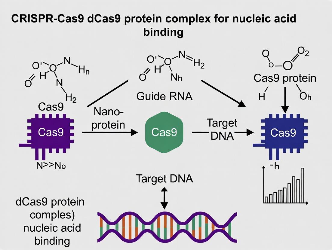
What is dCas9? Understanding the Core Mechanism of CRISPR Without Cutting
The CRISPR-Cas9 system, derived from a bacterial adaptive immune system, revolutionized genetic engineering with its precise DNA cleavage capability. The broader thesis of this field posits that by deactivating the nuclease function of Cas9, creating a catalytically "dead" Cas9 (dCas9), we can repurpose the system from a "molecular scissor" to a "programmable GPS." This dCas9 paradigm shift enables high-specificity nucleic acid binding without cleavage, unlocking transformative applications in transcriptional regulation, epigenetic editing, live-cell imaging, and diagnostics. This whitepaper provides an in-depth technical guide to the core principles, methodologies, and applications of dCas9 technology for researchers and drug development professionals.
Core Engineering: From Cas9 to dCas9
The conversion of wild-type Streptococcus pyogenes Cas9 (spCas9) to dCas9 is achieved through site-directed mutagenesis of two key catalytic residues in the RuvC (D10) and HNH (H840) nuclease domains. This generates a protein that maintains its guide RNA (gRNA)-programmed DNA-binding specificity but is incapable of generating double-strand breaks.
Table 1: Key Mutations for Generating Common dCas9 Variants
| Cas9 Origin | Wild-type Residues (RuvC / HNH) | dCas9 Mutations | Common Designation |
|---|---|---|---|
| S. pyogenes (spCas9) | D10 / H840 | D10A, H840A | spdCas9 |
| Staphylococcus aureus (saCas9) | D10 / N580 | D10A, N580A | sadCas9 |
| Campylobacter jejuni (cjCas9) | D8 / H559 | D8A, H559A | cjdCas9 |
Key Functional Modalities of the dCas9 Platform
dCas9 serves as a programmable DNA-binding scaffold. Its fusion to effector domains enables precise perturbation of genomic function.
Table 2: Primary dCas9-Effector Fusion Modalities and Applications
| Fusion Effector Domain | Resulting System | Primary Function | Key Application |
|---|---|---|---|
| Transcriptional Repressor (e.g., KRAB) | CRISPRi | Silences gene transcription | Loss-of-function studies, therapeutic suppression |
| Transcriptional Activator (e.g., VP64, p65AD) | CRISPRa | Activates gene transcription | Gain-of-function studies, gene therapy |
| Epigenetic Writer (e.g., DNMT3A, p300) | Epi-dCas9 | Adds DNA methylation or histone acetylation | Epigenetic reprogramming |
| Epigenetic Eraser (e.g., TET1, HDAC) | Epi-dCas9 | Removes DNA methylation or histone acetylation | Epigenetic reprogramming |
| Fluorescent Protein (e.g., GFP, mCherry) | dCas9-imaging | Binds to genomic loci | Live-cell chromatin imaging |
| Base Editor (e.g., TadA, rAPOBEC1) | dCas9-Base Editor | Catalyzes targeted point mutation | Gene correction without DSBs |
Diagram Title: dCas9 Functional Fusion Modalities & Outcomes
Experimental Protocols
Protocol: Establishment of a Stable dCas9-KRAB CRISPRi Cell Line
Objective: Generate a mammalian cell line stably expressing dCas9 fused to the Kruppel-associated box (KRAB) transcriptional repressor for programmable gene knockdown.
Materials: See "The Scientist's Toolkit" below. Procedure:
- Lentiviral Production:
- Co-transfect HEK293T cells with the following plasmids using a polyethylenimine (PEI) protocol:
- Packaging plasmid (psPAX2): 10 µg
- Envelope plasmid (pMD2.G): 5 µg
- Transfer plasmid (lenti-dCas9-KRAB-Puro): 15 µg
- At 48- and 72-hours post-transfection, harvest lentivirus-containing supernatant, filter through a 0.45 µm PES filter, and concentrate via ultracentrifugation (70,000 x g, 2 hrs at 4°C).
- Co-transfect HEK293T cells with the following plasmids using a polyethylenimine (PEI) protocol:
- Cell Line Transduction:
- Plate target cells (e.g., HEK293, K562) at 50% confluency in a 6-well plate.
- Add concentrated lentivirus and polybrene (8 µg/mL final concentration).
- Centrifuge at 800 x g for 30 mins at 32°C (spinoculation).
- Replace medium after 24 hours.
- Selection and Validation:
- At 48 hours post-transduction, add puromycin (concentration determined by kill curve, typically 1-5 µg/mL).
- Maintain selection for 5-7 days until control cells are dead.
- Validate dCas9-KRAB expression via western blot (anti-FLAG or anti-Cas9 antibody) and functional testing with a validated gRNA targeting a housekeeping gene (e.g., GAPDH), followed by qRT-PCR.
Protocol: Live-Cell Imaging with dCas9-EGFP
Objective: Visualize repetitive genomic loci in living cells using dCas9-EGFP and multiple, tiled gRNAs.
Procedure:
- gRNA Design and Cloning:
- Design 8-16 gRNAs targeting ~2 kb region of interest (e.g., telomere repeats, centromere).
- Clone gRNA arrays into a U6-driven expression plasmid (e.g., pMCB320 using Golden Gate assembly).
- Transient Transfection:
- Co-transfect a cell line stably expressing dCas9-EGFP (or transiently co-transfect dCas9-EGFP and gRNA plasmids) using a suitable method (lipofection, nucleofection).
- Use a total of 1-2 µg plasmid DNA per well of a 24-well glass-bottom imaging plate.
- Imaging and Analysis:
- 24-48 hours post-transfection, acquire images on a confocal or widefield fluorescence microscope with a high-NA objective (60x or 100x oil).
- Acquire Z-stacks (0.5 µm steps). Use appropriate filter sets for EGFP (Ex/Em: 488/510 nm).
- Process images (deconvolution, maximum intensity projection) and quantify locus brightness and number using software (e.g., Fiji/ImageJ).
Quantitative Data & Performance Metrics
Table 3: Performance Characteristics of Key dCas9 Systems
| System | Typical Efficiency (mRNA modulation) | On-Target Specificity (Key Metric) | Key Off-Target Concern | Reference (Recent) |
|---|---|---|---|---|
| CRISPRi (dCas9-KRAB) | 80-99% knockdown (strong promoters) | High (minimal off-target transcription) | Potential seed-mediated binding | (Horlbeck et al., Nat Biotechnol 2023) |
| CRISPRa (dCas9-VP64-p65AD) | 10-100 fold activation (varies widely) | Moderate | Promoter-specific gRNA design critical | (Gilbert et al., Cell 2023) |
| dCas9-p300 (Acetylation) | 5-30 fold increase in H3K27ac | Moderate (local spreading) | Histone acetylation can spread ~1kb | (Hilton et al., Nat Biotechnol 2023) |
| dCas9-TET1 (Demethylation) | 20-80% reduction in CpG methylation | High (within ~100bp) | Passive demethylation over time | (Nakamura et al., Science 2023) |
| dCas9-EGFP Imaging | Signal-to-Noise: 3-10 fold over background | High (requires tiled gRNAs) | Photobleaching, background signal | (Qin et al., Nucleic Acids Res 2024) |
Signaling Pathways in dCas9-Based Transcriptional Regulation
The dCas9-KRAB system recruits endogenous repressive machinery through a well-defined pathway.
Diagram Title: dCas9-KRAB Mediated Transcriptional Silencing Pathway
The Scientist's Toolkit: Essential Research Reagent Solutions
Table 4: Key Reagents for dCas9 Research
| Reagent / Material | Supplier Examples | Function in dCas9 Experiments |
|---|---|---|
| Lentiviral dCas9 Effector Plasmids | Addgene (pLX-sgRNA, lenti dCas9-KRAB-Puro), Sigma (MISSION CRISPRi) | Stable delivery and expression of dCas9-effector fusions in mammalian cells. |
| gRNA Cloning Kits | Synthego (gRNA Synthesis), IDT (Alt-R CRISPR-Cas9 gRNA), Takara Bio (Guide-it) | Fast and efficient generation of expression constructs for single or arrayed gRNAs. |
| Modified Synthetic gRNAs | Synthego, Trilink (CleanCap), IDT (Alt-R) | Chemically modified gRNAs (e.g., 2'-O-methyl, phosphorothioate) for enhanced stability and reduced immunogenicity. |
| Anti-Cas9/dCas9 Antibodies | Cell Signaling Tech (7A9-3A3), Abcam (ab191468), MilliporeSigma (8G8H7) | Validation of dCas9 protein expression via Western Blot, ELISA, or immunofluorescence. |
| dCas9-specific NGS Off-Target Kits | IDT (Alt-R CRISPR-Cas9 HTS Kit), Takara Bio (Guide-it Mutation Detection Kit) | Detection of potential off-target binding events for dCas9 systems, especially for epigenetic editors. |
| Live-Cell Imaging Dyes & Mountants | Thermo Fisher (ProLong Live), Tocris (Spinach RNA aptamer dyes) | Compatible reagents for long-term live imaging of dCas9-fluorescent protein fusions. |
| Epigenetic Modification Kits (qPCR/ELISA) | Active Motif (H3K27ac ELISA), Zymo Research (MethylFlash) | Quantitative validation of epigenetic changes induced by dCas9-effector fusions. |
The dCas9 paradigm represents a fundamental shift from destructive cleavage to programmable, nondestructive nucleic acid targeting. This GPS-like precision binding capability has spawned a vast toolkit for interrogating and manipulating genome function, regulation, and structure. For drug development, dCas9 systems offer unparalleled opportunities for targeted transcriptional modulation and epigenetic therapy with potentially safer profiles than nuclease-based approaches. As engineering advances—including improved specificity, compact variants, and novel effector domains—continue, the utility of dCas9 as a core platform for both basic research and therapeutic innovation will undoubtedly expand.
This whitepaper provides a technical analysis of the D10A and H840A mutations in Streptococcus pyogenes Cas9, which collectively convert the wild-type nuclease into a catalytically inactive "dead" Cas9 (dCas9). Framed within the broader thesis of utilizing dCas9 for precise nucleic acid binding without cleavage, this guide details the structural and mechanistic basis for the loss of nuclease function, supported by quantitative biochemical data, experimental protocols for validation, and essential research tools for the field.
The CRISPR-Cas9 system has revolutionized genetic engineering. The wild-type Cas9 protein induces double-strand breaks (DSBs) via two distinct nuclease domains: the RuvC-like domain cleaves the non-target strand, and the HNH-like domain cleaves the target strand. For applications requiring DNA binding without cleavage—such as transcriptional modulation, epigenetic editing, or live-cell imaging—the nuclease activity must be ablated. The combination of point mutations D10A in the RuvC domain and H840A in the HNH domain achieves this, producing dCas9, a foundational tool for precision genomic targeting.
Structural & Mechanistic Basis of Inactivation
Active Site Chemistry and Mutational Impact
The catalytic mechanisms of both nuclease domains rely on a set of conserved residues that coordinate divalent metal ions (typically Mg²⁺) essential for phosphodiester bond hydrolysis.
- RuvC Domain (D10A): The catalytic core of the RuvC domain features a DED motif (Asp10, Glu762, Asp986). Asp10 plays a critical role in positioning the catalytic metal ion. The D10A substitution replaces the negatively charged carboxylate side chain with a non-polar methyl group, disrupting metal ion binding and rendering the domain incapable of activating a water molecule for nucleophilic attack.
- HNH Domain (H840A): The HNH domain resembles a Mg²⁺-dependent endonuclease. His840 is part of a catalytic triad (HNH) that coordinates the metal ion and stabilizes the transition state. The H840A mutation removes the imidazole side chain, eliminating its metal-coordinating and proton-shuttling capabilities, thereby abolishing cleavage.
Table 1: Catalytic Residue Functions and Mutational Effects
| Domain | Wild-Type Residue | Proposed Role in Catalysis | Mutation | Consequence |
|---|---|---|---|---|
| RuvC | Aspartate 10 (D10) | Mg²⁺ ion coordination, nucleophile activation | D10A | Disrupts metal binding, abolishes non-target strand cleavage |
| HNH | Histidine 840 (H840) | Mg²⁺ ion coordination, transition state stabilization | H840A | Disrupts catalytic triad, abolishes target strand cleavage |
Quantitative Analysis of Cleavage Inactivation
Biochemical assays consistently demonstrate a near-total loss of nuclease activity in the double mutant.
Table 2: Quantitative Comparison of Nuclease Activity
| Cas9 Variant | In Vitro Cleavage Efficiency (%) | Cell-Based DSB Formation (Relative to WT) | Key Assay |
|---|---|---|---|
| Wild-Type (WT) Cas9 | 100 ± 5 | 1.0 | Plasmid linearization, SURVEYOR/T7E1 |
| D10A Single Mutant | <5 (Non-target strand intact) | <0.05 | Strand-specific cleavage assay |
| H840A Single Mutant | <5 (Target strand intact) | <0.05 | Strand-specific cleavage assay |
| D10A/H840A (dCas9) | Undetectable (<0.1) | Undetectable (<0.01) | Plasmid retention, sequencing |
Experimental Protocols for Validating dCas9 Activity
1In VitroCleavage Assay
Purpose: To quantitatively assess the loss of DNA cleavage activity. Materials: Purified WT Cas9, D10A, H840A, and D10A/H840A proteins; target plasmid DNA; sgRNA; reaction buffer (20 mM HEPES pH 7.5, 150 mM KCl, 10 mM MgCl₂, 1 mM DTT). Procedure:
- Form ribonucleoprotein (RNP) complexes by incubating 100 nM Cas9 protein with 120 nM sgRNA in reaction buffer for 10 min at 25°C.
- Add 10 nM target plasmid (containing the protospacer and PAM) to the RNP mixture.
- Incubate at 37°C for 1 hour.
- Stop the reaction with EDTA (50 mM final) and Proteinase K.
- Analyze products by agarose gel electrophoresis (0.8% gel). A successful inactivation will show only supercoiled/uncut plasmid for dCas9, compared to linearized product for WT.
Cell-Based Reporter Assay for DSB Detection
Purpose: To confirm the absence of double-strand breaks in living cells. Materials: HEK293T cells; GFP-based reporter plasmid (e.g., with an out-of-frame GFP gene that can be restored by Cas9-mediated DSB and error-prone repair); plasmids expressing WT or mutant Cas9 and sgRNA; flow cytometer. Procedure:
- Co-transfect cells with the GFP reporter plasmid, sgRNA expression plasmid, and a plasmid encoding WT, single mutant, or dCas9.
- Culture cells for 72 hours to allow for expression, cutting, and repair.
- Harvest cells and analyze GFP positivity via flow cytometry.
- Quantification: GFP+ percentage with dCas9 should be at baseline (comparable to negative control), while WT Cas9 will show a significant GFP+ population due to indel formation restoring the reading frame.
Experimental Validation Workflow for dCas9
The Scientist's Toolkit: Research Reagent Solutions
Table 3: Essential Materials for dCas9-Based Research
| Reagent/Material | Function & Application | Example/Supplier |
|---|---|---|
| dCas9 Expression Vectors | Delivery of catalytically inactive Cas9 (D10A/H840A) into cells for binding applications. | pLV-dCas9 (Addgene #52962), pcDNA-dCas9 |
| dCas9 Fusion Protein Systems | dCas9 linked to effector domains (e.g., VP64, p300, KRAB) for transcriptional control. | dCas9-VP64 (Activation), dCas9-KRAB (Repression) |
| Purified dCas9 Protein | For in vitro studies, SELEX, or direct delivery as RNP complexes. | Recombinant S. pyogenes dCas9 (NEB, Thermo Fisher) |
| sgRNA Scaffold Plasmids | For cloning and expression of guide RNAs complementary to the target of interest. | pGL3-U6-sgRNA (Addgene) |
| Synthetic sgRNAs | Chemically modified, high-purity guides for enhanced stability and RNP complex formation. | Custom synthesis (IDT, Synthego) |
| Nuclease Detection Kits | To confirm absence of cleavage (e.g., T7E1/SURVEYOR for residual activity). | SURVEYOR Mutation Detection Kit (IDT) |
| Positive Control (WT Cas9) | Essential control to validate that experimental conditions support cleavage. | WT SpCas9 expression plasmid or protein. |
| Next-Generation Sequencing (NGS) | Comprehensive assessment of off-target binding and verification of no indels. | Illumina-based amplicon sequencing. |
dCas9 in the Broader Thesis: Applications Beyond Cleavage
The creation of dCas9 via D10A/H840A mutations was the pivotal step that unlocked the potential of CRISPR as a programmable DNA-binding platform. This enables:
- CRISPRi/a: Transcriptional repression or activation.
- Epigenetic Editing: Targeted DNA methylation or histone modification.
- Live-Cell Imaging: Tagged dCas9 for visualization of genomic loci.
- High-Throughput Screens: dCas9-based functional genomics without genetic alteration.
dCas9 as a Platform for Diverse Applications
The strategic introduction of the D10A and H840A mutations disables the metal-dependent catalytic centers of the Cas9 nuclease domains through distinct but complementary mechanisms. The resulting dCas9 protein retains high-affinity, programmable DNA-binding capability, fulfilling a core requirement for the expanding field of cleavage-independent CRISPR technologies. This precise engineering underscores the principle that understanding and manipulating protein structure is fundamental to developing next-generation biotechnological tools.
Within the broader context of CRISPR-Cas9 research focused on nucleic acid binding without cleavage, the catalytically dead Cas9 (dCas9) system has become a cornerstone for precise genomic targeting. The fundamental efficacy of dCas9 as a programmable DNA-binding platform hinges on the precise assembly and function of its binding complex. This guide provides a technical deep-dive into the three core pillars governing this complex: the single-guide RNA (sgRNA), Protospacer Adjacent Motif (PAM) recognition, and the kinetics and thermodynamics of target site occupancy.
Core Components and Quantitative Parameters
The binding affinity and specificity of the dCas9 complex are governed by quantifiable interactions between its components and target DNA.
Table 1: Key Quantitative Parameters of the dCas9-sgRNA-DNA Complex
| Parameter | Typical Value/Range (S. pyogenes Cas9) | Description/Impact on Occupancy |
|---|---|---|
| PAM Sequence | 5'-NGG-3' | Strict requirement for initial DNA recognition; NGG is optimal for Sp-dCas9. |
| sgRNA Length (Spacer) | 20 nt | Standard length; truncation to 17-18 nt can reduce off-target binding. |
| On-target Dissociation Constant (Kd) | 0.1 - 1 nM | Measures binding affinity at the intended target site under optimal conditions. |
| Off-target Kd | > 10 nM | Weaker affinity for mismatched sites; varies with mismatch number/position. |
| Association Rate (kon) | ~10^5 M^-1s^-1 | Speed of complex formation; influenced by PAM recognition and R-loop formation. |
| Dissociation Rate (koff) | ~10^-4 s^-1 | Speed of complex dissociation; slower koff increases occupancy time. |
| Half-life (t1/2) On-target | ~60-90 minutes | Duration of stable binding; critical for prolonged effector function (e.g., repression). |
| GC Content (Optimal) | 40-60% | Impacts sgRNA DNA-binding stability; extremes can reduce occupancy. |
Table 2: Influence of sgRNA Mismatches on Target Occupancy
| Mismatch Position (from PAM) | Relative Binding Affinity (% of Perfect Match) | Impact on Occupancy |
|---|---|---|
| Distal (positions 18-20) | 60-90% | Moderate reduction; tolerated more than proximal mismatches. |
| Middle (positions 10-17) | 10-50% | Significant reduction in occupancy and specificity. |
| Proximal (positions 1-10, "Seed") | <5% | Severe or complete loss of binding and occupancy. |
| Multiple Mismatches (>3) | <1% | Near-total loss of specific complex formation. |
Detailed Experimental Protocols
Protocol: Electrophoretic Mobility Shift Assay (EMSA) for dCas9-sgRNA:DNA Binding Affinity (Kd)
Objective: Quantify the equilibrium dissociation constant (Kd) of the dCas9-sgRNA complex binding to target DNA.
Materials:
- Purified dCas9 protein (e.g., Sp-dCas9, with His-tag for purification).
- In vitro transcribed or synthesized target sgRNA.
- Target DNA duplex (30-50 bp, containing PAM and protospacer).
- Radiolabeled (γ-32P-ATP) or fluorescently labeled (e.g., Cy5) DNA probe.
- Binding Buffer: 20 mM HEPES pH 7.5, 100 mM KCl, 5 mM MgCl2, 1 mM DTT, 5% glycerol, 0.1 mg/mL BSA.
- Non-denaturing Polyacrylamide Gel (e.g., 6%).
- Gel Electrophoresis and Imaging System (Phosphorimager or fluorescence scanner).
Procedure:
- Complex Formation: Assemble 20 µL reactions with a constant, trace amount of labeled DNA probe (~0.1 nM) and increasing concentrations of pre-assembled dCas9-sgRNA ribonucleoprotein (RNP) complex (e.g., 0, 0.1, 0.5, 1, 5, 10, 50, 100 nM) in binding buffer. Incubate at 37°C for 30-60 minutes to reach equilibrium.
- Gel Loading: Add non-denaturing loading dye to each reaction. Do not heat.
- Electrophoresis: Load samples onto a pre-run 6% non-denaturing PAGE gel in 0.5x TBE buffer at 4°C. Run at 80-100 V for 60-90 minutes.
- Detection & Analysis: Expose gel to a phosphor screen or fluorescence scanner. Quantify the intensity of the free DNA band and the shifted RNP-DNA complex band for each RNP concentration.
- Kd Calculation: Fit the data (Fraction Bound vs. [RNP]) to a one-site specific binding model using software like Prism: Fraction Bound = Bmax * [RNP] / (Kd + [RNP]).
Protocol: Chromatin Immunoprecipitation-qPCR (ChIP-qPCR) forIn VivoTarget Site Occupancy
Objective: Measure the relative occupancy of dCas9-based fusion proteins at specific genomic loci in living cells.
Materials:
- Cells expressing dCas9 (or dCas9-fusion) and sgRNA.
- Formaldehyde for crosslinking.
- Cell Lysis & Sonication Buffers.
- Antibody specific to dCas9 (or epitope tag, e.g., anti-FLAG, anti-HA).
- Protein A/G Magnetic Beads.
- qPCR Primers for target and control (off-target) genomic sites.
- DNA Purification Kit.
Procedure:
- Crosslinking: Add 1% formaldehyde directly to cell culture medium for 10 min at room temperature. Quench with 125 mM glycine.
- Cell Lysis & Sonication: Lyse cells. Sonicate chromatin to shear DNA to ~200-500 bp fragments. Confirm fragment size by agarose gel.
- Immunoprecipitation: Clarify lysate. Incubate supernatant with antibody against dCas9/tag overnight at 4°C. Add Protein A/G beads for 2 hours. Wash beads extensively.
- Elution & Reverse Crosslinking: Elute chromatin from beads. Add NaCl and reverse crosslinks by heating at 65°C overnight.
- DNA Purification & qPCR: Purify DNA. Perform qPCR with primers for the target site and non-target control regions.
- Data Analysis: Calculate % Input or Fold Enrichment over a negative control locus (e.g., GAPDH promoter) for each sgRNA.
Key Diagrams
Diagram 1: dCas9-sgRNA Complex Assembly and Target Binding Pathway
Diagram 2: Experimental Workflow for Measuring dCas9 Binding and Occupancy
The Scientist's Toolkit: Research Reagent Solutions
Table 3: Essential Reagents for dCas9 Binding and Occupancy Studies
| Item | Example Product/Catalog | Function in Research |
|---|---|---|
| Catalytically Dead Cas9 (dCas9) | Purified Sp-dCas9 protein (e.g., Macrolab, IDT). | Core DNA-binding protein; scaffold for sgRNA and effector fusions. |
| sgRNA Synthesis Kit | In vitro Transcription Kit (e.g., NEB HiScribe) or synthetic sgRNA (IDT). | Produces the targeting component that directs dCas9 to specific DNA sequences. |
| dCas9 Expression Plasmid | px458-derived vector (Addgene #48138) with D10A/H840A mutations. | For stable or transient expression of dCas9 in mammalian cells. |
| sgRNA Expression Plasmid/Vector | U6-promoter driven sgRNA cloning vector (e.g., Addgene #41824). | For co-expression of sgRNA with dCas9 in cells. |
| Anti-dCas9 / Epitope Tag Antibody | Anti-FLAG M2 (Sigma), Anti-HA (Cell Signaling), Anti-Cas9 (7A9). | Critical for ChIP experiments to immunoprecipitate the dCas9 complex. |
| ChIP-grade Protein A/G Magnetic Beads | Magna ChIP Protein A/G beads (Millipore). | Efficient capture of antibody-bound dCas9-DNA complexes. |
| Fluorescent or Radioactive Nucleotides | Cy5-dCTP or γ-32P-ATP. | For labeling DNA probes in in vitro binding assays (EMSA). |
| Non-denaturing Gel Electrophoresis System | Mini-PROTEAN TGX system (Bio-Rad). | To separate bound from unbound DNA in EMSA. |
| Next-Generation Sequencing (NGS) Library Prep Kit | NEBNext Ultra II DNA Library Prep Kit. | For genome-wide occupancy analysis (ChIP-seq, GUIDE-seq). |
| dCas9-Specific Cell Line | HEK293T stably expressing dCas9 (e.g., from SLiM system). | Provides a consistent background for cellular occupancy and functional assays. |
CRISPR-Cas9 revolutionized genome editing via site-specific DNA cleavage. The development of the catalytically dead variant, dCas9, transformed the system into a versatile, programmable DNA- and RNA-binding platform without inducing double-strand breaks. This whitepaper examines the core advantages that underpin dCas9's utility in advanced research and therapeutic development: its unparalleled high-specificity binding, inherent reversibility, and powerful multiplexing potential. These attributes position dCas9-based technologies as foundational tools for transcriptional regulation, epigenetic editing, high-resolution imaging, and diagnostic applications, forming a critical thesis within the broader field of non-cleaving nucleic acid interrogation.
High-Specificity Binding: Mechanisms and Validation
The specificity of dCas9 is governed by the 20-nucleotide spacer sequence within the single-guide RNA (sgRNA) and the requisite Protospacer Adjacent Motif (PAM). Recent studies quantify specificity via genome-wide profiling techniques.
Table 1: Quantifying dCas9 Binding Specificity with ChIP-seq
| Metric | dCas9 (with perfect-match sgRNA) | dCas9 (with 1-2 bp mismatch sgRNA) | Method |
|---|---|---|---|
| On-target Peak Signal | 500 - 2000 reads per million (RPM) | < 50 RPM | ChIP-seq |
| Genome-wide Off-target Sites (≥ 20% of on-target signal) | 0 - 3 sites | > 15 sites | ChIP-seq, Digenome-seq |
| Primary Determinant | sgRNA complementarity & PAM (NGG for Sp-dCas9) | Mismatch tolerance at PAM-distal positions | N/A |
Experimental Protocol: Chromatin Immunoprecipitation Sequencing (ChIP-seq) for dCas9 Binding Specificity
- Cell Transfection: Introduce plasmids expressing dCas9 (fused to a tag, e.g., FLAG or HA) and a target-specific sgRNA into the cell line of interest.
- Crosslinking: Treat cells with 1% formaldehyde for 10 min at room temperature to fix protein-nucleic acid interactions.
- Cell Lysis & Chromatin Shearing: Lyse cells and sonicate chromatin to shear DNA into 200-500 bp fragments.
- Immunoprecipitation: Incubate lysate with antibody-coated beads specific to the dCas9 tag. Wash away non-specific binders.
- Reverse Crosslinking & Purification: Elute and reverse crosslinks at 65°C. Purify the co-immunoprecipitated DNA.
- Library Prep & Sequencing: Prepare a next-generation sequencing library from the purified DNA and perform high-throughput sequencing.
- Bioinformatic Analysis: Map sequencing reads to the reference genome. Call peaks to identify significant dCas9 binding sites. Compare to the intended target sequence and potential off-targets predicted by algorithms (e.g., Cas-OFFinder).
Diagram 1: dCas9 ChIP-seq Workflow for Specificity Profiling
Reversibility: Dynamic Control of Binding and Function
Unlike genetic editing, dCas9-mediated effects are inherently reversible upon degradation of the sgRNA or dCas9 protein, or via competitor sgRNA delivery. This is critical for safety and functional studies.
Table 2: Reversibility Kinetics of dCas9-Mediated Transcriptional Repression (CRISPRi)
| Intervention | Time to 50% Recovery of Basal Expression | Experimental Model |
|---|---|---|
| Doxycycline-induced sgRNA Transcript Turn-off | ~24-48 hours | HEK293T with inducible promoter |
| Delivery of Competitive, Non-functional sgRNA | ~12-24 hours | HeLa cells |
| dCas9 Protein Degradation (AID System) | < 6 hours | iPSCs with auxin-inducible degron tag |
Experimental Protocol: Measuring Reversibility of dCas9 CRISPR Interference (CRISPRi)
- Stable Cell Line Generation: Create a cell line stably expressing dCas9 fused to a KRAB transcriptional repression domain.
- Inducible sgRNA Delivery: Introduce an sgRNA targeting a gene of interest (e.g., a surface receptor) via a doxycycline-inducible vector.
- Repression Phase: Add doxycycline (1 µg/mL) for 72 hours to induce sgRNA expression and repress the target gene. Measure repression via qPCR or flow cytometry.
- Wash-Out/Intervention: Remove doxycycline to halt new sgRNA transcription. Alternatively, transfect a large excess of a "scrambled" non-targeting sgRNA plasmid to compete for dCas9 binding.
- Time-Course Monitoring: Harvest cells at 0, 12, 24, 48, and 72 hours post-intervention. Quantify target gene mRNA (by RT-qPCR) or protein expression to generate a recovery kinetic curve.
Diagram 2: Reversible Gene Repression via Inducible dCas9-KRAB
Multiplexing Potential: Parallel Interrogation of Genomic Loci
The facile programmability of sgRNAs enables simultaneous targeting of multiple genomic loci by co-expressing several sgRNAs from a single array or multiple vectors, a key advantage for studying polygenic traits and signaling networks.
Table 3: Multiplexing dCas9 Platforms and Their Capacities
| Platform | Typical Maximum Targets | Key Application | Delivery Method |
|---|---|---|---|
| Polycistronic tRNA-gRNA (PTG) Array | Up to 10-12 sgRNAs | Genome-scale CRISPRi/a screens | Lentiviral transduction |
| Csy4-based RNA Processing System | Up to 8 sgRNAs | Simultaneous imaging of multiple loci | Plasmid transfection |
| Multiple sgRNA Expression Vectors | 2-4 sgRNAs (co-transfection) | Combinatorial transcriptional programs | Lipid nanoparticle |
Experimental Protocol: Multiplexed Transcriptional Activation (CRISPRa) Using a PTG Array
- Array Design: Design oligonucleotides encoding your target sgRNA sequences. Each sgRNA is flanked by tRNA sequences (e.g., tRNA^Gly) for processing. Assemble these into a single polycistronic sequence via Golden Gate or Gibson assembly.
- Cloning: Clone the PTG array into a lentiviral vector expressing dCas9 fused to a strong transcriptional activator (e.g., VPR or p65AD).
- Virus Production & Transduction: Package lentiviral particles and transduce target cells at a low MOI (<0.3) to ensure single-copy integration.
- Selection & Validation: Apply antibiotic selection (e.g., puromycin) for 5-7 days. Harvest polyclonal populations. Validate multiplexed activation via RT-qPCR for all target genes and next-generation RNA-seq for unbiased transcriptome analysis.
Diagram 3: Multiplexed Gene Activation via a PTG sgRNA Array
The Scientist's Toolkit: Key Reagent Solutions
Table 4: Essential Research Reagents for dCas9 Nucleic Acid Binding Studies
| Reagent / Material | Provider Examples | Critical Function |
|---|---|---|
| Catalytically Dead Cas9 (dCas9) Expression Plasmids | Addgene (e.g., #47106, #99378), Thermo Fisher | Core protein scaffold for DNA binding without cleavage. |
| dCas9 Transcriptional Effector Fusions (KRAB, VPR, p300) | Addgene, Sigma-Aldrich | Enables functional outcomes like repression (CRISPRi) or activation (CRISPRa). |
| sgRNA Cloning Kits & Backbone Vectors | ToolGen, Synthego, Integrated DNA Technologies (IDT) | Facilitates rapid generation and cloning of target-specific sgRNA sequences. |
| Validated, Pre-designed sgRNA Libraries | Dharmacon (Horizon), MilliporeSigma | Off-the-shelf, quality-controlled sgRNA sets for genome-scale screens. |
| Anti-FLAG/HA Magnetic Beads for ChIP | MilliporeSigma, Cell Signaling Technology | Essential for chromatin immunoprecipitation assays to map dCas9 binding. |
| Lentiviral Packaging Mixes (for PTG Arrays) | Takara Bio, OriGene | Enables stable delivery of multiplexed sgRNA arrays into difficult-to-transfect cells. |
| dCas9-Specific Validated Antibodies | Abcam, Cell Signaling Technology, Diagenode | Critical for Western Blot validation and ChIP-grade immunoprecipitation. |
| Nucleofection/Kits for Primary Cell Delivery | Lonza, Nucleofector | High-efficiency delivery of ribonucleoprotein (RNP) complexes of dCas9 and sgRNA. |
Within the broader thesis on CRISPR-Cas9 and its catalytically dead variant (dCas9) for programmable nucleic acid binding without cleavage, three primary limitations consistently impede clinical and research translation: off-target binding, steric hindrance, and delivery challenges. This technical guide provides an in-depth analysis of these core constraints, supported by current experimental data and methodologies.
Off-Target Binding: Mechanisms and Mitigation
Off-target binding occurs when the dCas9-guide RNA (gRNA) ribonucleoprotein (RNP) complex binds to genomic loci with sequence homology to the intended target, leading to unintended transcriptional modulation or epigenetic editing.
Quantitative Analysis of Off-Target Rates
Recent studies (2023-2024) quantify off-target effects using high-throughput sequencing methods like GUIDE-seq, CIRCLE-seq, and Digenome-seq.
Table 1: Off-Target Profile Comparison Across dCas9 Engineering Strategies
| Strategy | Description | Average Reduction in Off-Target Binding vs. Wild-Type dCas9 (%) | Key Trade-off |
|---|---|---|---|
| High-Fidelity (HF1) dCas9 | Amino acid substitutions (N497A/R661A/Q695A/Q926A) to reduce non-specific backbone contacts. | 70-85% | Minor reduction (~25%) in on-target binding affinity. |
| Enhanced Specificity (eSpCas9) dCas9 | Mutations (K848A/K1003A/R1060A) to alter positive charge distribution, weakening non-canonical binding. | 65-80% | Can require higher expression levels for equivalent on-target effect. |
| Hyper-accurate (HypaCas9) dCas9 | Mutations (N692A/M694A/Q695A/H698A) to stabilize the gRNA-DNA heteroduplex recognition state. | 75-90% | Increased sensitivity to gRNA-DNA mismatch, especially at positions 18-20. |
| sgRNA Engineering (Truncated gRNAs) | Using gRNAs shortened by 2-3 nucleotides at the 5' end. | 50-70% | Significant reduction in on-target activity for some loci. |
| Protein-Directed Evolution (evoCas9) | Phage-assisted continuous evolution to select for specificity. | >90% | Can exhibit novel PAM requirements, limiting targetable sites. |
Experimental Protocol: GUIDE-seq for dCas9 Off-Target Detection
Objective: Genome-wide identification of dCas9-gRNA off-target binding sites. Reagents:
- Oligonucleotide Duplex: A 34-bp double-stranded oligonucleotide with phosphorothioate modifications, serving as the integration tag.
- dCas9-gRNA RNP Complex: Purified dCas9 protein complexed with in vitro transcribed target gRNA.
- NGS Library Prep Kit: For sequencing tag integration sites. Procedure:
- Co-deliver the oligonucleotide tag and dCas9-gRNA RNP into target cells via nucleofection.
- Allow 72 hours for tag integration at double-strand breaks generated by a co-transfected, catalytically active Cas9 (as a positive control) and, crucially, at dCas9 binding sites via endogenous repair mechanisms.
- Harvest genomic DNA and shear to ~500 bp fragments.
- Perform tag-specific PCR amplification to enrich fragments containing the integrated tag.
- Prepare an NGS library from the amplified product.
- Sequence and align reads to the reference genome. Off-target sites are identified as genomic loci flanking the integrated tag sequence. Note: For pure dCas9 applications, a catalytically active Cas9 nuclease is often included as a parallel control to distinguish binding from cleavage events. Advanced methods like Binding (rather than cutting) OUTPUT now adapt this for direct dCas9 binding detection.
Steric Hindrance in dCas9 Effector Recruitment
Steric hindrance refers to the physical blockade imposed by the large dCas9 protein (~160 kDa), which can impede the binding of endogenous transcription factors, RNA polymerase, or fused effector domains (activators, repressors, epigenomic modifiers).
Quantitative Impact on Transcriptional Modulation
The efficiency of dCas9-based transcriptional activation (CRISPRa) or repression (CRISPRi) is directly limited by steric constraints.
Table 2: Steric Limitations of Common dCas9-Effector Fusions
| Effector System | Total Size (kDa, approx.) | Max Observed Transcriptional Activation/Repression Fold-Change | Key Limitation |
|---|---|---|---|
| dCas9-VP64 (minimal activator) | ~190 | 5-50x activation | Limited by recruitment of single weak activator domain. |
| dCas9-p300 Core (histone acetyltransferase) | ~210 | 10-100x activation | Large size can hinder chromatin access; activity is locus-dependent. |
| dCas9-KRAB (repressor domain) | ~180 | 10-100x repression (mRNA reduction) | Dense chromatin can prevent KRAB domain from recruiting silencing machinery. |
| dCas9-DNMT3A (DNA methyltransferase) | ~220 | Can achieve ~80% methylation at CpG sites | Bulky enzyme; methylation spread is difficult to control precisely. |
| SunTag / Scaffold Systems (recruit multiple effectors) | >250+ | Up to 2000x activation (SunTag-VP64) | Extreme size exacerbates delivery challenges and can trigger immune response. |
Experimental Protocol: Chromatin Accessibility Assay (ATAC-seq) Post-dCas9 Binding
Objective: Assess changes in local chromatin architecture and nucleosome positioning upon dCas9-effector binding. Reagents:
- Hyperactive Tn5 Transposase: Pre-loaded with sequencing adapters.
- Cell Permeabilization Buffer: Contains digitonin or NP-40.
- Nuclei Isolation Buffer. Procedure:
- Transfect cells with dCas9-effector and target gRNA constructs. Include a dCas9-only control.
- After 48-72 hours, harvest and lyse cells to isolate nuclei.
- Treat nuclei with the adapter-loaded Tn5 transposase for 30 minutes at 37°C. Tn5 inserts adapters into open, nucleosome-free regions of DNA.
- Purify the transposed DNA and amplify it via PCR using barcoded primers.
- Sequence the library. Align reads to the reference genome.
- Analysis: Compare read density and fragment size distribution at the target locus between dCas9-effector samples and the dCas9-only control. A decrease in accessible fragments at the immediate target site indicates steric occlusion by dCas9. Peaks of accessibility may appear flanking the site if dCas9 binding displaces nucleosomes.
Delivery Challenges: In Vivo and In Vitro Hurdles
Efficient delivery of the large dCas9 protein and its gRNA, often with additional effector components, remains the paramount translational challenge.
Delivery Modality Efficiency Comparison
Table 3: Quantitative Delivery Efficiency for dCas9 Systems
| Delivery Method | Max Payload Size | Typical In Vitro Efficiency (HEK293T) | Typical In Vivo Efficiency (Mouse Liver) | Primary Challenge for dCas9 |
|---|---|---|---|---|
| Viral Vectors | ||||
| AAV (Adeno-Associated Virus) | ~4.7 kb | N/A (in vivo use) | ~10-40% hepatocyte transduction | Severely limited cargo capacity; requires split dCas9 systems. |
| Lentivirus (LV) | ~8-10 kb | >80% (transduction) | Variable (depends on tropism) | Random genomic integration risk; immunogenicity. |
| Non-Viral Vectors | ||||
| Lipid Nanoparticles (LNPs) | >10 kb | 70-90% (transfection) | 20-50% (hepatocytes, mRNA) | Packaging efficiency of RNP; systemic targeting beyond liver. |
| Electroporation (ex vivo) | N/A | 50-80% (primary T cells) | N/A | High cell mortality; not suitable for most in vivo applications. |
| Physical Methods | ||||
| Microinjection | N/A | >95% (per cell) | Not applicable | Low throughput, technically demanding. |
Experimental Protocol: Formulating LNPs for dCas9 mRNA/gRNA Co-Delivery
Objective: Formulate and test lipid nanoparticles for the co-delivery of dCas9 mRNA and sgRNA. Reagents (The Scientist's Toolkit): Table 4: Key Reagents for LNP Formulation
| Reagent | Function | Example/Composition |
|---|---|---|
| Ionizable Cationic Lipid | Encapsulates nucleic acids via electrostatic interaction; promotes endosomal escape. | DLin-MC3-DMA, SM-102, ALC-0315. |
| Phospholipid | Provides structural integrity to the LNP bilayer. | DSPC (1,2-distearoyl-sn-glycero-3-phosphocholine). |
| Cholesterol | Stabilizes the LNP structure and enhances fusogenicity. | Plant-derived cholesterol. |
| PEGylated Lipid | Modulates particle size, prevents aggregation, and influences pharmacokinetics. | DMG-PEG 2000 or ALC-0159. |
| dCas9 mRNA | Template for in vivo protein production. | Modified (N1-methylpseudouridine) for stability and reduced immunogenicity. |
| sgRNA | Guides dCas9 to target sequence. | Chemically modified (e.g., 2'-O-methyl, phosphorothioate) for stability. |
| Acidified Ethanol & Aqueous Buffer | Solvents for the microfluidic mixing process. | Ethanol (pH ~4.0) and Sodium Acetate buffer (pH 4.0). |
Procedure (Microfluidic Mixing):
- Prepare the Organic Phase: Dissolve ionizable lipid, phospholipid, cholesterol, and PEG-lipid in ethanol at a defined molar ratio (e.g., 50:10:38.5:1.5).
- Prepare the Aqueous Phase: Dilute dCas9 mRNA and sgRNA in a sodium acetate buffer (pH 4.0).
- Using a microfluidic mixer (e.g., NanoAssemblr), rapidly mix the organic and aqueous phases at a defined flow rate ratio (typically 3:1 aqueous:organic). The change in pH causes lipid precipitation and LNP formation.
- Dialyze the formed LNPs against a large volume of PBS (pH 7.4) for 24 hours to remove ethanol and raise the pH.
- Filter the LNP suspension through a 0.22 µm sterile filter.
- Characterize LNPs for size (dynamic light scattering, target 70-100 nm), polydispersity index (PDI), encapsulation efficiency (RiboGreen assay), and in vitro/in vivo activity.
Integrated Strategies for Overcoming Limitations
The future of dCas9 technology lies in integrated solutions. For example, using evolved, compact dCas9 variants (e.g., Staphylococcus aureus dCas9, ~105 kDa) packaged into AAV vectors with optimized gRNA scaffolds can simultaneously address steric hindrance and delivery constraints, while high-fidelity mutations mitigate off-target effects. Continuous iterative development across protein engineering, nucleic acid chemistry, and delivery vector design is essential to realize the full therapeutic potential of CRISPR-dCas9 systems.
dCas9 Applications: From Epigenetic Editing to Live-Cell Genomics
This whitepaper details the application of catalytically inactive dead Cas9 (dCas9) for programmable transcriptional regulation. Within the broader thesis of CRISPR-Cas9 dCas9 for nucleic acid binding without cleavage, this document focuses on the specific deployment of dCas9 as a scaffold to recruit effector domains that modulate gene expression—activating it (CRISPRa) or repressing it (CRISPRi). This represents a pivotal advancement beyond gene editing, enabling reversible, sequence-specific gene perturbation without altering the underlying DNA sequence, with immense implications for functional genomics, synthetic biology, and therapeutic development.
Core Mechanisms and Architectures
dCas9, engineered through point mutations (e.g., D10A and H840A in SpCas9) to inactivate its nuclease domains, retains its ability to bind DNA via guide RNA (gRNA) complementarity. This targeted binding event is exploited to tether transcriptional regulatory proteins to specific genomic loci.
- CRISPRi: dCas9 alone can cause steric hindrance, blocking transcription initiation or elongation. Enhanced repression is achieved by fusing dCas9 to transcriptional repressor domains, such as the Krüppel-associated box (KRAB) domain, which recruits heterochromatin-forming complexes to silence gene expression.
- CRISPRa: Activating gene expression requires recruitment of transcriptional activators. Initial designs fused dCas9 to single activator domains (e.g., VP64). Modern systems use synergistic multi-protein recruitment strategies, such as the SunTag system (where dCas9 recruits multiple copies of a peptide tag that in turn recruit activator proteins) or the SAM (Synergistic Activation Mediator) system (where a modified gRNA scaffold recruits cooperative activator complexes like MS2-p65-HSF1).
Quantitative Comparison of Major Systems
Table 1: Key dCas9 Transcriptional Regulatory Systems
| System Name | Core Architecture | Primary Effector Domain(s) | Typical Activation/Repression Fold-Change* | Key Advantage | Key Limitation |
|---|---|---|---|---|---|
| CRISPRi (KRAB) | dCas9-KRAB fusion | KRAB domain from Kox1 | Repression: 10-1000x (up to 99.9%) | Potent, consistent repression; minimal background. | Possible epigenetic memory of repression. |
| VP64 Fusion | dCas9-VP64 fusion | VP64 (4x VP16) | Activation: 2-10x | Simple, single-component system. | Weak activation for many mammalian genes. |
| SunTag | dCas9 + scFv-GCN4 array + Effector | VP64, p65-HSF1, etc. | Activation: 10-1000x+ | Strong, tunable activation via avidity effect. | Multi-component; larger payload. |
| SAM | dCas9-VP64 + MS2-p65-HSF1 gRNA | VP64 + MS2-p65-HSF1 | Activation: 10-1000x+ | Highly robust activation in mammalian cells. | Requires extended gRNA scaffold. |
| CRISPRoff/on | dCas9 + DNMT3A/3L + TET1 | DNA methyltransferase / demethylase | Stable repression/reactivation across cell divisions | Epigenetic memory; persistent effect without dCas9 presence. | Slower onset; potential off-target methylation. |
*Fold-change ranges are gene- and context-dependent and represent values reported in key literature.
Experimental Protocols
Protocol 1: Lentiviral Delivery for Genome-wide CRISPRi/a Screens
This protocol enables pooled, forward-genetic screens to identify genes involved in a phenotype.
- Library Design: Obtain or design a pooled lentiviral gRNA library targeting the genome (e.g., Brunello CRISPRko, Calabrese CRISPRi, or Dolcetto CRISPRa libraries).
- Virus Production: Co-transfect HEK293T cells with the library plasmid, a psPAX2 packaging plasmid, and a pMD2.G envelope plasmid using PEI transfection reagent.
- Target Cell Transduction: Infect target cells (e.g., K562, HeLa) at a low MOI (~0.3) to ensure single integration with appropriate polybrene (8 µg/mL). Spinoculate at 1000 x g for 1 hour at 32°C.
- Selection: Apply puromycin (dose determined by kill curve, typically 1-5 µg/mL) 24-48 hours post-transduction for 3-7 days to select for transduced cells.
- Phenotypic Application: Split cells and apply the phenotypic selection (e.g., drug treatment, FACS sorting).
- Genomic DNA Extraction & NGS: Harvest genomic DNA from pre-selection and post-selection populations. PCR amplify the integrated gRNA cassette with barcoded primers for multiplexing.
- Data Analysis: Sequence amplicons on an Illumina platform. Align reads to the library reference and use MAGeCK or similar algorithms to identify significantly enriched or depleted gRNAs.
Protocol 2: Targeted CRISPRa/i in Primary Cells using RNP Electroporation
This protocol allows for rapid, transient, and high-efficiency regulation in sensitive cell types.
- gRNA Synthesis: Chemically synthesize and anneal crRNA and tracrRNA, or use a single-guide RNA (sgRNA).
- RNP Complex Formation: Incubate purified dCas9-effector protein (e.g., dCas9-KRAB or dCas9-VPR) with a 3:1 molar ratio of sgRNA at room temperature for 10-20 minutes in nuclease-free buffer.
- Cell Preparation: Harvest and wash primary cells (e.g., T cells) in electroporation buffer. Count and resuspend at a high density (e.g., 50-100 million cells/mL).
- Electroporation: Mix cell suspension with pre-formed RNP complex and electroporate using a cell-type-specific program (e.g., Lonza 4D-Nucleofector, pulse code EO-115).
- Recovery & Analysis: Immediately transfer cells to pre-warmed culture medium. Assay for transcriptional changes via RT-qPCR 48-72 hours post-electroporation or assess phenotypic changes at later time points.
Visualizing Key Systems and Workflows
Title: Core Architectures of CRISPRi and CRISPRa Systems
Title: General Workflow for Targeted CRISPRa/i Experiments
The Scientist's Toolkit: Research Reagent Solutions
Table 2: Essential Materials for dCas9 Transcriptional Regulation Experiments
| Item | Example Product/Identifier | Function & Brief Explanation |
|---|---|---|
| dCas9-Effector Expression Plasmids | Addgene #110821 (dCas9-KRAB), #100000 (dCas9-VP64), #100000 (dCas9-VPR), #100000 (dCas9-SunTag) | Source of the dCas9 fusion protein. Each plasmid encodes a different effector (KRAB, VP64, etc.) for repression or activation. |
| gRNA Cloning Vector | Addgene #100000 (lentiGuide-Puro), #100000 (pXPR vectors) | Backbone for inserting target-specific 20-nt guide sequences. Contains the invariant gRNA scaffold. |
| Lentiviral Packaging Plasmids | Addgene #12260 (psPAX2), #12259 (pMD2.G) | Second-generation packaging system for producing lentiviral particles to deliver dCas9 and gRNA constructs. |
| Purified dCas9-Effector Protein | Custom from protein production core, or commercial suppliers (e.g., Thermo Fisher, Abcam). | Required for Ribonucleoprotein (RNP) delivery methods. Offers rapid action and reduced off-targets. |
| Synthetic gRNA | Chemically synthesized crRNA/tracrRNA duplex or sgRNA (IDT, Synthego). | High-purity RNA for RNP complexes or direct delivery. Enables rapid screening without cloning. |
| Transfection Reagent | Lipofectamine 3000 (Thermo), PEI MAX (Polysciences), Fugene (Promega). | For plasmid or RNP delivery into immortalized cell lines. Reagent choice is cell-type dependent. |
| Nucleofection Kit | Lonza P3 Primary Cell 4D-Nucleofector X Kit, Amaxa Cell Line Nucleofector Kit. | Specialized reagents and protocols for high-efficiency RNP or plasmid delivery into hard-to-transfect cells. |
| Selection Antibiotic | Puromycin, Blasticidin, Hygromycin B. | Selects for cells that have stably integrated lentiviral constructs. Dose must be titrated per cell line. |
| qPCR Assays | TaqMan Gene Expression Assays (Thermo), SYBR Green Master Mix + validated primers. | Gold-standard for quantifying changes in mRNA expression levels of target and control genes post-intervention. |
| NGS Library Prep Kit | Illumina Nextera XT, NEBNext Ultra II DNA Library Prep. | For preparing gRNA amplicons from genomic DNA for deep sequencing in pooled screens. |
Epigenome engineering represents a transformative approach for precise, programmable modification of gene expression without altering the underlying DNA sequence. This field is fundamentally enabled by the adaptation of the CRISPR-Cas9 system, specifically through the use of catalytically dead Cas9 (dCas9). dCas9 retains its ability to bind specific DNA sequences guided by a single guide RNA (sgRNA) but lacks endonuclease activity, thereby eliminating DNA cleavage. This programmable DNA-binding platform serves as a versatile scaffold for recruiting epigenetic effector domains to user-defined genomic loci. This whitepaper situates the targeting of DNA methyltransferases (DNMTs) and histone modifiers within this broader thesis, detailing the technical strategies, experimental protocols, and reagent tools essential for advancing therapeutic and research applications.
Core Epigenetic Targets & Effector Domains
The primary epigenetic marks engineered via dCas9 fusion systems include DNA methylation and a plethora of histone modifications. The table below summarizes key effector domains used to write or erase these marks.
Table 1: Key Epigenetic Effector Domains for dCas9 Fusion
| Epigenetic Mark | Function | Effector Domain (Writer) | Effector Domain (Eraser) | Catalytic Core Targeted |
|---|---|---|---|---|
| DNA Methylation | Transcriptional repression | DNMT3A (de novo) | TET1 (Ten-eleven translocation 1) | DNA methyltransferase |
| Maintenance | DNMT1 | - | - | |
| Histone H3K27 Acetylation | Transcriptional activation | p300 acetyltransferase core | - | Histone acetyltransferase (HAT) |
| Histone H3K9 Methylation | Transcriptional repression | SUV39H1 | KDM4A (Lysine demethylase 4A) | Histone methyltransferase / Demethylase |
| Histone H3K4 Methylation | Transcriptional activation | - | LSD1 (Lysine-specific demethylase 1A) | Demethylase |
| Histone H3K27 Methylation | Transcriptional repression | EZH2 (PRC2 subunit) | JMJD3 (KDM6B) | Histone methyltransferase / Demethylase |
Quantitative Data on Editing Efficiency & Stability
Recent studies provide quantitative metrics on the efficiency, specificity, and persistence of dCas9-mediated epigenetic editing.
Table 2: Quantitative Performance Metrics of Selected dCas9-Epigenetic Editor Fusions
| Fusion Construct | Target Gene/Locus | Max. Expression Change (Fold) | Editing Efficiency (% of alleles) | Persistence After Withdrawal | Key Reference (Year) |
|---|---|---|---|---|---|
| dCas9-DNMT3A/DNMT3L | MASPIN promoter | ~100-fold repression | 40-60% (methylation) | > 30 days (cell division-dependent) | Vojta et al., 2016 |
| dCas9-TET1 | FTO / BMI1 | Up to 10-fold activation | 50-80% (demethylation) | Several weeks | Liu et al., 2016 |
| dCas9-p300 Core | Myod1, IL1RN | 20- to 30-fold activation | N/A (measured by acetylation) | Transient (days) | Hilton et al., 2015 |
| dCas9-EZH2 | CXCR4 | ~5-fold repression | N/A (measured by H3K27me3) | > 15 days | O'Geen et al., 2017 |
| dCas9-LSD1 | Enhancer regions | 2- to 5-fold repression | N/A (measured by H3K4me2 loss) | Cell division-dependent | Kearns et al., 2015 |
Experimental Protocols
Protocol: Targeted DNA Methylation Using dCas9-DNMT3A Fusion
Objective: To induce de novo DNA methylation and transcriptional silencing of a specific gene promoter.
Materials: See "The Scientist's Toolkit" section. Workflow Diagram Title: Targeted DNA Methylation via dCas9-DNMT3A
Detailed Steps:
- sgRNA Design: Design two to four sgRNAs targeting the CpG island within the promoter region (e.g., -500 to +500 bp from TSS) of your gene of interest. Cloning-ready oligonucleotides should include appropriate overhangs for your chosen delivery vector (e.g., U6 promoter-containing plasmid).
- Vector Assembly: Clone the sgRNA sequence(s) into the sgRNA expression vector. The dCas9-DNMT3A (often fused with DNMT3L for stabilization) construct is typically obtained from addgene (e.g., pLV-dCas9-DNMT3A-DNMT3L). Ensure both constructs contain compatible selection markers (e.g., puromycin resistance on dCas9 fusion, ampicillin on sgRNA plasmid).
- Delivery: For mammalian cells, use lipofection (e.g., Lipofectamine 3000) or electroporation (e.g., Neon System) to co-transfect the dCas9-DNMT3A and sgRNA plasmids. For long-term studies, generate lentiviral particles for each construct and transduce target cells sequentially.
- Selection & Expansion: If using antibiotic selection, begin treatment (e.g., 1-2 µg/mL puromycin) 24 hours post-transfection. Maintain selection for 5-7 days to eliminate untransfected cells. Alternatively, FACS-sort cells based on a fluorescent marker (e.g., GFP).
- Post-Editing Culture: Culture the selected cell population for a minimum of 72-96 hours to allow for the accumulation of DNA methylation.
- Validation:
- Bisulfite Sequencing: Harvest genomic DNA. Treat with sodium bisulfite to convert unmethylated cytosines to uracil. PCR-amplify the targeted region and subject to Sanger or next-generation sequencing to quantify CpG methylation percentage.
- Expression Analysis: Isolate total RNA and perform RT-qPCR to measure transcript levels of the targeted gene. Normalize to housekeeping genes (e.g., GAPDH, ACTB). RNA-seq can assess genome-wide specificity.
Protocol: Targeted Histone Acetylation Using dCas9-p300
Objective: To recruit histone acetyltransferase activity for locus-specific gene activation.
Materials: See "The Scientist's Toolkit" section. Workflow Diagram Title: Gene Activation via dCas9-p300 Recruitment
Detailed Steps:
- Target Selection: Identify accessible enhancer or promoter regions upstream of your target gene using public chromatin accessibility data (e.g., ATAC-seq, DNase-seq).
- sgRNA Design: Design multiple sgRNAs (~3-5) targeting within a 200-500 bp window of the identified regulatory region.
- Delivery: Co-transfect the dCas9-p300 core fusion construct (e.g., pCMV-dCas9-p300 Core from Addgene #61357) and the sgRNA expression plasmid(s) into your cell line. Transient transfection is often sufficient due to the rapid and transient nature of acetylation.
- Incubation: Analyze cells 48-72 hours post-transfection.
- Validation:
- Chromatin Immunoprecipitation (ChIP): Crosslink cells with formaldehyde. Sonicate chromatin to ~200-500 bp fragments. Immunoprecipitate with an antibody against H3K27ac. Analyze enrichment at the target site via qPCR, comparing to a negative control locus.
- Expression Analysis: Perform RT-qPCR to measure target gene activation.
- Accessibility: Perform ATAC-seq to confirm increased chromatin openness at the targeted locus.
The Scientist's Toolkit: Research Reagent Solutions
Table 3: Essential Reagents for dCas9-Mediated Epigenome Engineering
| Reagent / Material | Supplier Examples | Function & Critical Notes |
|---|---|---|
| dCas9-Effector Plasmids | Addgene, Sigma-Aldrich | Source of pre-cloned, sequence-verified dCas9 fusions (e.g., dCas9-DNMT3A, dCas9-TET1, dCas9-p300). Key variables: promoter (EF1α, CMV), nuclear localization signals, fusion linker length. |
| sgRNA Cloning Vectors | Addgene (e.g., pU6-sgRNA) | Backbone for expressing sgRNA under RNA Pol III promoters (U6, H1). Contain restriction sites for oligo insertion and selection markers. |
| Delivery Reagents | Thermo Fisher (Lipofectamine), Lonza (Nucleofector) | Chemical transfection or electroporation kits optimized for your cell type. Critical for primary or difficult-to-transfect cells. |
| Lentiviral Packaging System | Addgene (psPAX2, pMD2.G), Sigma | For stable integration and long-term expression. psPAX2 (packaging), pMD2.G (VSV-G envelope), dCas9-effector & sgRNA transfer plasmids required. |
| Antibiotics for Selection | Thermo Fisher, Sigma | Puromycin, blasticidin, hygromycin for selecting successfully transduced/transfected populations. Must determine kill curve for each cell line. |
| Validated Antibodies for ChIP | Cell Signaling, Abcam, Diagenode | High-quality, ChIP-grade antibodies for histone marks (e.g., H3K27ac, H3K9me3, H3K4me3) and DNA methylation readers (e.g., MeCP2). |
| Bisulfite Conversion Kit | Qiagen (EpiTect), Zymo Research | For high-efficiency conversion of unmethylated cytosines prior to sequencing or pyrosequencing analysis of DNA methylation. |
| NGS Library Prep Kits | Illumina, NEB, KAPA | For preparing bisulfite-seq, ChIP-seq, or RNA-seq libraries to assess genome-wide editing specificity and efficacy. |
Within the broader thesis on CRISPR-Cas9-derived dead Cas9 (dCas9) for programmable nucleic acid binding without cleavage, the application of dCas9-fluorophore fusions for live-cell genomic imaging represents a pivotal advancement. This technology enables the direct visualization of specific genomic loci in real-time, providing unprecedented insights into nuclear architecture, chromatin dynamics, and gene expression processes. This technical guide details the core principles, methodologies, and reagent solutions essential for implementing dCas9-based loci tracking in research and drug development contexts.
Core Principles and System Components
dCas9, a catalytically inactive variant, retains its ability to bind DNA sequences specified by a guide RNA (gRNA). By fusing dCas9 to fluorescent proteins (FPs) or other fluorophores, researchers can tag and visualize specific genomic loci. The efficiency and signal-to-noise ratio depend on several factors, including the fusion design, gRNA architecture, and fluorophore brightness.
Quantitative Performance Metrics of Common dCas9-Fluorophore Systems
Table 1: Comparison of Common dCas9-Fluorophore Fusions for Live-Cell Imaging
| Fusion Protein | Fluorophore Type | Approx. Brightness (Relative to EGFP) | Common Target Loci | Typical Copy Number for Detection | Key Advantage |
|---|---|---|---|---|---|
| dCas9-EGFP | GFP variant | 1.0 (reference) | Telomeres, Satellites | ~30-40 | Standard, stable |
| dCas9-mCherry | RFP variant | 0.5 | MUC4, rRNA genes | ~50-60 | Red channel option |
| dCas9-SunTag* | scFv-EGFP | ~12-24 (multivalent) | Single-copy genes | 1-2 | High signal amplification |
| dCas9-HaloTag | Synthetic dye (e.g., JF549) | ~5-10 (dye-dependent) | Centromeres | ~15-20 | High photostability |
| dCas9-APEX2 | Enzymatic reporter | N/A (EM contrast) | Nuclear lamina-associated domains | N/A | Electron microscopy |
*SunTag system uses a peptide array (SunTag) fused to dCas9, which is bound by multiple single-chain antibody (scFv)-fluorophore fusions.
Detailed Experimental Protocol: Imaging a Repetitive Locus
This protocol outlines the steps for visualizing a repetitive genomic sequence, such as telomeres, using a dCas9-EGFP fusion in a human cell line.
Materials and Reagent Preparation
- Plasmid Constructs: pLV-dCas9-EGFP (Addgene #111169) and pU6-sgRNAexpressionvector.
- Cell Line: HeLa or U2OS cells.
- Transfection Reagent: Lipofectamine 3000.
- Culture Media: DMEM + 10% FBS.
- Imaging Chamber: Glass-bottom 35 mm dish.
- Microscope: Confocal or widefield with super-resolution capability.
Step-by-Step Methodology
Day 1: Cell Seeding
- Seed 2 x 10^5 cells into a glass-bottom imaging dish in complete medium. Allow to adhere overnight (37°C, 5% CO2).
Day 2: Plasmid Transfection
- Design and clone a gRNA targeting the repetitive TTAGGG telomeric sequence into the pU6 vector.
- Co-transfect 500 ng of pLV-dCas9-EGFP and 250 ng of the pU6-telomere-gRNA plasmid using Lipofectamine 3000 according to the manufacturer's protocol.
- Replace transfection mixture with fresh medium after 6 hours.
Day 3-4: Expression and Imaging
- Allow 24-48 hours for robust dCas9-EGFP and gRNA expression.
- Prior to imaging, replace medium with pre-warmed, phenol-red-free imaging medium.
- Microscope Settings:
- Excitation: 488 nm laser line.
- Emission filter: 500-550 nm bandpass.
- Use a 60x or 100x oil-immersion objective.
- For Z-stacks, acquire slices at 0.3 μm intervals.
- Minimize laser power and exposure time to reduce photobleaching.
Day 4: Data Analysis
- Use image analysis software (e.g., FIJI/ImageJ) to generate maximum intensity projections of Z-stacks.
- Identify foci as distinct puncta with intensity >3x the nuclear background. Quantify number per nucleus.
Title: Telomere Imaging Workflow with dCas9-EGFP
Advanced System: The SunTag Amplification Platform
For single-copy gene imaging, signal amplification is critical. The SunTag system employs a dCas9 fused to an array of peptide epitopes (GCN4), which are bound by multiple scFv-fluorophore fusions.
- Constructs: Express dCas9-SunTag (24xGCN4) and a separate scFv-EGFP protein.
- gRNAs: Use 4-8 gRNAs tiled across a 1-5 kb region of the target gene.
- Transfection: Co-deliver dCas9-SunTag plasmid, scFv-EGFP plasmid, and a pool of gRNA plasmids.
- Imaging: Image after 48 hours. The aggregated scFv-EGFP produces a bright punctum.
Title: SunTag Signal Amplification Logic
The Scientist's Toolkit: Key Research Reagent Solutions
Table 2: Essential Reagents for dCas9-Based Genomic Imaging
| Reagent / Material | Supplier/Example (Catalog #) | Function in Experiment |
|---|---|---|
| dCas9-FP Expression Plasmid | Addgene (#111169, #71237) | Source of the engineered dCas9-fluorophore fusion protein. |
| gRNA Cloning Vector | Addgene (#47108, #41824) | Backbone for expressing single or pooled gRNAs. |
| Lipofectamine 3000 | Thermo Fisher (L3000001) | High-efficiency transfection reagent for plasmid delivery. |
| HaloTag Ligand (e.g., JF549) | Promega (GA1110) / Janelia Farm dyes | Bright, photostable synthetic dye for HaloTag fusion imaging. |
| Glass-bottom Imaging Dishes | MatTek (P35G-1.5-14-C) | Optically superior substrate for high-resolution microscopy. |
| Phenol-red-free Medium | Gibco (21063029) | Reduces background autofluorescence during live imaging. |
| Anti-Bleach Reagent (e.g., Oxyrase) | Oxyrase, Inc. | Reduces photobleaching and phototoxicity during long acquisitions. |
| FIJI/ImageJ Software | Open Source | Critical open-source platform for image analysis and foci quantification. |
Data Analysis and Validation
Quantitative analysis is crucial. Key parameters include:
- Foci Intensity: Measure mean/peak intensity of puncta versus nuclear background.
- Specificity: Confirm loss of signal with a non-targeting gRNA or RNase treatment (destroys gRNA).
- Colocalization: For multi-color imaging, use Manders' coefficients to confirm co-localization with known markers (e.g., telomere FISH post-imaging).
Title: Image Analysis & Validation Pipeline
dCas9-fluorophore fusions have matured into a robust toolkit for loci tracking, directly supporting the thesis that dCas9 is a versatile platform for non-cleaving nucleic acid interrogation. Future advancements lie in improving multiplexing (via orthogonal Cas proteins), temporal resolution (with faster-folding FPs), and integration with transcriptomics. For drug development professionals, this technology offers a direct window into genomic positioning changes in response to therapeutics, enabling novel screens and mechanistic studies.
CRISPR-Cas9 has revolutionized genetics, and the catalytically dead variant (dCas9) is central to functional genomics. dCas9 binds DNA without inducing double-strand breaks, serving as a programmable platform for transcriptional modulation, epigenetic editing, and imaging. This whitepaper details high-throughput CRISPR screens using dCas9 effectors, framed within the broader thesis of utilizing dCas9 for precision nucleic acid binding and regulation. These screens enable genome-wide interrogation of gene function and regulatory elements, accelerating target discovery in biomedical research.
Core dCas9 Effector Systems for Screening
dCas9 is fused to effector domains to create precise perturbations. The primary systems used in high-throughput screens are:
dCas9-Transcriptional Regulators:
- CRISPRa (Activation): dCas9 fused to transcriptional activators (e.g., VPR, SAM system) upregulates gene expression.
- CRISPRi (Interference): dCas9 fused to repressors (e.g., KRAB, SID4x) downregulates gene expression.
dCas9-Epigenetic Editors:
- DNA Methylation: dCas9 fused to DNMT3A/3L induces DNA methylation.
- Histone Modification: dCas9 fused to writers (e.g., p300, PRDM9) or erasers (e.g., LSD1) alters chromatin marks.
dCas9-Based Imaging:
- Live-Cell Imaging: dCas9 fused to fluorescent proteins (e.g., GFP) enables tracking of genomic loci.
The choice of system dictates the biological question—from identifying essential genes and enhancers to mapping epigenetic memory.
Quantitative Comparison of Key dCas9 Effector Systems
The following table summarizes the performance characteristics of major dCas9-effector systems based on recent pooled screen data.
Table 1: Performance Metrics of dCas9-Effector Systems in High-Throughput Screens
| System | Primary Function | Typical Dynamic Range (Fold-Change) | Optimal sgRNA Length (nt) | Key Advantage | Primary Limitation |
|---|---|---|---|---|---|
| dCas9-KRAB (CRISPRi) | Transcriptional Repression | 5-100x knockdown | 20 | High specificity, minimal off-target effects | Repression efficiency can be gene-context dependent. |
| dCas9-VPR (CRISPRa) | Transcriptional Activation | 10-1000x upregulation | 20 | Robust activation of most genes | Higher off-target transcriptional noise. |
| dCas9-p300 Core | Histone Acetylation (H3K27ac) | 5-50x upregulation | 20-22 | Direct epigenetic manipulation; can activate from enhancers | Can induce broader local chromatin changes. |
| dCas9-DNMT3A/3L | DNA Methylation | 20-80% methylation at locus | 22-23 | Durable, heritable epigenetic silencing | Slower onset of effect; potential for spreading. |
| dCas9-SunTag (Imaging) | Genomic Loci Labeling | N/A (Signal-to-Noise) | 20 | High signal amplification for live-cell tracking | Not for functional perturbation; used for observation. |
Experimental Protocol: A Standard Pooled CRISPRi/a Screen
This protocol outlines a typical genome-wide screen using lentiviral sgRNA libraries.
A. Library Design and Cloning
- sgRNA Library Selection: Select a genome-scale library (e.g., Brunello for CRISPRko, Calabrese for CRISPRi, or SAM for CRISPRa). Libraries typically contain 4-10 sgRNAs per gene and 1000+ non-targeting controls.
- Library Amplification: Transform the plasmid library into electrocompetent E. coli (e.g., Endura cells) and plate on large bioassay dishes. Harvest plasmid DNA via maxiprep to maintain library diversity.
B. Lentiviral Production & Cell Line Engineering
- Virus Production: Co-transfect HEK293T cells with the sgRNA library plasmid, psPAX2 (packaging), and pMD2.G (VSV-G envelope) plasmids using PEI.
- Titration: Harvest virus at 48 and 72 hours. Transduce target cells (e.g., K562, RPE1) at a low MOI (<0.3) to ensure most cells receive a single sgRNA. Include puromycin selection to generate the screened pool.
C. Screening and Sequencing
- Phenotypic Application: Passage the pooled cell population under the selective condition (e.g., drug treatment, time course, FACS sorting for a marker). Maintain a representation of >500 cells per sgRNA at each passage.
- Genomic DNA Extraction & sgRNA Amplification: Harvest cells at baseline and endpoint timepoints. Extract gDNA (Qiagen Blood & Cell Culture DNA Maxi Kit). Perform PCR to amplify the integrated sgRNA cassette using barcoded primers for multiplexing.
- Next-Generation Sequencing (NGS): Pool PCR products and sequence on an Illumina platform (MiSeq/NextSeq) to obtain sgRNA counts per condition.
D. Data Analysis
- Read Alignment & Count Normalization: Align reads to the reference sgRNA library. Normalize counts using counts-per-million (CPM) or DESeq2 median-of-ratios.
- Enrichment/Depletion Scoring: Use specialized algorithms (MAGeCK, PinAPL-Py) to compare endpoint vs. baseline sgRNA abundances, calculating robust z-scores, p-values, and false discovery rates (FDR) for each gene.
Essential Research Reagent Solutions
Table 2: The Scientist's Toolkit for dCas9 Screens
| Reagent/Material | Function & Key Consideration |
|---|---|
| Validated dCas9 Effector Cell Line | Stable cell line expressing dCas9-KRAB or dCas9-VPR. Essential for consistent screen performance. |
| Genome-Scale sgRNA Library | Pre-cloned, sequenced-validated pooled library (e.g., from Addgene). Defines the screen's genomic coverage. |
| Lentiviral Packaging Plasmids (psPAX2, pMD2.G) | Required for producing safe, high-titer lentivirus to deliver the sgRNA library. |
| Polybrene (Hexadimethrine Bromide) | Enhances viral transduction efficiency in many cell types. |
| Puromycin Dihydrochloride | Selects for cells successfully transduced with the sgRNA library. Critical step concentration must be pre-determined. |
| Next-Generation Sequencing Platform | Illumina MiSeq/NextSeq for deep sequencing of sgRNA representations. |
| Bioinformatics Pipeline (MAGeCK) | Open-source software for the statistical analysis of CRISPR screen data. Identifies significantly enriched/depleted genes. |
Visualized Workflows and Pathways
The advent of catalytically inactive Streptococcus pyogenes Cas9 (dCas9) has transformed CRISPR technology from a genome-editing tool into a precise, programmable nucleic acid-binding platform. This paradigm shift is central to a broader thesis: that dCas9-based effector systems represent the most versatile scaffold for achieving targeted transcriptional regulation, epigenetic remodeling, and—critically—the interrogation and manipulation of non-coding RNAs (ncRNAs) and specific genomic alleles. Targeting ncRNAs, which lack open reading frames, and achieving allele-specific discrimination, which requires single-nucleotide resolution, are formidable challenges for traditional small-molecule and biologic therapies. dCas9, fused to appropriate effector domains, provides a genetically encoded solution to these problems, enabling the functional dissection and therapeutic targeting of previously "undruggable" regulatory layers of the genome.
Targeting Non-Coding RNAs with dCas9 Systems
2.1 Core Strategies dCas9 can target ncRNAs in two primary modalities: (1) DNA-targeting to regulate the transcription of ncRNA genes (e.g., promoters of miRNAs or lncRNAs), and (2) RNA-targeting via dCas13 or dCas9 with PAM-presenting oligonucleotides (PAMmers) to bind mature ncRNA transcripts directly.
Table 1: dCas9-based Platforms for ncRNA Manipulation
| Target ncRNA Class | dCas9 System | Fused Effector | Primary Function | Key Application |
|---|---|---|---|---|
| miRNA Gene (DNA) | dCas9 | KRAB (Repressor) | Silences primary miRNA transcript | Knockdown of oncogenic miRNA cluster (e.g., miR-17-92) |
| miRNA Gene (DNA) | dCas9 | p300 activator | Activates transcription | Overexpression of tumor-suppressive miRNA (e.g., let-7) |
| Mature lncRNA (RNA) | dCas13d | ADAR2 (A->I edit) | Alters RNA structure & stability | Disruption of lncRNA-protein interaction (e.g., MALAT1) |
| Nuclear lncRNA (RNA) | dCas9 + PAMmer | GFP | Live imaging & tracking | Subcellular localization studies of NEAT1 |
2.2 Detailed Protocol: Repression of an Oncogenic lncRNA via dCas9-KRAB
- Objective: Transcriptional silencing of the lncRNA H19 in a cancer cell line.
- Materials:
- Plasmid Construct: pLV-dCas9-KRAB-P2A-BlastR.
- sgRNA Expression Vector: pLKO.1-sgRNA targeting the H19 promoter or enhancer region.
- Cell Line: HepG2 hepatocellular carcinoma cells.
- Reagents: Lipofectamine 3000, blasticidin (10 µg/mL), puromycin (2 µg/mL), TRIzol.
- Procedure:
- Design: Design three sgRNAs within 500 bp upstream of the H19 transcription start site (TSS) using validated design tools (e.g., CHOPCHOP).
- Co-transfection: Co-transfect HepG2 cells with pLV-dCas9-KRAB and pLKO.1-sgRNA vectors using Lipofectamine 3000.
- Selection: 48 hours post-transfection, begin selection with blasticidin and puromycin for 7 days to generate a stable pool.
- Validation: Harvest RNA using TRIzol. Quantify H19 expression via RT-qPCR. Assess downstream phenotypic effects (e.g., proliferation assay, Annexin V staining).
Allele-Specific Regulation with dCas9
3.1 Principle of Specificity Allele-specific targeting leverages single-nucleotide polymorphisms (SNPs), mutations, or structural variations to discriminate between alleles. The specificity is dictated by the protospacer adjacent motif (PAM) and the seed region (8-12 bases proximal to PAM) of the sgRNA. A mismatch in the seed region, particularly adjacent to the PAM, severely disrupts dCas9 binding.
Table 2: Quantitative Efficacy of Allele-Specific Targeting
| Target Allele (Disease) | Wild-type Sequence | Mutant Sequence (sgRNA target) | Specificity Ratio (Mutant:WT effect) | dCas9 Effector |
|---|---|---|---|---|
| Huntingtin (mHTT) | CAG repeat (≤26) | Expanded CAG repeat (≥40) | 3:1 (Transcript repression) | dCas9-KRAB |
| APOE4 (Alzheimer's) | rs429358 C (Arg112) | rs429358 T (Cys112) | 10:1 (Histone demethylation) | dCas9-LSD1 |
| SNCA (Parkinson's) | rs356168 (G) | rs356168 (A) | 5:1 (Transcriptional activation) | dCas9-VPR |
3.2 Detailed Protocol: Silencing Mutant HTT Allele with dCas9-KRAB
- Objective: Preferentially repress the mutant huntingtin (mHTT) allele in patient-derived fibroblasts.
- Materials:
- Cell Line: Human fibroblasts heterozygous for the HTT CAG expansion (GM04281).
- RNP Complex: Recombinant dCas9-KRAB protein and in-vitro transcribed sgRNA targeting the expanded CAG repeat (PAM: CAG-NGG). An sgRNA with a mismatch for the wild-type allele is designed in parallel.
- Delivery: Neon Transfection System (Thermo Fisher).
- Assay: Allele-specific digital droplet PCR (ddPCR) probes.
- Procedure:
- RNP Formation: Complex 20 pmol of dCas9-KRAB protein with 60 pmol of sgRNA (3:1 molar ratio) in buffer for 10 min at 25°C.
- Electroporation: Electroporate 2x10^5 fibroblasts with the RNP complex using the Neon system (1 pulse, 1400V, 20ms).
- Incubation: Culture cells for 72 hours.
- Analysis: Extract genomic DNA and RNA. Use ddPCR with allele-discriminating probes to quantify HTT genomic occupancy (ChIP-dPCR) and allele-specific mRNA expression.
The Scientist's Toolkit: Essential Research Reagents
Table 3: Key Reagent Solutions for dCas9-ncRNA/Allele Research
| Reagent / Material | Function & Purpose |
|---|---|
| dCas9-KRAB Fusion Vector | Central tool for transcriptional repression; enables stable, long-term gene silencing. |
| dCas9-p300 Core Activator | Catalyzes histone H3K27 acetylation for robust, sustained transcriptional activation. |
| Chemically Modified sgRNA | Incorporates 2'-O-methyl 3' phosphorothioate analogs; enhances stability and reduces immune responses in primary cells. |
| Off-target Prediction Software | In-silico tools (e.g., Cas-OFFinder) are critical for assessing and minimizing off-target binding, especially for allele-specific work. |
| Allele-Specific qPCR/Digital PCR Assays | Gold standard for quantifying on-target efficacy versus off-target allele effects. |
| dCas13d-ADAR2 Fusion System | Enables precise A-to-I editing on RNA transcripts for modulating ncRNA function without permanent genomic change. |
| PAMmer Oligonucleotides | Synthetic DNA oligos that provide a PAM for dCas9 binding to RNA, enabling targeting of mature RNA species. |
Visualizing Core Concepts & Workflows
Diagram Title: Two Strategies for Targeting Non-Coding RNAs
Diagram Title: Allele-Specific Targeting Experimental Workflow
Diagram Title: dCas9 Effector Mechanisms for Gene Regulation
Optimizing dCas9 Systems: Solutions for Efficiency, Specificity, and Delivery
The adaptation of CRISPR-Cas9 into a programmable DNA-binding tool, via the use of a catalytically dead Cas9 (dCas9), has revolutionized functional genomics and therapeutic targeting. The core efficacy of dCas9 systems for transcriptional regulation, epigenetic editing, and live-cell imaging hinges on its binding efficiency and residence time at the target genomic locus. This is fundamentally governed by the design and architecture of the single guide RNA (sgRNA). This whitepaper provides an in-depth technical guide to optimizing sgRNA design and engineering novel architectures to maximize binding efficiency for dCas9-based applications, a critical focus for drug development professionals aiming to develop precise, off-target-minimized interventions.
Core sgRNA Design Rules for Enhanced Binding
The sgRNA is a chimeric RNA comprising a ~20 nt spacer sequence (crRNA) and a scaffold (tracrRNA). Optimal binding is dictated by both spacer sequence composition and scaffold stability.
Spacer Sequence Determinants
Recent high-throughput studies have quantified the impact of nucleotide composition and epigenetic context on dCas9 binding efficiency.
Table 1: Quantitative Impact of Spacer Sequence Features on dCas9 Binding Efficiency
| Feature | Optimal Characteristic | Relative Binding Affinity Impact (vs. Average) | Key Reference (Method) |
|---|---|---|---|
| GC Content | 40-60% | +20-40% | (Doench et al., 2016, FACS-seq) |
| 5' Terminal Nucleotide | G or C (for U6 promoter) | +15-25% | (Wang et al., 2019, ChIP-seq) |
| Poly-T Tracts | Avoid >4 consecutive T's | -50% (can cause premature termination) | (Larson et al., 2013, Fluorescence Assay) |
| Secondary Structure | Low ΔG in seed region (nt 1-12) | +30% (unstructured) | (Kocak et al., 2019, SHAPE-MaP) |
| Epigenetic Context | Target site in open chromatin (DNase I hypersensitive) | + Up to 300% | (Horlbeck et al., 2016, Flow Cytometry) |
| Off-Target Mismatch Profile | Avoid seeds with low-energy mismatch tolerance (nt 2-5, 18-20) | Drastically improves specificity | (Hsu et al., 2013, GUIDE-seq) |
Experimental Protocol: Measuring Binding Efficiency via ChIP-qPCR
- Objective: Quantify dCas9 occupancy at a specific genomic locus.
- Materials: Cells expressing dCas9 and sgRNA of interest, formaldehyde, glycine, cell lysis buffer, sonicator, protein A/G beads, anti-Cas9 antibody, qPCR reagents.
- Procedure:
- Crosslink cells with 1% formaldehyde for 10 min at RT. Quench with 125mM glycine.
- Lyse cells and isolate nuclei. Sonicate chromatin to ~200-500 bp fragments.
- Immunoprecipitate dCas9-DNA complexes with anti-Cas9 antibody overnight at 4°C.
- Wash beads, reverse crosslinks, and purify DNA.
- Perform qPCR for the target locus and a control (non-target) locus.
- Calculate % input and fold-enrichment over a non-targeting sgRNA control.
Scaffold Engineering for Stability and RNP Formation
The scaffold's secondary and tertiary structure is crucial for Cas9 interaction. Engineering can enhance stability and binding kinetics.
Table 2: Engineered sgRNA Scaffold Modifications
| Modification Type | Description | Functional Outcome | Key Study |
|---|---|---|---|
| Stem Loop Extensions | Insertion of structured RNA motifs (e.g., MS2, PP7, boxB) at the tetraloop or stem loop 2. | Enables recruitment of effector proteins (activators, repressors) without impairing binding; can slightly stabilize RNP. | (Zalatan et al., 2015) |
| Thermostabilizing Mutations | Introduction of G-C pairs in stem loop 1 or 3. | Increases sgRNA half-life in vivo; improves binding efficiency in slow-dividing cells. | (Kiani et al., 2015) |
| Truncated Scaffolds | Use of minimal, ~67-nt sgRNA scaffolds (e.g., trugRNA). | Can reduce steric hindrance in fused dCas9-effector proteins, improving activity for some fusions. | (Kocak et al., 2019) |
| Chemical Modifications | 2'-O-methyl-3'-phosphonoacetate (MP) at 5' and 3' termini. | Protects from exonuclease degradation; significantly enhances serum stability for ex vivo delivery. | (Rahdar et al., 2015) |
Architectural Engineering: Beyond the Standard sgRNA
Novel architectures decouple the targeting and effector-recruitment functions to enhance multiplexing and efficiency.
Multiplexing Systems: Scaffold RNA (scRNA)
scRNAs act as a programmable docking platform for multiple effector proteins, separate from the targeting sgRNA.
Title: scRNA System for Multiplexed Effector Recruitment
Experimental Protocol: Validating scRNA/dCas9 Binding via EMSA
- Objective: Confirm in vitro assembly and binding of dCas9:sgRNA:scRNA complex.
- Materials: Purified dCas9 protein, in vitro transcribed sgRNA and scRNA, target DNA oligo, Cy5-labeled non-target DNA, native PAGE gel, EMSA buffer.
- Procedure:
- Pre-incubate dCas9 (100 nM) with sgRNA (120 nM) for 10 min at 37°C to form RNP.
- Add scRNA (150 nM) and incubate for further 10 min.
- Add target DNA (50 nM) and incubate for 15 min.
- Load samples on a 6% native PAGE gel in 0.5x TBE. Run at 100V for 60-90 min at 4°C.
- Visualize using stain for RNA/DNA or via fluorescent label. A mobility shift indicates complex formation.
Synergistic Binding with Tandem Guides
Using two adjacent sgRNAs with linked dCas9-effector fusions can dramatically increase local effector concentration and residence time.
Title: Synergistic Binding with Tandem dCas9-sgRNA Complexes
The Scientist's Toolkit: Essential Research Reagents
Table 3: Key Reagent Solutions for sgRNA/dCas9 Binding Studies
| Reagent / Material | Function & Description | Example Product/Code |
|---|---|---|
| Nuclease-Deficient Cas9 (dCas9) | Core DNA-binding protein. Catalytic mutations (D10A, H840A for SpCas9) abolish cleavage while retaining binding. | Addgene #47316 (pNL-Sp-dCas9). |
| U6-sgRNA Expression Vector | Backbone for sgRNA cloning and expression from RNA Pol III U6 promoter. Contains scaffold and BsaI site for spacer insertion. | Addgene #48138 (pSpCas9(BB)-2A-Puro). |
| Chemically Modified sgRNA | Synthetic sgRNA with terminal modifications (e.g., 2'-O-Methyl, Phosphorothioates) for enhanced stability in delivery. | Synthego "CRISPR EZ" sgRNAs. |
| Anti-Cas9 ChIP-Grade Antibody | High-specificity antibody for chromatin immunoprecipitation of dCas9-DNA complexes. | Diagenode C15200208. |
| Gel Shift Assay Kit | For EMSA to analyze dCas9-sgRNA-DNA complex formation in vitro. | Thermo Fisher Scientific E33075. |
| SHAPE-MaP Reagent (1M7) | Chemical probe to interrogate sgRNA secondary structure in solution, informing design. | Merck 900279. |
| dCas9 Lentiviral System | For stable, inducible expression of dCas9 in hard-to-transfect cell lines. | Takara Bio 631844 (Lenti dCas9-VP64_Blast). |
| High-Fidelity DNA Polymerase | For amplifying genomic targets for cloning as sgRNA spacers and for qPCR amplicons in ChIP. | NEB Q5 Hot-Start. |
This whitepaper serves as a technical guide within a broader thesis on CRISPR-dCas9 systems for programmable nucleic acid binding without cleavage. The primary application of catalytically dead Cas9 (dCas9) is as a targeting module for gene regulation (CRISPRi/a), epigenome editing, base editing, and live-imaging. However, the binding fidelity of the wild-type (WT) dCas9 protein remains a critical concern, as off-target binding can lead to erroneous experimental results and significant safety risks in therapeutic contexts. This document provides an in-depth analysis of engineered high-fidelity dCas9 variants and the essential validation protocols required to confirm their specificity.
High-Fidelity dCas9 Variants: Mechanisms and Performance
High-fidelity variants are engineered through structure-guided mutagenesis to reduce non-specific interactions with DNA, primarily by altering electrostatic interactions between the protein and the DNA phosphate backbone. The following table summarizes key variants, their mutations, and their characterized improvements over WT dCas9.
Table 1: High-Fidelity dCas9 Variants for Reduced Off-Target Binding
| Variant Name | Key Mutations (from S. pyogenes Cas9) | Proposed Mechanism | Reported Reduction in Off-Target Binding (vs. WT dCas9) | Primary Citation |
|---|---|---|---|---|
| dCas9-HF1 | N497A, R661A, Q695A, Q926A | Disrupts non-specific interactions with the DNA phosphate backbone. | ~70-90% reduction in off-target sites detected by ChIP-seq. | Kleinstiver et al., 2016 (Nature) |
| HypaCas9 (dCas9) | N692A, M694A, Q695A, H698A | Stabilizes the REC3 domain in a non-DNA binding conformation, increasing proofreading. | >85% reduction in detectable off-target binding (via GUIDE-seq). | Chen et al., 2017 (Nature) |
| evoCas9 (dCas9) | M495V, Y515N, K526E, R661Q | Evolved for fidelity; mutations alter positive charge distribution in DNA interface. | ~93% reduction in off-target binding events (CIRCLE-seq). | Casini et al., 2018 (Nature Biotechnology) |
| SuperFi-Cas9 (dCas9) | Y515N, R661Q | Slows down kinetics of binding at mismatched targets, enhancing discrimination. | ~500-fold improved specificity for mismatches at positions 18-20. | Bravo et al., 2022 (Science) |
These variants trade a degree of on-target activity for greatly enhanced specificity. The choice of variant depends on the application, required on-target efficiency, and the necessary level of fidelity.
Core Validation Protocols for Assessing dCas9 Fidelity
Rigorous validation is mandatory when deploying any dCas9 system. Below are detailed methodologies for key profiling techniques.
In Vitro Profiling: CIRCLE-seq
CIRCLE-seq is a highly sensitive, in vitro method for genome-wide off-target site identification.
- Principle: Genomic DNA is circularized, digested with Cas9-sgRNA complexes, and sequenced to reveal all potential cleavage (or high-affinity binding, for dCas9) sites.
- Protocol:
- Genomic DNA Isolation & Shearing: Extract high-molecular-weight gDNA and shear to ~300 bp.
- Circularization: Use ssDNA ligase to circularize sheared fragments. Linear DNA is digested with exonuclease.
- In Vitro Cleavage/Binding Reaction: Incubate circularized DNA library with purified dCas9 protein (e.g., dCas9-HF1) and the sgRNA of interest. Note: For dCas9 binding-only assays, a nuclease-deficient version is used, and subsequent steps detect bound/accessed DNA via other means (e.g., chemical footprinting), or a nickase version (nCas9) may be used to generate a strand break for detection.
- Library Preparation & Sequencing: Linearize the cut circles, add adapters, and perform high-throughput sequencing.
- Bioinformatics Analysis: Map sequences to the reference genome to identify all sites of dCas9-sgRNA interaction.
Cell-Based Profiling: GUIDE-seq
GUIDE-seq is an unbiased, cell-based method to detect off-target DNA double-strand breaks, adaptable for dCas9 binding if paired with a nicking enzyme.
- Principle: A double-stranded oligonucleotide tag is integrated into double-strand breaks (DSBs) generated by Cas9. Sequencing tags reveal off-target sites.
- Protocol for dCas9-Fused Nickase (for binding proxy):
- Cell Transfection: Co-transfect cells with three plasmids: a) dCas9-nickase fusion + sgRNA, b) a plasmid expressing the donor oligonucleotide (dsODN).
- Genomic DNA Extraction & Shearing: Harvest cells after 48-72 hours, extract gDNA, and shear.
- Library Preparation & Enrichment: Prepare a sequencing library. Use PCR with one primer specific to the integrated dsODN tag to enrich for off-target sites.
- Sequencing & Analysis: Perform deep sequencing and map reads to the genome to identify nickase-mediated tag integration sites, representing high-affinity dCas9 binding loci.
Direct Binding Profiling: ChIP-seq
Chromatin Immunoprecipitation followed by sequencing (ChIP-seq) directly maps dCas9 binding sites in their cellular chromatin context.
- Principle: An antibody against dCas9 (or an epitope tag like HA or FLAG) immunoprecipitates protein-DNA complexes.
- Detailed Protocol:
- Cell Fixation & Lysis: Express epitope-tagged dCas9 variant and sgRNA in cells. Cross-link with formaldehyde (1% for 10 min at RT). Quench with glycine, lyse cells, and sonicate chromatin to ~200-500 bp fragments.
- Immunoprecipitation: Incubate lysate with antibody-coated magnetic beads (e.g., anti-FLAG M2). Wash extensively.
- Elution & Reverse Cross-linking: Elute bound complexes, reverse cross-links with heat and NaCl, and purify DNA.
- Library Prep & Sequencing: Construct a sequencing library from the purified DNA and the input control. Sequence.
- Peak Calling: Use tools like MACS2 to identify significant peaks of dCas9 enrichment compared to input and control sgRNA samples.
Diagram Title: dCas9 Specificity Validation Multi-Method Workflow
The Scientist's Toolkit: Essential Research Reagents
Table 2: Key Reagent Solutions for dCas9 Fidelity Research
| Item | Function/Benefit | Example/Note |
|---|---|---|
| High-Fidelity dCas9 Plasmids | Expression vectors for HF1, HypaCas9, evoCas9, etc. Critical for initial screening. | Available from Addgene (non-profit repository). |
| Purified High-Fidelity dCas9 Protein | Essential for in vitro assays (CIRCLE-seq, SELEX, EMSA). Ensures no cellular interference. | Commercial sources (e.g., Thermo Fisher, IDT) or in-house purification from E. coli. |
| ChIP-Grade Anti-Cas9/Tag Antibodies | For ChIP-seq validation. High specificity and affinity are required. | Anti-FLAG M2 (Sigma), Anti-HA (C29F4, Cell Signaling), Anti-Cas9 (7A9-3A3, Cell Signaling). |
| dsODN Donor for GUIDE-seq | Double-stranded oligodeoxynucleotide tag for integration at DNA break sites. | Designed as per original protocol; HPLC-purified. |
| Next-Generation Sequencing Library Prep Kits | For preparing sequencing libraries from ChIP, GUIDE-seq, or CIRCLE-seq DNA. | Kits from Illumina (TruSeq), NEB (NEBNext), or Takara Bio. |
| Genomic DNA Isolation Kits (Sonication-ready) | To obtain high-quality, high-MW DNA for CIRCLE-seq and other assays. | Qiagen Genomic-tip, Monarch HMW DNA Extraction Kit. |
| Control sgRNA Plasmids/RNA | Non-targeting or targeting a known safe-harbor locus. Essential for benchmarking. | e.g., sgRNA targeting AAVS1 locus or scrambled sequence. |
| Positive Control (WT dCas9) | Wild-type dCas9 is the baseline for comparing fidelity improvements. | Must be used in parallel with high-fidelity variants. |
The mitigation of off-target effects is paramount for the research and therapeutic application of dCas9 technologies. Implementing a combination of engineered high-fidelity dCas9 variants (such as dCas9-HF1 or dHypaCas9) with a tiered validation protocol—starting with in silico prediction, followed by targeted NGS and culminating in unbiased genome-wide methods like ChIP-seq—provides a robust framework for ensuring data integrity and patient safety. The choice of variant and validation depth should be scaled to the application's risk profile, from basic research to clinical development.
Diagram Title: Thesis Context: Solving Off-Target Binding for dCas9 Applications
The fusion of effector domains to catalytically dead Cas9 (dCas9) has revolutionized programmable transcriptional regulation and epigenome editing. However, the steric blockade—physical interference between the dCas9-effector complex, nucleosomes, and the basal transcriptional machinery—remains a primary constraint on efficiency. This guide details strategies for optimizing effector domain and linker placement to overcome this barrier, a core challenge within the broader thesis of enhancing CRISPR-dCas9 systems for high-fidelity nucleic acid binding applications.
Quantitative Analysis of Effector Placement & Linker Attributes
Recent studies quantify the impact of fusion topology and linker design on functional output. Key data are synthesized below.
Table 1: Functional Output of Effector Domains Fused to dCas9 at Different Termini
| Effector Domain (Function) | Fusion Position | Model System | Relative Activity (% of Max) | Key Finding | Citation (Year) |
|---|---|---|---|---|---|
| p65 AD (Activation) | C-terminus | HEK293T | 100% | Standard position for VP64-p65-Rta (VPR) activator. | Chavez et al., 2015 |
| p65 AD | N-terminus | HEK293T | 15-30% | Severely reduced activity; possible occlusion of guide RNA binding. | Tanenbaum et al., 2014 |
| KRAB (Repression) | C-terminus | HEK293T | 100% | Effective repression when placed C-terminal. | Gilbert et al., 2013 |
| KRAB | N-terminus | HEK293T | 80-95% | Retains strong repression, more tolerant than activators. | Yeo et al., 2018 |
| DNMT3A (Methylation) | C-terminus | Mouse ESCs | 100% | Optimal for DNA methylation. | Liu et al., 2016 |
| DNMT3A | N-terminus | Mouse ESCs | ~40% | Suboptimal but functional. | Vojta et al., 2016 |
| TET1 CD (Demethylation) | Internal (SunTag) | HEK293T | >200%* | Multiplexed recruitment via SunTag surpasses direct fusion. | Morita et al., 2016 |
*Relative to a single, direct C-terminal fusion.
Table 2: Impact of Linker Composition and Length on Effector Activity
| Linker Type | Sequence/Properties | Optimal Length (AA) | Effect on Activity vs. Rigid/Short Linker | Primary Use Case |
|---|---|---|---|---|
| Flexible (GS-rich) | (GGGGS)n | n=3-5 (15-25 AA) | Increase up to 3x | Connecting effectors to dCas9; separating domains in multimers. |
| Rigid (Alpha-helical) | (EAAAK)n | n=2-3 (10-15 AA) | Moderate increase (1.5-2x) | Maintaining domain separation in crowded environments. |
| Cleavable | PVGLRT (Nuclear Localization) | N/A | Context-dependent | Intracellular release of effector. |
| Long Disordered | XTEN (864 AA) | >400 AA | Can significantly reduce steric hindrance | Large epigenetic modifiers (e.g., p300). |
Experimental Protocols for Evaluating Steric Blockade
Protocol 1: Systematic Fusion Topology Screening Objective: Compare effector activity when fused to dCas9 N-terminus, C-terminus, or internally.
- Cloning: Generate a set of lentiviral transfer plasmids encoding:
- dCas9-Effector (C-term).
- Effector-dCas9 (N-term).
- dCas9 with a compatible peptide array (e.g., SunTag) and a separate scFv-Effector plasmid.
- Cell Assay: Co-transfect HEK293T cells in 24-well plates with:
- 500 ng of dCas9-effector construct.
- 100 ng of guide RNA plasmid targeting a luciferase reporter gene's promoter.
- 50 ng of luciferase reporter plasmid.
- Quantification: Harvest cells 48h post-transfection. Measure luciferase activity (activation or repression) via luminescence assay. Normalize to protein concentration or constitutively expressed Renilla luciferase.
- Analysis: Express activity of each construct relative to the strongest performer to identify optimal topology.
Protocol 2: Linker Length Optimization via Golden Gate Assembly Objective: Test a series of flexible linkers of varying lengths.
- Design: Assemble a library of double-stranded DNA linkers encoding (GGGGS)n repeats, where n = 1, 2, 3, 4, 5, 6.
- Golden Gate Reaction: Set up a BsaI-HFv2 Golden Gate assembly to clone each linker variant between a constant dCas9 module and a constant effector module (e.g., p65 AD) in a single step.
- Validation: Sequence confirm clones and express purified proteins from E. coli to verify integrity via SDS-PAGE.
- Functional Test: Perform a standardized transcriptional activation assay (as in Protocol 1) with each variant. Plot linker length versus activity to determine the optimum.
Visualization of Strategic Configurations and Workflows
Title: dCas9 Effector Fusion Topologies and Steric Factors
Title: Optimization Workflow for dCas9 Effector Constructs
The Scientist's Toolkit: Research Reagent Solutions
Table 3: Essential Reagents for dCas9-Effector Engineering
| Reagent/Material | Function/Benefit | Example Vendor/Catalog |
|---|---|---|
| Modular Cloning Toolkit (MoClo) | Enables rapid, standardized assembly of multiple effector and linker variants. | Addgene (Kit #1000000044) |
| SunTag Scaffold System | Recruits multiple copies of an effector via scFv, amplifying signal and mitigating steric issues. | Addgene (plasmid #60910) |
| dCas9 Expression Plasmids (N/C-terminal tags) | Backbones with standardized cloning sites for N- or C-terminal fusions. | Addgene (e.g., #61425, #61422) |
| Chromatin Immunoprecipitation (ChIP) Kit | Validates dCas9-effector binding and chromatin modification at target loci. | Diagenode (C01010051) |
| Reporter Cell Lines (e.g., HEK293T GFP/Luc) | Provides a quantitative, rapid readout for effector activity during screening. | System Biosciences (TR010PA-1) |
| Size-Exclusion Chromatography (SEC) Column | Analyzes the size and monodispersity of purified dCas9-effector fusion proteins. | Cytiva (Superdex 200 Increase) |
| Gibson Assembly or Golden Gate Master Mix | Facilitates seamless and efficient cloning of large effector and linker sequences. | NEB (Gibson #E2611, Golden Gate #BsaI-HFv2) |
| sgRNA Cloning Kit | Streamlines the generation of targeting guides for functional testing. | Synthego (Custom Synthesis) |
Within the burgeoning field of CRISPR-Cas9-derived technologies, the catalytically dead Cas9 (dCas9) system has emerged as a pivotal tool for precise transcriptional modulation, epigenetic editing, and nucleic acid imaging without inducing double-strand breaks. The efficacy of these applications is fundamentally contingent upon the efficient and cell-specific delivery of large dCas9 effector complexes. This whitepaper provides a technical guide to optimizing delivery vectors for dCas9 systems, focusing on the critical evaluation of viral and non-viral platforms for both in vitro and in vivo applications.
Viral Vectors: High-Efficiency Delivery Platforms
Viral vectors leverage evolved mechanisms for cellular entry and gene transduction, offering high delivery efficiency.
Key Viral Vector Classes
- Adeno-Associated Viruses (AAVs): The most widely used vector for in vivo dCas9 delivery due to low immunogenicity, sustained expression, and a broad range of serotypes for tropism targeting. The primary limitation is the ~4.7 kb packaging capacity, necessitating the use of split-dCas9 systems or smaller orthologs (e.g., SaCas9).
- Lentiviruses (LVs): Integrate into the host genome, enabling stable, long-term expression suitable for in vitro studies and ex vivo cell engineering (e.g., CAR-T cells). They accommodate larger cargoes (~8-10 kb) but pose insertional mutagenesis risks.
- Adenoviruses (Ads): High transduction efficiency in dividing and non-dividing cells with very large cargo capacity (>30 kb). Their strong immunogenicity can limit repeated administration in vivo but can be advantageous for vaccine development.
Quantitative Comparison of Viral Vectors
Table 1: Key Properties of Viral Vectors for dCas9 Delivery
| Vector | Packaging Capacity | Immunogenicity | Integration | Primary Use Case for dCas9 | Typical In Vitro Titer | Typical In Vivo Dose (gene copy/kg) |
|---|---|---|---|---|---|---|
| AAV | ~4.7 kb | Low | Rare, non-integrative | In vivo transcriptional modulation | 10^12 – 10^13 vg/mL | 10^11 – 10^13 vg |
| Lentivirus | ~8-10 kb | Moderate | Yes (random) | In vitro/ex vivo stable cell line generation | 10^7 – 10^9 TU/mL | Limited (ex vivo focus) |
| Adenovirus | >30 kb | High | No | In vivo for transient, high-level expression | 10^10 – 10^11 PFU/mL | 10^10 – 10^12 vp |
Protocol: AAV Production for In Vivo dCas9 Delivery
Method: Triple-transfection in HEK293T cells.
- Plasmids: Co-transfect cells with: i) Rep/Cap plasmid (serotype-specific, e.g., AAV9 for systemic delivery), ii) Adenoviral helper plasmid (pHelper), and iii) ITR-flanked transgene plasmid encoding the dCas9-effector fusion.
- Harvest: 72 hours post-transfection, pellet cells and lysate via freeze-thaw cycles.
- Purification: Purify virus from clarified lysate using iodixanol density gradient ultracentrifugation.
- Concentration & Buffer Exchange: Concentrate using Amicon centrifugal filters and exchange into PBS + 0.001% Pluronic F-68.
- Titration: Quantify vector genomes (vg/mL) via digital droplet PCR using ITR-specific primers.
Non-Viral Vectors: Flexibility and Safety
Non-viral vectors offer advantages in safety, cargo flexibility, and manufacturing, though often with lower transfection efficiency in vivo.
Key Non-Viral Platforms
- Lipid Nanoparticles (LNPs): The leading platform for systemic in vivo delivery of nucleic acids. Modern ionizable lipids enable efficient encapsulation and endosomal escape of mRNA encoding dCas9-effector proteins or ribonucleoprotein (RNP) complexes.
- Electroporation/Nucleofection: A highly efficient in vitro and ex vivo method for direct delivery of dCas9 RNP complexes or plasmid DNA, especially in hard-to-transfect primary cells.
- Polymeric Nanoparticles: Cationic polymers (e.g., PEI) can condense nucleic acids but often have higher cytotoxicity. New biodegradable polymers are under development.
- Cell-Penetrating Peptides (CPPs): Can facilitate the direct delivery of pre-assembled dCas9 RNP complexes, enabling rapid, transient activity with minimal off-target genomic integration.
Quantitative Comparison of Non-Viral Methods
Table 2: Key Properties of Non-Viral Delivery Methods for dCas9
| Method | Typical Cargo | Efficiency (In Vitro) | Efficiency (In Vivo) | Toxicity | Onset/Duration |
|---|---|---|---|---|---|
| LNP (mRNA) | dCas9 mRNA + sgRNA | High (cell-type dependent) | Moderate-High (liver tropism) | Low | Fast onset (h), transient (days) |
| LNP (RNP) | dCas9 RNP Complex | Moderate | Low-Moderate | Low | Very fast onset (<1h), very transient |
| Nucleofection | Plasmid, mRNA, RNP | Very High (primary cells) | Not applicable | Moderate | Varies by cargo |
| Polymeric NPs | Plasmid DNA | Moderate-High | Low-Moderate | Moderate-High | Delayed onset, days |
| CPPs | dCas9 RNP | Low-Moderate | Low | Low | Immediate, transient (hours) |
Protocol: LNP Formulation for dCas9 mRNA Delivery
Method: Microfluidic mixing.
- Lipid Stock Preparation: Dissolve ionizable lipid (e.g., DLin-MC3-DMA), DSPC, cholesterol, and PEG-lipid in ethanol at molar ratios (e.g., 50:10:38.5:1.5).
- Aqueous Phase Preparation: Dilute purified dCas9 mRNA in citrate buffer (pH 4.0).
- Mixing: Using a microfluidic device (e.g., NanoAssemblr), rapidly mix the ethanolic lipid stream with the aqueous mRNA stream at a fixed flow rate ratio (typically 3:1, aqueous:ethanol).
- Dialyze & Filter: Dialyze the formed LNPs against PBS (pH 7.4) for 24h to remove ethanol and buffer exchange. Sterile filter through a 0.22 µm membrane.
- Characterization: Measure particle size (Z-average, ~80-100 nm) via DLS, polydispersity index (<0.2), encapsulation efficiency (>90%) by RiboGreen assay, and zeta potential (near neutral).
The Scientist's Toolkit: Research Reagent Solutions
Table 3: Essential Materials for dCas9 Delivery Optimization
| Item | Function & Application | Example Product/Catalog |
|---|---|---|
| AAVpro Purification Kit | Purification of AAV vectors from cell lysates via gradient centrifugation. | Takara Bio, 6233 |
| Lenti-X Concentrator | Rapid concentration of lentiviral supernatants without ultracentrifugation. | Takara Bio, 631231 |
| Lipofectamine MessengerMAX | Transfection reagent optimized for mRNA delivery in vitro. | Thermo Fisher, LMRNA001 |
| Ionizable Lipid (SM-102) | Critical component of modern LNPs for efficient in vivo mRNA delivery. | MedChemExpress, HY-135151 |
| Neon Transfection System | Electroporation device for high-efficiency RNP delivery into primary cells. | Thermo Fisher, MPK5000 |
| Cas9 Protein (dCas9) | Recombinant catalytically dead Cas9 for RNP complex assembly. | IDT, 1081058 |
| Polyethylenimine (PEI) | High molecular weight polymer for transient plasmid transfection in vitro. | Polysciences, 23966 |
| AAVance Titering Kit | AAV genome quantification via SYBR Green qPCR. | Takara Bio, 6666 |
Diagrams
Delivery Vector Selection Workflow
LNP Delivery & Intracellular dCas9-mRNA Pathway
Within the broader research context of CRISPR-Cas9/dCas9 for nucleic acid binding without cleavage, the ability to simultaneously regulate multiple genetic loci is paramount. This technical guide explores advanced multiplexing strategies, focusing on arrayed single guide RNA (sgRNA) constructs and systems enabling coordinated transcriptional regulation. These approaches are foundational for constructing sophisticated synthetic gene networks, modeling polygenic diseases, and identifying combinatorial therapeutic targets.
Catalytically dead Cas9 (dCas9) serves as a programmable DNA-binding platform. When fused to effector domains (e.g., transcriptional activators like VP64, or repressors like KRAB), it enables precise locus-specific regulation without inducing double-strand breaks. Multiplexing, the concurrent targeting of multiple genomic sites, exponentially increases the utility of this technology for complex biological interrogation and engineering.
Core Strategies for sgRNA Multiplexing
Effective multiplexing requires the coordinated delivery and expression of multiple sgRNAs. Current strategies are compared in Table 1.
Table 1: Quantitative Comparison of sgRNA Multiplexing Strategies
| Strategy | Typical Capacity (sgRNAs) | Key Advantage | Key Limitation | Common Delivery Method |
|---|---|---|---|---|
| Tandem Promoters | 2-4 | Simple design, defined stoichiometry | Limited scalability, large construct size | Plasmid, Virus |
| tRNA-sgRNA Arrays | Up to 16+ | High efficiency of processing via endogenous RNases | Variable processing efficiency between sites | Plasmid, Virus |
| Csy4-sgRNA Arrays | Up to 16+ | Precise, rapid cleavage by Csy4 ribonuclease | Requires co-expression of Csy4 protein | Plasmid |
| Self-cleaving Ribozyme Arrays | Up to 10+ | Protein-independent processing | Can have lower processing efficiency in eukaryotic cells | Plasmid, Virus |
| Multiple sgRNA Expression Vectors | Limited by transfection | Flexibility in mixing & matching | Transfection/co-infection inefficiency | Multiple Plasmids |
| All-in-One Polystronic Transcripts | 5-10 | Single transcript regulation | Can be challenging to optimize | Lentivirus, AAV |
Systems for Coordinated Regulation
Beyond simple delivery, coordination of dCas9 effector activity across loci is critical.
3.1. Logic-Gated Regulation: Split dCas9-effector systems or chemically induced dimerization domains allow for AND, OR, and NOT logic gates in response to small molecules or endogenous signals. 3.2. Inducible and Tunable Systems: dCas9-effector fusions under control of inducible promoters (doxycycline, rapamycin) or the use of degraded-based domains (DHFR, auxin-inducible degron) enable temporal control. 3.3. Scaffold Recruitment Systems: Systems like dCas9-SunTag or dCas9-p65-HSF1 allow recruitment of multiple copies of an effector protein to a single sgRNA, amplifying the regulatory signal.
Detailed Experimental Protocols
Protocol 4.1: Construction of a tRNA-gRNA Array for Lentiviral Delivery Objective: To create a lentiviral vector expressing a polycistronic array of 5 sgRNAs targeting distinct genomic loci for simultaneous transcriptional repression (CRISPRi). Materials: BsmBI restriction enzyme, T4 DNA Ligase, synthesized oligonucleotides encoding sgRNA spacers, pLV hU6-sgRNA-hUbC-dCas9-KRAB backbone (Addgene #71236), DH5α competent cells. Procedure:
- Design: Design sgRNA spacer sequences (20-nt) for your target loci. Flank each spacer sequence with 5'-GGAA and 3'-AACCA for tRNAGly processing. Assemble the final array sequence:
U6 promoter - [tRNA*Gly* - sgRNA scaffold - spacer] x5. - Cloning: Digest the backbone vector with BsmBI (37°C, 2 hours). Gel-purify the linearized vector.
- Annealing & Phosphorylation: For each spacer, anneal complementary oligonucleotides. Phosphorylate the annealed duplexes with T4 PNK.
- Golden Gate Assembly: Set up a Golden Gate reaction using BsmBI (cycling: 37°C 5 min, 16°C 10 min, for 30 cycles). The tRNA-sgRNA units are designed with BsmBI sites such that ligation only reforms the functional array.
- Transformation & Validation: Transform into DH5α cells. Isolate plasmid DNA from colonies. Validate by Sanger sequencing using primers spanning the entire array.
Protocol 4.2: Evaluating Multiplexed CRISPRa with a SunTag System Objective: To activate a panel of 3 endogenous genes simultaneously and measure mRNA output. Materials: HEK293T cells, Lipofectamine 3000, plasmid encoding dCas9-10xGCN4_v4 (SunTag), plasmid encoding scFv-sfGFP-VP64 (activator), plasmid encoding a 3-sgRNA array (Csy4-based), TRIzol, qRT-PCR reagents. Procedure:
- Transfection: Seed HEK293T cells in a 6-well plate. Co-transfect 1 µg dCas9-SunTag plasmid, 1 µg scFv-VP64 plasmid, and 1 µg sgRNA array plasmid using Lipofectamine 3000.
- Harvest: 48 hours post-transfection, harvest cells with TRIzol and isolate total RNA.
- Analysis: Perform cDNA synthesis. Conduct qPCR for each of the 3 target genes and 2 housekeeping genes (e.g., GAPDH, ACTB).
- Data Interpretation: Calculate fold-change (2^-ΔΔCt) relative to cells transfected with a non-targeting sgRNA control array. Successful multiplexed activation is indicated by >5-fold increase for all three targets.
Diagrams
Title: Workflow for Implementing sgRNA Array Multiplexing
Title: AND Logic Gate Using Split-dCas9 and Dimerization
The Scientist's Toolkit: Key Research Reagent Solutions
Table 2: Essential Materials for Multiplexed dCas9 Studies
| Item & Example Source | Function in Multiplexing Experiments |
|---|---|
| dCas9-KRAB Plasmid (Addgene #71236) | Core repressor scaffold for CRISPRi. Enables stable, long-term knockdown of multiple genes when combined with sgRNA arrays. |
| dCas9-VPR Plasmid (Addgene #63798) | Potent activator scaffold (VP64-p65-Rta) for CRISPRa. Used for multiplexed gene activation screens. |
| dCas9-SunTag System (Addgene #60903, #60910) | Amplification scaffold. A single sgRNA recruits multiple activator/repressor proteins, enhancing efficacy in arrayed designs. |
| BsmBI-v2 Restriction Enzyme (NEB #E0735) | Type IIS enzyme essential for Golden Gate assembly of sgRNA arrays into standard (e.g., lentiguide) backbones. |
| tRNA Scaffold Oligo Pools (Custom Synthesis) | Pre-designed DNA fragments encoding tRNA-sgRNA units for rapid array construction via overlap extension PCR or synthesis. |
| LentiArray CRISPR Libraries (Dharmacon) | Pre-built, arrayed lentiviral sgRNA libraries targeting gene families or pathways for pooled or arrayed multiplex screens. |
| Csy4 Nuclease (His-tagged) | Recombinant ribonuclease for in vitro or co-expression processing of Csy4-based sgRNA arrays for precise RNP generation. |
| Doxycycline-inducible dCas9-Effector Lines (CLS) | Stable cell lines allowing temporal control over multiplexed regulation, critical for studying dynamic processes. |
| Multiplexed qPCR Assay Kits (Fluidigm) | For high-throughput validation of transcriptional changes across many target genes from a single cDNA sample. |
Validating dCas9 Function: Benchmarks and Comparison to Alternative Technologies
The deployment of nuclease-dead CRISPR-Cas9 (dCas9) for programmable nucleic acid binding has revolutionized functional genomics and therapeutic targeting. dCas9, fused to effector domains, enables precise transcriptional modulation (CRISPRa/i), epigenetic editing, and genomic imaging without introducing double-strand breaks. Validating the specificity, efficiency, and functional outcomes of these interventions is paramount. This guide details three essential, orthogonal validation tiers: ChIP-qPCR (target engagement), RNA-seq (transcriptomic outcome), and Phenotypic Readouts (functional consequence).
Chromatin Immunoprecipitation Quantitative PCR (ChIP-qPCR)
Purpose: To quantitatively assess the binding efficiency and specificity of dCas9-effector fusions to intended genomic loci and to evaluate resultant histone modifications.
Detailed Protocol:
- Crosslinking: Treat cells (~1x10⁷) with 1% formaldehyde for 10 min at RT. Quench with 125mM glycine.
- Lysis & Sonication: Lyse cells in SDS lysis buffer. Sonicate chromatin to 200-500 bp fragments (validated by agarose gel).
- Immunoprecipitation: Dilute lysate in ChIP dilution buffer. Incubate overnight at 4°C with:
- Test Antibody: Anti-FLAG (for dCas9-FLAG), Anti-V5, or histone modification-specific antibodies (e.g., H3K27ac, H3K9me3).
- Control: Species-matched IgG.
- Beads: Pre-blocked Protein A/G magnetic beads.
- Washes & Elution: Wash sequentially with Low Salt, High Salt, LiCl, and TE buffers. Elute complexes in fresh elution buffer (1% SDS, 100mM NaHCO₃).
- Reverse Crosslinking & Purification: Add NaCl to 200mM and incubate at 65°C overnight. Treat with Proteinase K, then purify DNA with spin columns.
- qPCR Analysis: Perform qPCR on target and non-target (control) genomic regions. Calculate % Input and Fold Enrichment relative to IgG control.
Key Data Presentation (Example):
| Target Locus | dCas9-VP64 % Input (Mean ± SD) | IgG Control % Input | Fold Enrichment | p-value vs IgG |
|---|---|---|---|---|
| Gene A Promoter | 2.5 ± 0.3 | 0.1 | 25.0 | <0.001 |
| Gene B Enhancer | 1.8 ± 0.2 | 0.15 | 12.0 | <0.001 |
| Off-target Site 1 | 0.2 ± 0.1 | 0.12 | 1.7 | 0.15 |
RNA Sequencing (RNA-seq)
Purpose: To capture genome-wide transcriptional changes elicited by dCas9-mediated interventions, identifying both on-target and unexpected off-target effects.
Detailed Protocol:
- Library Preparation (Poly-A Selection):
- Extract total RNA using TRIzol/column-based methods. Assess RNA Integrity Number (RIN > 8.5).
- Purify poly-A mRNA using oligo-dT magnetic beads.
- Fragment mRNA (∼200 nt) using divalent cations at elevated temperature.
- Generate double-stranded cDNA. Perform end repair, A-tailing, and adapter ligation.
- Amplify library with index primers for multiplexing (∼12-15 PCR cycles).
- Sequencing & Analysis:
- Sequence on a platform (e.g., Illumina NovaSeq) for >30 million 150bp paired-end reads per sample.
- Pipeline: FastQC (quality control) → Trimmomatic (adapter trimming) → HISAT2/STAR (alignment to reference genome) → featureCounts (gene quantification) → DESeq2 (differential expression analysis).
Key Data Presentation (Example Output):
| Gene ID | Log2 Fold Change (Treatment vs. Control) | Adjusted p-value | Gene Function | Validation Status |
|---|---|---|---|---|
| Target Gene X | +3.45 | 1.2E-12 | Transcription Factor | ChIP-qPCR confirmed |
| Off-target Gene Y | -1.98 | 0.03 | Metabolic Enzyme | Requires orthogonal validation |
| Pathway Z Member | +2.10 | 0.001 | Cell Signaling | Novel downstream effect |
Title: RNA-seq Experimental & Computational Workflow
Phenotypic Readouts
Purpose: To link molecular perturbations to tangible biological or disease-relevant functional changes.
Common Assays & Protocols:
- Cell Viability/Proliferation (MTT Assay):
- Seed cells in 96-well plates post-transfection/transduction.
- At assay timepoint, add MTT reagent (0.5 mg/mL final).
- Incubate 2-4 hrs at 37°C.
- Solubilize formazan crystals with DMSO or SDS buffer.
- Measure absorbance at 570 nm. Normalize to untreated controls.
- Flow Cytometry for Surface Markers:
- Harvest cells, wash with PBS.
- Incubate with fluorescent-conjugated antibody for 30 min on ice.
- Wash, resuspend in buffer containing viability dye (e.g., DAPI).
- Acquire data on flow cytometer. Analyze using FlowJo software.
- Migration/Invasion (Boyden Chamber):
- Seed serum-starved cells in Matrigel-coated (invasion) or uncoated (migration) upper chamber.
- Place complete medium in lower chamber as chemoattractant.
- Incubate 24-48 hrs. Scrape non-migrated cells from top.
- Fix and stain migrated cells on membrane bottom.
- Image and count 5 random fields per well.
Key Data Presentation (Example):
| Phenotypic Assay | Control (Mean) | dCas9-SAM Treatment (Mean) | % Change | p-value |
|---|---|---|---|---|
| Viability (OD570nm) | 1.00 ± 0.08 | 0.65 ± 0.06 | -35% | <0.005 |
| % CD44+ Cells | 15.2 ± 2.1 | 42.5 ± 3.8 | +180% | <0.001 |
| Invaded Cells/Field | 50 ± 8 | 22 ± 5 | -56% | <0.01 |
The Scientist's Toolkit: Research Reagent Solutions
| Reagent / Material | Function in dCas9 Validation |
|---|---|
| dCas9 Effector Plasmids (e.g., pLV-dCas9-VP64, pXPR_502) | Delivery vector for dCas9-transcriptional activator/inhibitor fusion. |
| Validated ChIP-Grade Antibodies (Anti-FLAG M2, Anti-H3K27ac) | Target-specific immunoprecipitation of dCas9 fusion or histone marks. |
| Magnetic Protein A/G Beads | Efficient capture and purification of antibody-chromatin complexes. |
| RNase Inhibitor & High-RIN RNA Kits | Preserve RNA integrity for accurate transcriptome analysis. |
| Stranded mRNA-seq Library Prep Kit (e.g., Illumina TruSeq) | Generate high-complexity, directionally preserved sequencing libraries. |
| Cell Viability Assay Kit (MTT, CellTiter-Glo) | Quantify metabolic activity as a proxy for proliferation/cell health. |
| Flow Cytometry Antibody Panels | Multiplexed detection of surface/intracellular proteins for phenotyping. |
| Matrigel (Basement Membrane Matrix) | Simulate extracellular matrix for cell invasion assays. |
| qPCR Probes/Primers (TaqMan, SYBR Green) | Quantify ChIP DNA or validate RNA-seq hits with high sensitivity. |
Title: Three-Tiered Orthogonal Validation Cascade
Within the expanding field of CRISPR-Cas9 research, the catalytically dead variant (dCas9) has emerged as a transformative tool for targeted nucleic acid binding without cleavage. Its applications range from transcriptional regulation (CRISPRi/a) and epigenetic editing to live-cell imaging. A central challenge in this domain is the quantitative distinction between the mere physical occupancy of dCas9 at a target locus and its subsequent biological effect (e.g., gene repression, activation, or chromatin modification). This guide provides a technical framework for researchers to dissect this critical relationship, a prerequisite for robust experimental design and therapeutic development.
Core Concepts: Binding vs. Function
- Occupancy (Binding): The physical presence and residence time of dCas9, often fused to an effector domain, at a specific genomic locus. This is a pharmacokinetic measure.
- Biological Effect (Function): The downstream phenotypic outcome resulting from occupancy, such as changes in mRNA transcript levels, protein expression, or cellular state. This is a pharmacodynamic measure.
The relationship is not always linear. High occupancy does not guarantee a strong functional output, which can be influenced by chromatin context, cooperativity, effector domain efficiency, and kinetic parameters.
Quantitative Methodologies for Measuring Occupancy
These techniques quantify dCas9 binding directly.
Chromatin Immunoprecipitation Quantitative PCR (ChIP-qPCR)
Protocol:
- Crosslinking: Treat cells expressing dCas9-fusion protein with 1% formaldehyde for 10 min at room temperature. Quench with 125 mM glycine.
- Cell Lysis & Sonication: Lyse cells in SDS lysis buffer. Sonicate chromatin to shear DNA to 200-1000 bp fragments. Confirm fragment size by agarose gel.
- Immunoprecipitation: Incubate lysate with antibody specific to the tag on dCas9 (e.g., anti-HA, anti-FLAG) or dCas9 itself, coupled to magnetic protein A/G beads. Include an isotype control IgG.
- Washing & Elution: Wash beads sequentially with Low Salt, High Salt, LiCl, and TE buffers. Elute complexes in elution buffer (1% SDS, 0.1M NaHCO3).
- Reverse Crosslinking & DNA Purification: Add NaCl to 200 mM and incubate at 65°C overnight. Treat with Proteinase K, then purify DNA using a silica-membrane column.
- qPCR Analysis: Perform qPCR on purified DNA using primers flanking the target site and a control, non-target genomic region. Calculate % input or fold enrichment over control.
Cleavage Under Targets and Release Using Nuclease (CUT&RUN) or CUT&Tag
Protocol (CUT&RUN):
- Permeabilization: Harvest cells, wash, and permeabilize with Digitonin-containing buffer.
- Antibody Binding: Incubate cells with primary antibody against dCas9 (or tag) overnight at 4°C.
- pA-MNase Binding: Wash and incubate with Protein A-Micrococcal Nuclease (pA-MNase) conjugate.
- Targeted Cleavage: Activate MNase by adding CaCl₂ to a final concentration of 2 mM. Incubate on ice for 30 min.
- Reaction Stop & DNA Release: Stop reaction with EGTA-based STOP buffer. Release cleaved DNA fragments by incubating at 37°C.
- DNA Purification & Analysis: Purify DNA and analyze by qPCR (for specific loci) or next-generation sequencing (for genome-wide profiling).
Live-Cell Imaging (e.g., dCas9-EGFP)
Protocol:
- Cell Line Preparation: Generate a stable cell line expressing dCas9-EGFP and a stably integrated, MS2-repeat tagged reporter locus.
- Imaging: Perform time-lapse confocal microscopy to visualize dCas9-EGFP foci colocalizing with the reporter locus (labeled with MCP-mCherry).
- Quantification: Use fluorescence intensity analysis and fluorescence recovery after photobleaching (FRAP) to quantify binding kinetics and residence time.
Quantitative Data Table: Occupancy Methods
| Method | Measured Parameter | Resolution | Throughput | Key Advantage | Key Limitation |
|---|---|---|---|---|---|
| ChIP-qPCR | DNA enrichment at target | Locus-specific | Low-Moderate | Gold standard, quantitative | Requires high-quality antibody |
| CUT&RUN-seq | Genome-wide binding profile | ~100-200 bp | High | Low background, high signal-to-noise | Requires cell permeabilization |
| CUT&Tag-seq | Genome-wide binding profile | ~100-200 bp | High | Works well with low cell numbers | Optimized protocols needed for dCas9 |
| FRAP/Imaging | Binding kinetics, residence time | Single-cell | Low | Live-cell, dynamic data | Requires specialized reporter locus |
Quantitative Methodologies for Measuring Biological Effect
These techniques measure the downstream consequences of dCas9 occupancy.
Quantitative Reverse Transcription PCR (RT-qPCR)
Protocol:
- RNA Extraction: Isolate total RNA from treated/transfected cells 48-72h post-dCas9 delivery using a column-based kit with DNase I treatment.
- cDNA Synthesis: Use 1 µg total RNA and random hexamer/oligo-dT primers with a reverse transcriptase enzyme.
- qPCR: Perform qPCR using gene-specific primers for the target transcript and at least two stable reference genes (e.g., GAPDH, ACTB).
- Analysis: Calculate ΔΔCq to determine fold-change in gene expression relative to a control sample (e.g., non-targeting sgRNA).
RNA Sequencing (RNA-seq)
Protocol:
- Library Preparation: Following poly-A selection or ribosomal RNA depletion, generate cDNA libraries from RNA samples (biological triplicates recommended).
- Sequencing: Perform paired-end sequencing on an Illumina platform to a depth of 20-40 million reads per sample.
- Bioinformatic Analysis: Align reads to the reference genome, quantify gene-level counts (e.g., using Salmon or featureCounts), and perform differential expression analysis (e.g., using DESeq2).
Phenotypic Assays
- Flow Cytometry: For fluorescent reporter knock-down/activation.
- Cell Viability/Proliferation Assays: (e.g., CellTiter-Glo) for essential gene repression studies.
- Differentiation Markers: Western blot or immunofluorescence for lineage-specific changes.
Quantitative Data Table: Functional Readouts
| Method | Measured Parameter | Information Gained | Key Advantage | Key Limitation |
|---|---|---|---|---|
| RT-qPCR | mRNA level of target gene | Specific transcript change | Highly sensitive, quantitative, low cost | Limited to known, primer-accessible targets |
| RNA-seq | Genome-wide transcriptome | All expression changes, isoforms | Unbiased, discovery-driven | Higher cost, complex bioinformatics |
| Reporter Assay | Fluorescent/ Luminescent signal | Direct functional output | High-throughput, quantifiable | May not reflect endogenous context |
| Phenotypic Assay | Cell growth, morphology, etc. | Integrated biological outcome | Directly relevant to biology | Multifactorial, may have indirect causes |
Integrated Experimental Workflow to Correlate Occupancy and Effect
Title: Integrated Workflow to Link dCas9 Binding to Function
Key Signaling Pathways Involving dCas9 Effector Domains
Title: Core dCas9-Effector Transcriptional Pathways
The Scientist's Toolkit: Research Reagent Solutions
| Item | Function/Application | Example/Note |
|---|---|---|
| dCas9 Expression Vector | Stable expression of dCas9 fused to effector domain (KRAB, VPR, etc.) | pLV hUbC-dCas9-KRAB, Addgene #71236 |
| sgRNA Cloning Backbone | For expression of single-guide RNA targeting specific loci | pLKO.5-sgRNA, Addgene #57822 |
| Anti-dCas9/ Tag Antibody | Critical for ChIP, CUT&RUN, Western Blot. | Anti-FLAG M2 (Sigma), Anti-HA.11 (BioLegend) |
| Protein A/G-MNase (pA-MNase) | Enzyme conjugate for targeted chromatin cleavage in CUT&RUN. | Available from commercial kits (e.g., EpiCypher). |
| High-Sensitivity DNA/RNA Kits | For purification of low-abundance nucleic acids from IP or low-cell-number samples. | QIAseq UltraLow Input kits, NEBNext Ultra II FS. |
| Validated qPCR Primers | For specific amplification of target locus (ChIP-DNA) or transcript (cDNA). | Design using Primer-BLAST, validate efficiency. |
| Stable Reference Genes | For normalization in RT-qPCR experiments. | GAPDH, ACTB, HPRT1 (must be validated per cell type). |
| Nuclease-Free sgRNA Controls | Non-targeting/scrambled sgRNA to control for off-target binding and non-specific effects. | Essential for baseline correction. |
| Live-Cell Imaging Reporter | Fluorescently tagged, MS2-repeats containing locus for visualizing dCas9 binding in real time. | Requires stable cell line engineering. |
Precise quantification and distinction between dCas9 occupancy and biological function are non-trivial but essential for advancing CRISPR-dCas9 technologies from research tools toward predictable therapeutic modalities. By employing the parallel and quantitative methodologies outlined here—ChIP/CUT&RUN for occupancy and RNA-seq/functional assays for effect—researchers can build rigorous, dose-response models of dCas9 effector activity. This disciplined approach is fundamental for optimizing sgRNA design, effector domain choice, and delivery strategies, ultimately enabling the rational engineering of predictable genetic and epigenetic perturbations.
Within the broader research thesis on CRISPR-Cas9, the advent of catalytically dead Cas9 (dCas9) has revolutionized programmable nucleic acid binding without cleavage. This capability places dCas9 in direct comparison with earlier engineered platforms: Transcription Activator-Like Effectors (TALEs) and Zinc Finger Proteins (ZFPs). This whitepaper provides an in-depth, technical comparison of these three technologies, focusing on their specificity, ease of use, and application in research and drug development.
Zinc Finger Proteins (ZFPs)
ZFPs are synthetic proteins created by fusing multiple zinc finger domains, each recognizing a specific 3-bp DNA sequence. Custom arrays are engineered to target longer sequences.
Transcription Activator-Like Effectors (TALEs)
TALEs are derived from plant pathogenic bacteria. Their DNA-binding domain consists of highly conserved repeats, where each repeat recognizes a single nucleotide. The key residues at positions 12 and 13 (Repeat Variable Diresidues, RVDs) determine specificity (e.g., NI for A, NG for T, HD for C, NN for G).
dCas9
dCas9 is a modified version of the Streptococcus pyogenes Cas9 nuclease, where point mutations (e.g., D10A and H840A) inactivate its cleavage function while preserving its ability to bind DNA guided by a single-guide RNA (sgRNA).
Quantitative Comparison Table
Table 1: Core Characteristics and Performance Metrics
| Feature | Zinc Finger Proteins (ZFPs) | TALEs | dCas9 |
|---|---|---|---|
| Molecular Nature | Protein-based | Protein-based | RNA-guided Protein (dCas9-sgRNA complex) |
| Targeting Rule | 3 bp per zinc finger domain | 1 bp per TALE repeat | ~20 bp guided by sgRNA seed sequence |
| Typical Target Length | 9-18 bp | 12-20 bp | 20 bp + NGG PAM |
| Design Specificity | Complex context effects | Simple code (RVD-to-nucleotide) | Simple Watson-Crick base pairing |
| Assembly/Cloning Difficulty | High (modular assembly prone to errors) | Moderate (Golden Gate cloning of repeats) | Very Low (synthesize oligo for sgRNA) |
| Time to Reagent (for new target) | Weeks to months | 1-2 weeks | < 1 week |
| Off-Target Binding Risk | Moderate-High (context-dependent affinity) | Low (linear recognition) | Moderate (tolerates mismatches, especially 5' end) |
| Multiplexing Capacity | Low (difficult protein fusion) | Moderate (large construct size) | Very High (multiple sgRNAs) |
| Size (aa, approx.) | ~300-600 aa | ~700-1200 aa | ~1368 aa (Sp-dCas9) |
| Ease of Screening | Low | Moderate | High (deep sequencing of guide libraries) |
| Typical Delivery Method | Viral vectors, mRNA | Viral vectors, mRNA | Plasmid, RNP, Viral vectors |
Table 2: Experimental and Application-Specific Data
| Parameter | ZFPs | TALEs | dCas9 |
|---|---|---|---|
| Binding Affancy (Kd) | ~ nM range, variable | Sub-nM to nM range | ~ 0.1 - 10 nM (PAM-dependent) |
| Success Rate for de novo Design | ~30-50% | >90% | >90% (for binding) |
| Relative Cost for New Target | Very High | High | Low |
| Common Fusion Partners | Transcriptional repressors (KRAB), activators (VP64), nucleases (FokI) | Transcriptional repressors/activators, nucleases (FokI), epigen editors | Transcriptional modulators, epigen editors (p300, DNMT3A), base editors, recruiters. |
| Primary Research Use | Gene regulation, rare gene editing (with FokI) | Gene regulation, precise gene editing (with FokI) | Gene regulation (CRISPRi/a), epigenome editing, live imaging, target enrichment. |
Detailed Experimental Protocols
Protocol 1: Assessing Target Occupancy and Specificity (ChIP-qPCR)
This protocol is applicable for dCas9, TALE-, or ZFP-based transcriptional activators/repressors.
Materials:
- Cells expressing the DNA-binding platform (dCas9-fusion, TALE-fusion, ZFP-fusion) and appropriate guide/target.
- Crosslinking Buffer (1% formaldehyde in PBS).
- Glycine Solution (1.25 M).
- Cell Lysis Buffer I/II.
- Sonication Equipment (Bioruptor or equivalent).
- Protein A/G Magnetic Beads.
- Antibody specific to the epitope tag on the dCas9/TALE/ZFP protein.
- Elution Buffer (1% SDS, 0.1M NaHCO3).
- DNA Purification Kit (PCR cleanup).
- qPCR Primers for on-target and predicted off-target sites.
Procedure:
- Crosslink: Treat ~10^7 cells with Crosslinking Buffer for 10 min at RT. Quench with Glycine.
- Lysis: Wash cells, resuspend in Lysis Buffer I, incubate on ice. Pellet, resuspend in Lysis Buffer II, incubate.
- Sonication: Sonicate lysate to shear DNA to 200-500 bp fragments. Clarify by centrifugation.
- Immunoprecipitation: Pre-clear lysate. Incubate supernatant with antibody-bound beads overnight at 4°C.
- Washes: Wash beads sequentially with Low Salt, High Salt, LiCl, and TE buffers.
- Elution & Reverse Crosslink: Elute chromatin in Elution Buffer. Add NaCl and heat at 65°C overnight.
- DNA Recovery: Treat with RNase A and Proteinase K. Purify DNA using a PCR cleanup kit.
- Analysis: Perform qPCR with site-specific primers. Calculate enrichment (% Input or fold over control IgG).
Protocol 2: High-Throughput Off-Target Assessment (GUIDE-seq for dCas9)
Adapted for dCas9 binding, not cleavage.
Materials:
- dCas9 expression plasmid and sgRNA.
- GUIDE-seq Oligo: 5’-phosphorylated, 3’-blocked, dsDNA oligo (e.g., 5’-pAATTAACCTTCAAAATTATTATTCGTGTCATCCGTCCTTTTAAGTACTCCATATAGGAACATCGATGTACGTAGTC-3BioTEG).
- Nucleofection system (e.g., Lonza 4D-Nucleofector).
- Genomic DNA extraction kit.
- PCR reagents for tag-specific amplification.
- Next-generation sequencing platform.
Procedure:
- Co-delivery: Co-nucleofect cells with the dCas9-sgRNA ribonucleoprotein (RNP) complex and the GUIDE-seq oligo.
- Culture: Allow cells to recover for 72 hours.
- Genomic DNA Extraction: Harvest cells and extract high-molecular-weight gDNA.
- Tag-Specific Amplification: Perform nested PCR using primers specific to the integrated GUIDE-seq oligo tag to enrich for off-target sites.
- Sequencing Library Prep: Prepare sequencing libraries from the amplified products.
- Bioinformatic Analysis: Map sequencing reads to the reference genome to identify genomic sites where the GUIDE-seq tag integrated, indicating dCas9 binding events (both on- and off-target).
Visualizations
Title: Technology Selection Initiates Experimental Workflow
Title: Specificity Drivers and Off-Target Causes by Platform
The Scientist's Toolkit: Research Reagent Solutions
Table 3: Essential Materials for dCas9/TALE/ZFP Binding Studies
| Item | Function / Description | Example Supplier/Catalog |
|---|---|---|
| dCas9 Expression Plasmid | Expresses catalytically dead S. pyogenes Cas9 (D10A, H840A mutations) with optional nuclear localization signals (NLS) and epitope tags (e.g., HA, FLAG). | Addgene #47106 (pNL-dCas9) |
| TALE Assembly Kit | Modular kit for efficient Golden Gate assembly of TALE repeat arrays targeting a custom DNA sequence. | Addgene (#1000000024) Kit #TALE Toolbox |
| ZFP Design Service/Kit | Commercial service for designing and constructing validated zinc finger arrays due to high design complexity. | Sigma-Aldrich (CompoZr) |
| sgRNA Cloning Vector | Backbone for inserting a 20-nt guide sequence via BbsI or BsaI restriction sites, often with a U6 promoter. | Addgene #41824 (pSpCas9(BB)) |
| GUIDE-seq Oligo | Double-stranded, end-protected oligonucleotide tag for genome-wide identification of nuclease (or dCas9) binding sites. | Integrated DNA Technologies (Custom) |
| Anti-FLAG M2 Magnetic Beads | For chromatin immunoprecipitation (ChIP) of FLAG-tagged dCas9, TALE, or ZFP fusion proteins. | Sigma-Aldrich M8823 |
| Nucleofector Kit & Device | Electroporation-based system for high-efficiency delivery of RNP complexes or plasmids into hard-to-transfect cells (e.g., primary cells). | Lonza |
| HaloTag Ligand (Janelia Fluor 646) | For live-cell imaging of HaloTag-fused dCas9 to visualize genomic locus dynamics. | Promega GAF646 |
| CRISPRa/i Activation/Repression Pools | Lentiviral libraries of sgRNAs targeting gene promoters for large-scale gain/loss-of-function screens using dCas9. | Dharmacon (Edit-R), MilliporeSigma |
| Targeted DNA Methylation Kit (dCas9-DNMT3A) | All-in-one system for targeted CpG methylation using a dCas9-DNMT3A fusion for epigenetic silencing studies. | Thermo Fisher Scientific (Custom) |
CRISPR-Cas9 technology has revolutionized genetic engineering. The canonical CRISPR-Cas9 system, utilizing the catalytically active Cas9 nuclease, creates double-strand breaks (DSBs) in target DNA, enabling gene knockout via non-homologous end joining (NHEJ) or precise editing via homology-directed repair (HDR). In contrast, a catalytically dead Cas9 (dCas9), generated through point mutations (e.g., D10A and H840A in Streptococcus pyogenes Cas9), lacks endonuclease activity but retains sequence-specific DNA-binding capability. This whitepaper, framed within broader research on dCas9 for nucleic acid binding without cleavage, provides an in-depth technical guide for choosing between cleavage and binding applications, targeting researchers and drug development professionals.
Core Mechanisms: Cleavage vs. Binding
Catalytic CRISPR-Cas9: The wild-type Cas9 protein complexed with a single guide RNA (sgRNA) induces site-specific DSBs. The repair of these breaks is the foundation of gene editing.
dCas9-Based Technologies: dCas9 serves as a programmable DNA-binding platform. By fusing it with various effector domains, it can be repurposed for multiple applications without altering the DNA sequence, including:
- Transcriptional Regulation: dCas9 fused to transcriptional activators (e.g., VP64, p65AD) or repressors (e.g., KRAB) enables CRISPRa (activation) or CRISPRi (interference).
- Epigenetic Remodeling: Fusions with epigenetic modifiers (e.g., DNMT3A for DNA methylation, TET1 for demethylation, p300 for histone acetylation) allow locus-specific epigenetic editing.
- Genomic Imaging & Labeling: dCas9 fused to fluorescent proteins (e.g., GFP) enables visualization of specific genomic loci in live cells.
- High-Throughput Screens: dCas9-based CRISPRi/a screens allow functional genomics studies without inducing DNA damage.
- Base Editing: While not a pure binding application, base editors (e.g., BE4, ABE8.20) utilize dCas9 or nickase Cas9 fused to a deaminase enzyme for direct, irreversible point mutation without DSBs.
Comparative Analysis: Key Decision Factors
The choice between catalytic CRISPR and dCas9 platforms depends on the experimental or therapeutic goal. The following table summarizes the primary quantitative and qualitative differences.
Table 1: Comparative Analysis of Catalytic CRISPR-Cas9 vs. dCas9 Platforms
| Parameter | Catalytic CRISPR-Cas9 | dCas9-Based Technologies |
|---|---|---|
| Primary Action | Creates Double-Strand Breaks (DSBs) | Programmable DNA binding without cleavage |
| Key Outcomes | Gene knockout (indels), precise insertion/replacement (HDR) | Reversible transcriptional modulation, epigenetic marking, imaging, base editing |
| Genetic Outcome | Permanent sequence alteration | Typically reversible (except base editing) |
| Cellular Repair Pathways Engaged | NHEJ, HDR, MMEJ | Not applicable (no DSBs) |
| Major Risk Factors | Off-target indels, chromosomal translocations, p53 activation, complex karyotypic abnormalities | Off-target binding (can affect transcription of non-target genes), immunogenicity of fusion proteins |
| Therapeutic Context | Best for diseases requiring permanent gene correction (e.g., monogenic disorders like sickle cell anemia) | Best for diseases requiring regulation of gene expression (e.g., upregulating fetal hemoglobin, silencing oncogenes) or epigenetic reprogramming |
| Typical Editing/Modulation Efficiency | HDR: 1-20% (varies greatly by cell type); NHEJ: 20-80% | CRISPRi/a: 50-90% repression/activation (highly dependent on effector and locus); Base Editing: 10-50% |
| Indel Formation Rate | High (20-80% via NHEJ) | Negligible (by design) |
| Throughput for Functional Screens | Excellent for loss-of-function (KO) screens; can confound results with mixed indels and p53 responses. | Superior for CRISPRi/a screens; provides consistent, reversible knock-down without genotoxic stress. |
When to Choose dCas9 Binding Over Catalytic Cleavage
Choose dCas9-based systems when:
- The goal is transcriptional modulation or epigenetic marking, not permanent sequence change.
- Reversibility is desired or safer (e.g., in regenerative medicine or functional studies).
- Minimizing genotoxic risk is critical (e.g., in therapeutic contexts where DSB-associated risks like chromosomal rearrangements are unacceptable).
- Performing large-scale genetic screens where uniform, reversible gene perturbation is preferred over a mix of indels.
- Visualizing genomic loci in living cells.
- Installing specific point mutations without DSBs using base editors (a specialized dCas9/nCas9 application).
Choose Catalytic CRISPR-Cas9 when:
- Permanent gene knockout via indel formation is the objective.
- Precise gene insertion or correction via HDR is required.
- The target is non-DNA (e.g., RNA targeting with Cas13, which has catalytic RNA cleavage activity).
Detailed Experimental Protocols
Protocol 5.1: CRISPR Interference (CRISPRi) for Gene Repression
Objective: To achieve robust, reversible knockdown of a target gene in mammalian cells. Materials: See "The Scientist's Toolkit" below. Method:
- Design sgRNAs: Design 3-5 sgRNAs targeting the transcriptional start site (TSS) or promoter region (within -50 to +300 bp relative to TSS) of your gene of interest. Use established algorithms (e.g., CHOPCHOP, CRISPick).
- Clone sgRNAs: Clone sgRNA sequences into a lentiviral dCas9-KRAB expression vector (e.g., lenti sgRNA(MS2)_zeo backbone + lenti dCas9-KRAB).
- Produce Lentivirus: Co-transfect 293T cells with the sgRNA plasmid, psPAX2 (packaging), and pMD2.G (envelope) plasmids using PEI transfection reagent. Harvest virus-containing supernatant at 48 and 72 hours.
- Transduce Target Cells: Infect target cells with viral supernatant in the presence of 8 µg/mL polybrene. Spinoculate at 800 x g for 30-60 minutes at 32°C if needed.
- Selection and Validation: Begin antibiotic selection (e.g., Zeocin for the sgRNA vector, Blasticidin for dCas9-KRAB) 48 hours post-transduction. Maintain selection for 5-7 days.
- Assay Repression: Harvest polyclonal or single-cell clones. Assess mRNA levels via qRT-PCR (70-95% repression expected) 10-14 days post-selection.
Protocol 5.2: CRISPR-Based Base Editing (C->T Conversion)
Objective: To install a specific C•G to T•A point mutation without creating DSBs. Materials: See "The Scientist's Toolkit." Method:
- sgRNA Design: Design an sgRNA to position the target C within the editing window (typically positions 4-8, counting the PAM as 21-23) of the BE4 base editor. The sgRNA should also avoid creating undesired editable C's within the window.
- Delivery: For mammalian cells, transfect the BE4 plasmid (or mRNA) and sgRNA expression plasmid (or synthetic sgRNA) using a method suitable for your cell line (e.g., Lipofectamine 3000 for HEK293T, nucleofection for primary cells).
- Harvest and Analyze: Harvest genomic DNA 3-7 days post-transfection. Analyze target site editing efficiency by Sanger sequencing followed by decomposition tracing (e.g., using EditR or BEAT) or next-generation sequencing (10-50% efficiency typical).
Visualizing Key Concepts
Title: Decision Workflow for Choosing CRISPR Platforms
The Scientist's Toolkit: Essential Research Reagents
Table 2: Key Reagents for dCas9 and Catalytic CRISPR Experiments
| Reagent / Material | Function / Description | Example Product/Catalog # (Representative) |
|---|---|---|
| dCas9 Expression Vector | Expresses catalytically dead Cas9 (D10A, H840A mutations). Backbone for fusion proteins. | pAC1542-dCas9 (Addgene #48217) |
| dCas9-KRAB Fusion Vector | Expresses dCas9 fused to the KRAB transcriptional repressor domain for CRISPRi. | lenti dCas9-KRAB (Addgene #89567) |
| dCas9-VPR Fusion Vector | Expresses dCas9 fused to the VPR transcriptional activator (VP64-p65-Rta) for CRISPRa. | pHAGE dCas9-VPR (Addgene #96918) |
| Base Editor Plasmid (BE4) | Expresses a dCas9- or nCas9- fused cytidine deaminase, uracil glycosylase inhibitor (UGI), for C->T editing. | pCMV_BE4 (Addgene #100802) |
| Lentiviral Packaging Mix | Plasmid mix (psPAX2, pMD2.G) for producing lentiviral particles to deliver CRISPR components. | psPAX2 (Addgene #12260), pMD2.G (Addgene #12259) |
| Polyethylenimine (PEI) | High-efficiency transfection reagent for plasmid DNA in 293T cells during virus production. | Linear PEI, MW 25,000 (Polysciences #23966) |
| Next-Generation Sequencing Kit | For deep sequencing of target loci to quantify editing efficiency and profile off-target effects. | Illumina MiSeq Reagent Kit v3 |
| T7 Endonuclease I / Surveyor Nuclease | Detects indels caused by catalytic CRISPR by cleaving mismatched heteroduplex DNA. | Surveyor Mutation Detection Kit (IDT) |
| sgRNA Synthesis Kit | For in vitro transcription of sgRNAs for direct RNP delivery or mRNA co-transfection. | HiScribe T7 Quick High Yield Kit (NEB #E2050) |
| Lipofectamine CRISPRMAX | A lipid-based transfection reagent optimized for Cas9/sgRNA ribonucleoprotein (RNP) delivery. | Thermo Fisher Scientific #CMAX00008 |
This whitepares the evolving landscape of CRISPR-derived technologies for precision genetic manipulation, focusing on catalytically dead Cas9 (dCas9), Cas13 systems, and base editors. Framed within the broader thesis of utilizing dCas9 for programmable nucleic acid binding without cleavage, this guide provides a technical comparison of these platforms, detailing their mechanisms, applications, and experimental protocols for research and therapeutic development.
The foundational CRISPR-Cas9 system revolutionized genetics by enabling targeted DNA double-strand breaks. The development of dCas9, through point mutations (e.g., D10A and H840A in SpCas9) that abolish nuclease activity, transformed the system into a programmable DNA-binding platform. This innovation unlocked functions beyond cleavage, including transcriptional regulation, epigenetic modification, and live-cell imaging. Concurrently, CRISPR-Cas13 systems (e.g., Cas13a, Cas13d) target RNA, offering knockdown, editing, and detection capabilities. Base Editors (BEs), fusions of dCas9 or nickase Cas9 with deaminase enzymes, enable direct, irreversible conversion of one DNA base pair to another without double-strand breaks. This document provides an in-depth comparison of these three platforms.
Mechanism & Core Components
dCas9 Platforms
dCas9 serves as a scaffold. Its function is dictated by effector proteins fused to it.
- Mechanism: Guided by a single-guide RNA (sgRNA) to a DNA sequence via Watson-Crick base pairing, dCas9 binds but does not cut. Fused effectors (activators, repressors, epigenetic modifiers) then modulate the locus.
- Key Variants: dSpCas9, dSaCas9. Newer engineered variants (e.g., dCas9-SunTag, dCas9-Mini) allow for modular recruitment of multiple effectors or improved delivery.
CRISPR-Cas13 Platforms
Cas13 proteins are RNA-guided RNA-targeting effectors with collateral RNase activity upon target recognition.
- Mechanism: Upon binding to a target RNA sequence specified by its crRNA, Cas13 undergoes a conformational change, activating its HEPN (Higher Eukaryotes and Prokaryotes Nucleotide-binding) domain. This leads to cleavage of the target RNA and non-specific degradation of nearby collateral RNA.
- Key Variants: Cas13a (LshCas13a), Cas13b (PspCas13b), Cas13d (RfxCas13d). Cas13d is favored for its compact size and high specificity.
Base Editor Platforms
Base Editors are fusion proteins combining a DNA-targeting component with a nucleobase deaminase.
- Mechanism:
- Cytosine Base Editors (CBEs): Fuse dCas9 (or nickase Cas9) with a cytidine deaminase (e.g., APOBEC1). They convert C•G to T•A within a narrow editing window (~5 nucleotides).
- Adenine Base Editors (ABEs): Fuse dCas9/nickase Cas9 with an engineered adenine deaminase (e.g., TadA). They convert A•T to G•C.
- Recent advances include Dual Base Editors and Prime Editors, which offer broader editing capabilities.
Quantitative Comparison Table
Table 1: Core Platform Characteristics
| Feature | dCas9 Systems | CRISPR-Cas13 Systems | Base Editors |
|---|---|---|---|
| Primary Target | DNA | RNA | DNA (C•G to T•A or A•T to G•C) |
| Catalytic Activity | None (scaffold) | RNA cleavage (target & collateral) | Deamination (C→U or A→I) |
| Primary Output | Binding, Regulation, Epigenetics | RNA knockdown, detection, editing | Point mutation without DSBs |
| PAM/PFS Requirement | Yes (PAM, specific to Cas9) | Yes (PFS, varies by Cas13 type) | Yes (inherited from Cas9/nCas9) |
| Editing Window | N/A | N/A (binds/cleaves RNA) | ~3-5 nucleotides (protospacer positions 4-9) |
| Off-Target Effects | DNA binding off-targets | RNA cleavage off-targets (collateral activity) | DNA/RNA off-target deamination |
| Typical Efficiency | High binding (>80%), variable effector function | High RNA knockdown (>70-90%) | Variable (10-70%, depends on locus & BE) |
| Delivery Size | ~4.2 kb (SpdCas9) | ~3.0-4.0 kb (Cas13d) | ~5.0-6.5 kb (BE construct) |
Table 2: Therapeutic & Research Applications
| Application | dCas9 Suitability | Cas13 Suitability | Base Editor Suitability |
|---|---|---|---|
| Gene Knockout | Poor (requires fused nuclease) | Not applicable (RNA target) | Not applicable |
| Transcriptional Activation | Excellent (dCas9-VPR) | Not applicable | Not applicable |
| Transcriptional Repression | Excellent (dCas9-KRAB) | Excellent (via RNA knockdown) | Poor |
| RNA Knockdown | Possible (via sgRNA design) | Excellent (native function) | Not applicable |
| Point Mutation Correction | Poor | Not applicable | Excellent |
| Epigenetic Editing | Excellent (dCas9-DNMT3A, etc.) | Not applicable | Not applicable |
| Live Nucleic Acid Imaging | Excellent (dCas9-fluorophores) | Good (Cas13-fluorophores) | Poor |
| Diagnostic Detection | Possible (complex) | Excellent (SHERLOCK, collateral activity) | Not applicable |
Detailed Experimental Protocols
Protocol: dCas9-Mediated Transcriptional Activation (dCas9-VPR)
Aim: To upregulate gene expression of a target endogenous gene. Materials: See "The Scientist's Toolkit" (Section 6). Workflow:
- Design & Cloning: Design sgRNAs targeting promoter regions (200 bp upstream of TSS). Clone sgRNA sequence into delivery vector (e.g., lentiGuide-Puro). Prepare dCas9-VPR expression plasmid.
- Cell Transfection: Seed HEK293T cells in a 6-well plate. Co-transfect with 1 µg dCas9-VPR plasmid and 1 µg sgRNA plasmid using polyethylenimine (PEI).
- Selection & Expansion: 48h post-transfection, add puromycin (1-2 µg/mL) for sgRNA selection. Maintain for 3-5 days.
- Validation:
- qRT-PCR: 72h post-transfection, extract RNA, synthesize cDNA, perform qPCR with primers for target gene and housekeeping control.
- Western Blot: 96-120h post-transfection, assess protein level increase.
Protocol: Cas13d-Mediated RNA Knockdown
Aim: To achieve targeted degradation of a specific mRNA transcript. Workflow:
- crRNA Design: Design crRNAs targeting exonic regions of the mRNA. Avoid seed regions with high homology to other transcripts.
- RNP Assembly: For high efficiency, assemble ribonucleoprotein (RNP) complexes in vitro: incubate 2 µL of 60 µM purified RfxCas13d protein with 2 µL of 60 µM synthetic crRNA in NEBuffer 3.1 for 10 min at 25°C.
- Delivery: Transfect the pre-assembled RNP complex into target cells (e.g., HeLa) using a lipofection reagent optimized for RNP delivery.
- Analysis: Harvest cells 48h post-delivery. Isolate RNA and quantify knockdown efficiency via RT-qPCR. Always include a non-targeting crRNA control.
Protocol: Base Editing (BE4max) in Mammalian Cells
Aim: To install a specific C•G to T•A point mutation. Workflow:
- sgRNA Design: Design sgRNAs placing the target cytidine within the editing window (positions 4-9, counting the PAM as 21-23). Use online tools (BE-Hive, BE-Design) to predict efficiency and purity.
- Plasmids: Use BE4max plasmid (Addgene #112093) and a U6-sgRNA expression plasmid.
- Transfection: Seed HEK293T cells to ~70% confluency. Transfect with 1 µg BE4max and 0.5 µg sgRNA plasmid.
- Harvest & Analysis: Harvest genomic DNA 72h post-transfection.
- PCR & Sanger Sequencing: PCR amplify the target region. Submit for Sanger sequencing. Analyze the chromatogram for base conversion using Inference of CRISPR Edits (ICE) or EditR software.
- High-Throughput Sequencing: For quantitative, unbiased analysis, perform amplicon sequencing (Illumina MiSeq).
Visualizations
The Scientist's Toolkit: Research Reagent Solutions
Table 3: Essential Reagents for Featured Protocols
| Reagent | Function/Description | Example Supplier/Catalog |
|---|---|---|
| dCas9-VPR Plasmid | All-in-one expression vector for transcriptional activation. | Addgene #63798 |
| lentiGuide-Puro | Lentiviral sgRNA expression backbone for stable selection. | Addgene #52963 |
| RfxCas13d (Ruminococcus flavefaciens) | Compact, efficient Cas13 protein for RNA targeting. | Addgene #138147 |
| BE4max Plasmid | Optimized cytosine base editor (CBE) for mammalian cells. | Addgene #112093 |
| PEI Max (Polyethylenimine) | High-efficiency, low-cost transfection reagent for plasmids. | Polysciences #24765 |
| Lipofectamine CRISPRMAX | Lipid transfection reagent optimized for RNP delivery. | Thermo Fisher #CMAX00008 |
| KAPA HiFi HotStart ReadyMix | High-fidelity PCR enzyme for amplicon generation for sequencing. | Roche #7958935001 |
| Sanger Sequencing Service | Validation of edits and integrations. | Genewiz, Eurofins |
| Next-Generation Sequencing Service | Quantitative, unbiased analysis of editing outcomes (amplicon-seq). | Illumina MiSeq, IDT xGen |
| EditR Software | Open-source tool for analyzing Sanger sequencing chromatograms from base editing. | https://moriaritylab.shinyapps.io/editr_v10/ |
Conclusion
dCas9 has evolved from a simple component of the CRISPR-Cas9 system into a versatile cornerstone for precision molecular biology. It enables researchers to interrogate and manipulate the genome without permanent DNA breaks, opening avenues in functional genomics, epigenetic therapy, and diagnostic imaging. Success hinges on understanding its foundational binding mechanics, strategically applying fused effector domains, rigorously optimizing for specificity and efficiency, and employing appropriate validation benchmarks. While challenges in delivery and off-target occupancy persist, ongoing development of high-fidelity variants, compact systems, and novel effectors continues to expand its utility. The future of dCas9 lies in sophisticated multiplexed interventions, single-cell analyses, and clinically translatable platforms for treating diseases driven by dysregulated transcription, positioning it as a fundamental tool for the next generation of targeted genetic medicine.