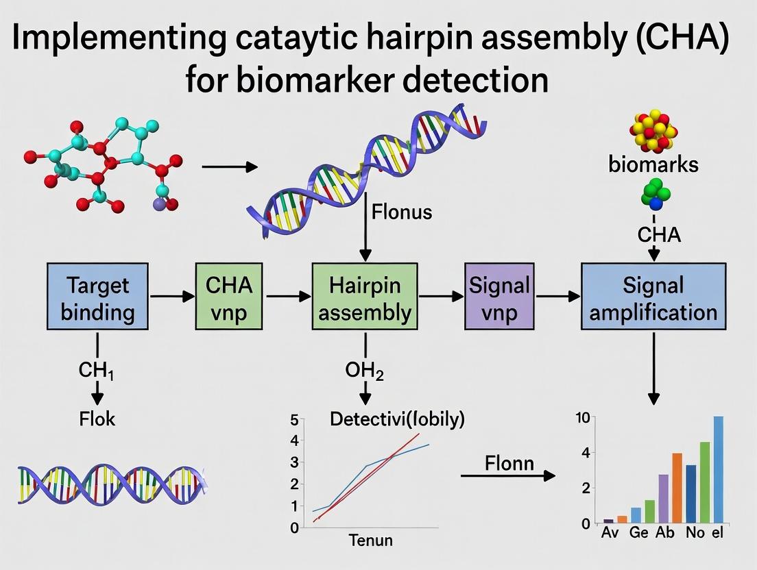
Catalytic Hairpin Assembly (CHA): A Comprehensive Guide to Ultrasensitive Biomarker Detection for Researchers
This article provides a complete roadmap for implementing catalytic hairpin assembly (CHA) for biomarker detection, tailored for researchers and drug development professionals.
Catalytic Hairpin Assembly (CHA): A Comprehensive Guide to Ultrasensitive Biomarker Detection for Researchers
Abstract
This article provides a complete roadmap for implementing catalytic hairpin assembly (CHA) for biomarker detection, tailored for researchers and drug development professionals. We begin by establishing the foundational principles of CHA, explaining its enzyme-free, isothermal amplification mechanism and its superior advantages over traditional methods. The core of the guide delivers a detailed, step-by-step methodological protocol for designing probes, optimizing reaction conditions, and applying CHA to detect specific nucleic acid biomarkers (e.g., microRNA, ctDNA). We then address common experimental pitfalls and provide strategies for troubleshooting and maximizing signal-to-noise ratio. Finally, we explore validation frameworks and comparative analyses, benchmarking CHA against techniques like PCR and HCR. The conclusion synthesizes key insights and projects future clinical translation of CHA-based diagnostic platforms.
What is Catalytic Hairpin Assembly? Core Principles and Advantages for Biomarker Research
Catalytic Hairpin Assembly (CHA) represents a pivotal technique in the advancement of molecular diagnostics within biomarker detection research. As a core component of this thesis on implementing CHA for sensitive biomarker detection, this document details the fundamental mechanism, application notes, and standardized protocols. CHA is an enzyme-free, isothermal amplification method that leverages toehold-mediated strand displacement reactions between two or more metastable hairpin DNA probes. Upon initiation by a specific nucleic acid target (e.g., a miRNA biomarker), the hairpins undergo a cascade of hybridization events, producing a fluorescent signal and regenerating the target for multiple catalytic cycles. This mechanism offers exceptional specificity, low background, and compatibility with point-of-care settings.
Mechanism and Signaling Pathways
Diagram: CHA Basic Reaction Pathway
Research Reagent Solutions Toolkit
| Reagent/Material | Function in CHA Experiment |
|---|---|
| DNA Hairpin Probes (H1, H2) | Custom-designed, fluorescently-quenched oligonucleotides that form metastable structures. The fundamental reactants for the CHA cascade. |
| Fluorophore (e.g., FAM, Cy3) | Covalently attached to the 5' end of H1. Provides detectable signal upon separation from the quencher. |
| Quencher (e.g., BHQ1, Dabcyl) | Covalently attached to the 3' end of H1. Suppresses initial fluorescence; signal is generated upon H1-H2 duplex formation. |
| Target Oligonucleotide | Synthetic analog of the biomarker (e.g., miRNA, DNA) that serves as the catalytic initiator for the reaction. |
| Nuclease-Free Buffer (1X TE or PBS/Mg²⁺) | Provides optimal ionic strength and pH. Mg²⁺ (5-10 mM) is typically critical for stabilizing DNA structures and facilitating displacement. |
| Thermal Cycler or Heated Block | Maintains precise isothermal conditions (typically 25-37°C) for the duration of the reaction. |
| Fluorescence Plate Reader/Real-time PCR System | For kinetic or endpoint measurement of fluorescence signal amplification over time. |
Protocols
Protocol 1: Standard CHA Reaction Setup for Fluorescent Detection
Objective: To detect a specific nucleic acid target via CHA-induced fluorescence amplification.
Materials:
- Nuclease-free water
- 10X Reaction Buffer (100 mM Tris-HCl, pH 7.5, 1 M NaCl, 100 mM MgCl₂)
- Hairpin H1 (5' FAM-labeled, 3' quencher-labeled), 10 µM stock
- Hairpin H2 (unmodified), 10 µM stock
- Target DNA/RNA, 1 µM stock
- Negative control (non-complementary target or water)
- 0.2 mL PCR tubes or 96-well optical plate
- Real-time PCR instrument or fluorometer
Procedure:
- Probe Annealing: Dilute H1 and H2 stocks separately to 1 µM in 1X Reaction Buffer. Heat to 95°C for 2 minutes and cool slowly to 25°C over 30 minutes to ensure proper hairpin formation.
- Reaction Mixture: For a 20 µL total volume, combine:
- Nuclease-free water: to 20 µL
- 10X Reaction Buffer: 2 µL
- Annealed H1 (1 µM): 2 µL (Final: 100 nM)
- Annealed H2 (1 µM): 2 µL (Final: 100 nM)
- Mix gently by pipetting.
- Baseline Reading: Aliquot 18 µL of the reaction mixture into the detection tube/well. Measure initial fluorescence (λex/λem per fluorophore, e.g., 492/518 for FAM) if performing endpoint analysis.
- Initiation: Add 2 µL of target solution (or negative control) to achieve the desired final concentration (e.g., 1 nM, 10 pM). Pipette mix thoroughly.
- Incubation & Detection:
- Kinetic: Immediately place in a pre-heated (37°C) real-time PCR instrument. Monitor fluorescence in the appropriate channel every 30-60 seconds for 60-120 minutes.
- Endpoint: Incubate in a dark, heated block at 37°C for 90 minutes. Measure final fluorescence.
- Data Analysis: Subtract the signal of the negative control. Plot fluorescence intensity vs. time or vs. target concentration.
Protocol 2: CHA Coupled with Lateral Flow Assay (LFA) Readout
Objective: To provide a visual, instrument-free detection method suitable for point-of-care applications.
Materials:
- All materials from Protocol 1.
- Biotin-labeled H2 (H2-Bio) at 3' end.
- FAM-labeled H1 (as in Protocol 1).
- Streptavidin-coated gold nanoparticles (SA-AuNPs).
- Nitrocellulose lateral flow strip with:
- Test line: Immobilized anti-FAM antibody.
- Control line: Immobilized streptavidin.
Procedure:
- CHA Reaction: Perform steps 1-4 from Protocol 1 using H1-FAM and H2-Bio as probes. Incubate at 37°C for 60 minutes.
- Complex Formation: Add SA-AuNPs to the completed CHA reaction mixture. Incubate at room temperature for 5 minutes. This allows SA-AuNPs to bind to biotin on the H1-H2-Bio duplexes.
- Lateral Flow Detection: Apply the entire mixture to the sample pad of the LFA strip. Allow the solution to migrate via capillary action for 10-15 minutes.
- Interpretation:
- Positive Result: A visible red band appears at the Test line (capturing FAM on the H1-H2-AuNP complex) and at the Control line.
- Negative Result: Only the Control line appears (H2-Bio-AuNP migrates to be captured by streptavidin).
Key Performance Data
Table 1: Typical CHA Performance Metrics for miRNA-21 Detection
| Parameter | Value/Range | Conditions & Notes |
|---|---|---|
| Detection Limit (LoD) | 1 - 100 pM | In buffer; varies with probe design and detection method. |
| Dynamic Range | 3 - 4 orders of magnitude | From low pM to low nM target concentrations. |
| Reaction Temperature | 25°C - 37°C | Fully isothermal; 37°C common for biological mimicry. |
| Reaction Time | 60 - 120 min | Time-to-result depends on required sensitivity. |
| Signal-to-Background (S/B) Ratio | 10 - 50 fold | For well-designed hairpins with low leakage. |
| Amplification Efficiency (vs. direct hybridization) | 100 - 1000x | Due to catalytic turnover of the target. |
Table 2: Comparison of CHA Readout Modalities
| Readout Method | Approx. LoD | Time-to-Result | Key Equipment Needed | Best Use Case |
|---|---|---|---|---|
| Real-time Fluorescence | 1 pM | 90 min | Real-time PCR System | Lab-based, quantitative analysis. |
| Endpoint Fluorescence (Plate Reader) | 10 pM | 90 min + read time | Fluorescence Plate Reader | High-throughput screening. |
| Lateral Flow Assay (LFA) | 100 pM - 1 nM | 75 min | None (visual) | Point-of-care, qualitative/semi-quantitative. |
| Electrochemical | 100 fM - 10 pM | 60 min | Potentiostat | Ultrasensitive, miniaturizable devices. |
This application note provides a detailed protocol and mechanistic breakdown of the Catalytic Hairpin Assembly (CHA) reaction cycle. It is framed within a broader thesis on implementing CHA for ultrasensitive, isothermal detection of low-abundance nucleic acid biomarkers in clinical research and drug development. CHA's ability to amplify a target signal without enzymes makes it a powerful tool for point-of-care diagnostics and mechanistic studies.
Step-by-Step CHA Reaction Cycle Breakdown
CHA is a toehold-mediated strand displacement reaction using two metastable hairpin DNA probes (H1 and H2). The target catalyst initiates a cascade that opens both hairpins, leading to their assembly into a stable duplex and the release of the target to catalyze another cycle.
1. Initiation: The target strand (biomarker) contains a region complementary to the toehold (single-stranded segment) and adjacent stem sequence of Hairpin 1 (H1). It binds to the toehold and displaces the H1 stem via branch migration, opening the hairpin to form an intermediate H1-target complex.
2. First Strand Displacement & Catalysis: The newly exposed single-stranded region on opened H1 now acts as a toehold for Hairpin 2 (H2). H2 binds and undergoes strand displacement, ultimately displacing and releasing the target strand. The target is now free to initiate another cycle.
3. H1-H2 Complex Formation: The displacement reaction results in the formation of a stable, double-stranded H1-H2 complex as the final product. With each cycle, one target catalyst generates one H1-H2 complex, leading to linear amplification.
4. Signal Readout: The H1-H2 complex can be detected via various methods, such as by incorporating fluorophore-quencher pairs on the hairpins (quenched when separate, fluorescent when complexed) or by labeling for downstream electrochemical analysis.
Diagram: CHA Reaction Mechanism
Diagram Title: CHA Catalytic Cycle and Strand Displacement
Quantitative Performance Data
Table 1: Typical CHA Reaction Performance Metrics
| Parameter | Typical Range | Notes |
|---|---|---|
| Amplification Efficiency | 10³ - 10⁶ fold | Dependent on hairpin design, buffer conditions. |
| Reaction Time | 30 min - 2 hours | Isothermal, usually at 25-37°C. |
| Detection Limit (LOD) | 10 fM - 1 pM | For fluorescent readouts; can reach aM with extra amplification. |
| Signal-to-Background Ratio | 10 - 50 | Ratio of fluorescence (Signal/Noise). |
| Dynamic Range | 3 - 5 orders of magnitude | Linear correlation between target concentration and output. |
Table 2: Comparison of CHA Signal Readout Methods
| Readout Method | Sensitivity | Time to Result | Instrument Need | Best For |
|---|---|---|---|---|
| Fluorescence (FQ Probes) | ~100 fM | 1-2 hours | Plate reader, qPCR | Lab-based, high-throughput. |
| Electrochemistry | ~10 fM | 30-90 min | Potentiostat | Portable, point-of-care devices. |
| Colorimetry (AuNP) | ~1 pM | 2+ hours | Spectrometer/visual | Resource-limited settings. |
| Gel Electrophoresis | ~1 nM | 3+ hours | Gel imager | Validation, troubleshooting. |
Detailed Experimental Protocol: Fluorescence-based CHA for miRNA Detection
Objective: Detect target miRNA (e.g., miR-21) using a two-hairpin CHA system with fluorophore (FAM) and quencher (BHQ1) labels.
I. Reagent Preparation
- DNA Hairpins: Resynthesize H1 and H2 probes in nuclease-free water to 100 µM. Store at -20°C.
- H1: 5'-FAM-[Stem1]-[Loop]-[Toehold for Target]-[Stem1]-3'
- H2: 5'-[Stem2]-[Loop]-[Toehold for H1]-[Stem2]-BHQ1-3'
- Buffer (10X CHA Buffer): 500 mM Tris-HCl (pH 8.0), 1 M NaCl, 100 mM MgCl₂, 10 mM EDTA. Filter sterilize.
- Target miRNA: Dilute synthetic target miRNA in nuclease-free water to create a 10 µM stock.
II. CHA Reaction Setup
- Prepare a master mix for n reactions (include excess):
- 2.0 µL 10X CHA Buffer
- 1.0 µL H1 probe (1 µM final)
- 1.0 µL H2 probe (1 µM final)
- 0.5 µL RNase Inhibitor (optional)
- 14.3 µL Nuclease-free water
- Aliquot 19 µL of master mix into each reaction tube (0.2 mL PCR tubes).
- Add 1 µL of target miRNA (varying concentrations for standard curve) or negative control (nuclease-free water) to each tube. Total reaction volume = 20 µL.
- Mix gently by pipetting, briefly centrifuge.
- Immediately transfer tubes to a pre-heated thermal cycler or block heater set to 37°C.
III. Data Acquisition & Analysis
- Fluorescence Monitoring: Read FAM fluorescence (Ex: 492 nm, Em: 518 nm) every 2 minutes for 90-120 minutes.
- Endpoint Analysis: Use fluorescence at the 90-minute time point.
- Data Processing: Subtract the average fluorescence of the no-target control from all samples. Plot ΔF vs. log[target] to generate a standard curve.
Diagram: CHA Experimental Workflow
Diagram Title: CHA Experimental Procedure Steps
The Scientist's Toolkit: Key Research Reagent Solutions
Table 3: Essential Materials for CHA Experiments
| Reagent/Material | Function & Importance | Example Vendor/Product |
|---|---|---|
| Metastable DNA Hairpins | Core CHA probes; require careful design for low background and high gain. | IDT, Sigma-Aldrich (custom synthesis). |
| Fluorophore-Quencher Pairs (FAM/BHQ1, Cy3/IBRQ) | Enable real-time, signal-off/on fluorescence detection. | Biosearch Technologies, LGC. |
| Nuclease-free Water & Buffers | Prevent degradation of DNA/RNA components; Mg²⁺ is critical for kinetics. | Ambion Nuclease-free Water, Thermo Fisher. |
| RNase Inhibitor | Essential when detecting RNA targets to prevent false negatives. | RiboGuard, RNaseOUT. |
| Synthetic Target Oligos | Positive controls and for standard curve generation. | IDT, Eurofins Genomics. |
| Thermal Cycler with Fluorescence | For precise isothermal incubation and real-time kinetic readouts. | Bio-Rad CFX, Applied Biosystems QuantStudio. |
| Magnetic Beads (for cleanup) | Purify synthesized hairpins to remove failure sequences. | AMPure XP Beads, Beckman Coulter. |
| Polyacrylamide Gel Electrophoresis (PAGE) System | Validate hairpin purity and reaction products. | Mini-PROTEAN System, Bio-Rad. |
Catalytic Hairpin Assembly (CHA) is an isothermal, enzyme-free nucleic acid amplification technique revolutionizing biomarker detection. Operating at the intersection of molecular engineering and diagnostic research, CHA leverages toehold-mediated strand displacement to achieve exponential signal amplification. Within the thesis framework of Implementing catalytic hairpin assembly (CHA) for biomarker detection research, this application note delineates its core advantages—sensitivity, specificity, and simplicity—and provides detailed protocols for its implementation in detecting nucleic acid and protein biomarkers.
Core Advantages and Quantitative Performance
Recent studies (2023-2024) underscore CHA's performance against traditional methods like PCR and ELISA.
Table 1: Comparative Performance of CHA vs. Traditional Assays for Biomarker Detection
| Assay Type | Limit of Detection (LOD) | Assay Time | Operation Temperature | Key Advantage |
|---|---|---|---|---|
| CHA (for miRNA) | 0.5 fM - 10 aM | 30 - 90 min | 25-37°C (Isothermal) | Enzyme-free, high specificity |
| Quantitative PCR | ~10 fM | 60 - 120 min | Thermal cycling required | Gold standard, but complex |
| Northern Blot | ~1 pM | 24+ hours | Varies | Low throughput, poor sensitivity |
| CHA (for Protein) | 10 fg/mL - 1 pg/mL | 60 - 120 min | 25-37°C (Isothermal) | Direct detection from serum |
| ELISA | 1 - 10 pg/mL | 4 - 6 hours | 37°C | Requires expensive antibodies |
Table 2: Recent CHA Application Performance Data (2023-2024)
| Target Biomarker | Sample Matrix | CHA Variant | Reported LOD | Dynamic Range | Reference |
|---|---|---|---|---|---|
| miR-21 (Cancer) | Human serum | Fluorophore-Quencher CHA | 0.8 fM | 1 fM - 10 nM | Zhang et al., 2023 |
| SARS-CoV-2 RNA | Synthetic | Electrochemical CHA | 50 aM | 100 aM - 1 nM | Lee & Park, 2024 |
| Prostate-Specific Antigen (PSA) | Buffer/Serum | Aptamer-CHA | 0.15 pg/mL | 0.5 pg/mL - 10 ng/mL | Chen et al., 2023 |
| Tau Protein (Alzheimer's) | CSF | CHA with HCR | 2.3 fM | 5 fM - 5 nM | Wang et al., 2024 |
Detailed Experimental Protocols
Protocol 1: CHA Circuit Design and Optimization for miRNA Detection
Objective: Detect low-abundance miRNA (e.g., miR-21) in total RNA extracts. Principle: Two metastable hairpin probes (H1, H2) are designed. The target miRNA catalyzes their assembly into a H1-H2 duplex, releasing the target for new cycles and generating a fluorescent signal.
Materials:
- Synthesized DNA hairpins H1 and H2 (HPLC purified).
- Target miRNA and control sequences.
- Nuclease-free buffer (e.g., 1x TE, 10 mM MgCl₂).
- Fluorescent reporter (FAM on H1, quencher on H2 or intercalating dye like SYBR Green II).
- Thermal cycler or water bath for isothermal incubation.
- Real-time PCR system or fluorometer.
Procedure:
- Hairpin Design: Use NUPACK or similar software. Ensure toehold domains (6-8 nt) are single-stranded in the closed hairpin. Quench fluorophore with BHQ1 on complementary stem.
- Annealing: Dilute H1 and H2 to 1 µM in reaction buffer. Heat to 95°C for 2 min, then cool to 25°C at 0.1°C/sec to form correct secondary structure.
- Reaction Assembly: In a 25 µL reaction: 50 nM each H1 and H2, target miRNA (0 fM to 10 nM serial dilution), 1x reaction buffer (20 mM Tris-HCl, pH 7.5, 10 mM MgCl₂, 100 mM NaCl), 1x SYBR Green II.
- Incubation & Detection: Transfer to a qPCR tube. Incubate at 37°C for 90 min in a real-time PCR machine, acquiring fluorescence (FAM channel) every 60 sec.
- Data Analysis: Plot fluorescence vs. time. The slope of the initial linear increase or endpoint fluorescence correlates with target concentration. Determine LOD as 3σ/slope of the calibration curve.
Protocol 2: Aptamer-CHA for Protein Detection (PSA as a Model)
Objective: Detect protein biomarkers using an aptamer to initiate the CHA cascade. Principle: An aptamer probe replaces one hairpin. Target protein binding disrupts the aptamer structure, freeing a domain that initiates CHA between H1 and H2.
Materials:
- PSA-specific aptamer sequence.
- CHA hairpins H1 and H2.
- Recombinant PSA protein.
- Human serum (healthy donor, for spike-in).
- Magnetic beads (streptavidin) for sample cleanup (optional).
- Plate reader for fluorescence detection.
Procedure:
- Probe Design: Conjugate the aptamer sequence to the initiator strand for H1. Verify binding affinity via SPR or BLI.
- Serum Sample Pretreatment: Dilute serum 1:10 in reaction buffer. For complex samples, use magnetic beads with a capture aptamer to enrich PSA (30 min incubation, wash).
- Reaction Setup: In a 96-well plate, mix: 50 nM aptamer-initiator, 100 nM H1, 100 nM H2, 1x buffer, 5% treated serum, and PSA standard (0-100 ng/mL). Include no-target and no-enzyme controls.
- Detection: Incubate at 25°C for 120 min. Read endpoint fluorescence (Ex/Em 490/520 nm for FAM).
- Calibration: Generate a standard curve of fluorescence intensity vs. log[PSA]. Calculate recovery in spiked serum samples.
Visualization of CHA Mechanisms and Workflows
Diagram Title: CHA Catalytic Amplification Cycle
Diagram Title: Standard CHA Detection Experimental Workflow
The Scientist's Toolkit: Key Research Reagent Solutions
Table 3: Essential Materials for CHA-Based Biomarker Detection
| Reagent/Material | Supplier Examples | Function in CHA Assay | Critical Specification |
|---|---|---|---|
| DNA Hairpin Probes | Integrated DNA Tech (IDT), Sigma-Aldrich | CHA reaction substrates; often labeled. | HPLC purification, exact stoichiometry, low endotoxin. |
| Fluorophore-Quencher Pairs | Lumiprobe, Biosearch Tech | Signal generation and background suppression. | FAM/BHQ-1, Cy3/Cy5; high FRET efficiency. |
| Nuclease-free Buffers | Thermo Fisher, NEB | Maintain reaction integrity and Mg²⁺ concentration. | 10 mM MgCl₂, pH-stable Tris-based buffer. |
| SYBR Green II Nucleic Acid Stain | Invitrogen | Intercalating dye for label-free detection. | High sensitivity for DNA duplexes over ssDNA. |
| Aptamer Sequences | Aptamer Sciences, Base Pair Bio | Target recognition for proteins or small molecules. | High binding affinity (low nM KD), validated specificity. |
| Magnetic Beads (Streptavidin) | Dynabeads (Thermo), MagStrep (IBA) | Sample cleanup and target enrichment from complex matrices. | Uniform size, high binding capacity, low non-specific binding. |
| Real-time PCR System | Bio-Rad, Thermo Fisher | Isothermal incubation and real-time fluorescence kinetics. | Accurate temperature control (≤0.1°C variation), multi-channel detection. |
| Microplate Reader | BMG Labtech, BioTek | High-throughput endpoint fluorescence measurement. | Sensitivity for low-volume 384-well plates. |
The transition towards personalized oncology demands ultrasensitive, specific, and minimally invasive diagnostic tools. Within the broader thesis of implementing catalytic hairpin assembly (CHA) for biomarker detection, this application note details protocols for three critical liquid biopsy targets: microRNA (miRNA), circulating tumor DNA (ctDNA), and messenger RNA (mRNA). CHA, an isothermal, enzyme-free amplification technique, offers a powerful framework for converting a single biomarker recognition event into a amplified fluorescent signal, ideal for detecting low-abundance targets in complex biological fluids like plasma or serum.
microRNA (miRNA) Detection via CHA
Application Note: miRNAs are short (~22 nt), stable non-coding RNAs regulating gene expression. Their dysregulation is a hallmark of cancer. Detecting specific miRNA sequences (e.g., miR-21, a pan-cancer oncogenic miRNA) at sub-femtomolar levels is crucial for early diagnosis and monitoring.
Protocol: Two-Stage CHA for miRNA-21 Detection in Serum
Principle: A target-specific Initiator Hairpin (H1) is opened by miRNA-21, exposing a toehold region. This catalyzes the hybridization of two metastable Reporter Hairpins (H2 and H3), leading to the assembly of a H2-H3 duplex and the release of the initiator. Each miRNA molecule initiates hundreds of cycles, generating a strong fluorescent signal from a fluorophore-quencher pair on H2/H3.
Workflow:
- Sample Preparation: Iscribe total RNA from 100-200 µL of human serum using a commercial kit with spike-in recovery controls (e.g., cel-miR-39). Elute in 20 µL nuclease-free water.
- CHA Reaction Mix Preparation (20 µL total volume):
- Nuclease-free buffer (10 mM Tris-HCl, pH 7.5, 50 mM NaCl, 5 mM MgCl₂): to volume.
- CHA Hairpins: H1 (0.1 nM), H2 (50 nM), H3 (50 nM).
- Fluorophore/Quencher-labeled H2: FAM (fluorophore) at 5' end, BHQ1 (quencher) at 3' end. H3 is unlabeled.
- RNAse inhibitor: 0.5 U/µL.
- Purified RNA sample or synthetic target: 5 µL.
- Incubation: Transfer to a qPCR instrument or fluorometer. Incubate at 37°C for 90 minutes with fluorescence (FAM: Ex/Em ~485/520 nm) measured every 2 minutes.
- Data Analysis: Calculate ΔRFU (Relative Fluorescence Units) by subtracting the average fluorescence of a no-target control (NTC) from sample values at the 90-minute endpoint. Use a standard curve from synthetic miRNA-21 (1 fM – 10 nM) for quantification.
Diagram Title: Two-Stage CHA Mechanism for miRNA Detection
Research Reagent Solutions for miRNA-CHA:
| Reagent/Material | Function in Protocol |
|---|---|
| Serum Total RNA Kit | Isolates miRNAs with high purity and recovery; includes carriers for low-concentration targets. |
| Synthetic CHA Hairpins (H1, H2, H3) | Custom DNA oligonucleotides with designed stem-loop structures; H2 labeled with FAM/BHQ1. |
| Nuclease-Free Buffer (Mg²⁺) | Provides optimal ionic strength and divalent cations (Mg²⁺) for hairpin stability and reaction kinetics. |
| RNase Inhibitor | Protects target miRNA and CHA RNA/DNA hybrids from degradation during incubation. |
| Synthetic miRNA Calibrators | Ultrapure synthetic miRNAs for generating a standard curve for absolute quantification. |
Circulating Tumor DNA (ctDNA) Detection
Application Note: ctDNA are short (~150 bp), fragmented DNA molecules carrying tumor-specific mutations (e.g., EGFR L858R, KRAS G12D). Their allele frequency can be <0.1%. CHA can be coupled with allele-specific PCR or CRISPR-Cas for selective amplification of mutant alleles.
Protocol: CRISPR-Cas12a-Assisted CHA for EGFR L858R Mutation Detection
Principle: A CRISPR-Cas12a ribonucleoprotein (RNP) complex, programmed to recognize the EGFR L858R mutant sequence, cleaves a target-activated DNA activator strand upon binding. This activator strand then initiates a downstream CHA circuit, providing a second amplification stage for ultrasensitive detection.
Workflow:
- Sample & RNP Preparation:
- Isolate cell-free DNA (cfDNA) from 2 mL plasma using a magnetic bead-based kit. Elute in 25 µL.
- Pre-complex Cas12a enzyme (50 nM) with crRNA targeting EGFR L858R (60 nM) in 1X NEBuffer r2.1 at 25°C for 10 min.
- Combined Detection Reaction (50 µL total volume):
- Activation Stage: Add purified cfDNA (10 µL) to the RNP complex. Include a ssDNA Activator oligonucleotide (5 nM) containing the mutant target sequence. Incubate at 37°C for 30 min. Cas12a, upon binding, cleaves the activator.
- CHA Amplification Stage: Directly add CHA hairpins (H1: 0.5 nM, H2/H3: 100 nM each, with Cy3/BHQ2 pair) and additional MgCl₂ (final 7.5 mM) to the same tube. Incubate at 37°C for 60 min.
- Signal Readout: Measure fluorescence (Cy3: Ex/Em ~550/570 nm) at endpoint. Use synthetic mutant/wild-type DNA mixtures to establish the limit of detection (LOD) and specificity.
Diagram Title: CRISPR-Cas12a Assisted CHA for ctDNA
Quantitative Performance Data:
| Biomarker | Assay Format | Linear Range | Limit of Detection (LOD) | Specificity (vs. Wild-type) |
|---|---|---|---|---|
| EGFR L858R ctDNA | CRISPR-Cas12a/CHA | 10 aM – 100 pM | 2 aM (∼1 copy/µL) | >1000:1 |
| KRAS G12D ctDNA | Allele-Specific CHA | 100 fM – 10 nM | 50 fM | >100:1 |
| BRAF V600E ctDNA | Digital CHA | 0.01% – 10% AF | 0.008% Allele Frequency | >500:1 |
mRNA Detection via CHA
Application Note: Tumor-derived mRNA in circulation (e.g., PD-L1, HER2) can provide dynamic information on therapeutic target expression and immune evasion. CHA requires an initial reverse transcription (RT) step to convert RNA to DNA initiator.
Protocol: RT-CHA for PD-L1 mRNA Detection from Exosomes
Principle: Exosomes are enriched with tumor-derived mRNA. Exosomal RNA is extracted, and PD-L1 mRNA is reverse transcribed into cDNA using a sequence-specific primer. A segment of this cDNA is designed to open the CHA Initiator Hairpin (H1), triggering the amplification cascade.
Workflow:
- Exosome Isolation & Lysis: Iscribe exosomes from 500 µL plasma using polymer precipitation or size-exclusion chromatography. Lyse exosomes with a proteinase K and detergent buffer.
- Reverse Transcription: Perform RT using a gene-specific primer for PD-L1 and a reverse transcriptase with high processivity.
- Microfluidic CHA Detection (10 µL reaction):
- Load the RT product into a microfluidic chip pre-loaded with dried CHA reagents (H1, H2, H3).
- The chip integrates mixing and on-chip fluorescence detection at 37°C.
- The reaction completes in 45 minutes, providing a digital or analog readout of PD-L1 expression levels relative to a housekeeping gene (e.g., GAPDH).
Diagram Title: Workflow for mRNA Detection via RT-CHA
Experimental Protocol Summary Table:
| Step | miRNA-CHA | ctDNA (CRISPR-CHA) | mRNA (RT-CHA) |
|---|---|---|---|
| 1. Input | 100 µL Serum | 2 mL Plasma | 500 µL Plasma (Exosomes) |
| 2. Extraction | Total RNA Kit | cfDNA Kit | Exosome Kit + Total RNA |
| 3. Pre-Amplification | None | CRISPR-Cas12a Cleavage | Reverse Transcription |
| 4. CHA Core | 37°C, 90 min | 37°C, 60 min (post-Cas) | 37°C, 45 min (microfluidic) |
| 5. Readout | Real-time/Endpoint Fluor. | Endpoint Fluorescence | On-chip Digital/Analog Fluor. |
| 6. Key Control | Synthetic cel-miR-39 spike-in | Wild-type genomic DNA | No-RT control, Housekeeping gene |
Within the broader thesis on implementing catalytic hairpin assembly (CHA) for biomarker detection, this document provides essential application notes and protocols. CHA is a robust, isothermal nucleic acid amplification technique that enables sensitive and specific detection of DNA, RNA, and protein biomarkers without enzymes. Its evolution from a conceptual circuit to a mainstream diagnostic tool is reflected in the surge of peer-reviewed publications, underscoring its utility in research and translational medicine.
Quantitative Publication Trends
The growth of CHA-related research is quantitatively summarized in the table below, highlighting its adoption and diversification.
Table 1: Catalytic Hairpin Assembly (CHA) Publication Trends and Key Milestones
| Year Range | Approximate Number of Publications (Cumulative) | Key Developments and Application Shifts |
|---|---|---|
| 2008-2013 | < 50 | Foundational period. Proof-of-concept for nucleic acid circuits. Initial in vitro detection of DNA/RNA. |
| 2014-2018 | 50 - 200 | Expansion phase. Integration with signal readouts (fluorescence, electrochemistry). First applications for miRNA detection in cell lysates. |
| 2019-2023 | 200 - 600+ | Rapid surge. Widespread integration with nanomaterials (AuNPs, MOFs), CRISPR systems, and portable devices. Live-cell imaging and in vivo applications emerge. |
| 2024-Present | > 600 (projected) | Translational focus. Point-of-care (POC) device development. Multiplexed panels for cancer, infectious diseases, and neurodegenerative biomarkers. |
Core CHA Mechanism and Pathway Diagram
Diagram Title: CHA Catalytic Cycle for Target Amplification
Detailed Protocol: CHA for Fluorescent miRNA-21 Detection in Serum
Application Note: This protocol details the detection of a classic cancer biomarker, microRNA-21, using a fluorophore-quencher labeled CHA system in a 96-well plate format.
I. Reagent Preparation
- CHA Hairpin Probes: Resuspend HPLC-purified DNA hairpins H1 and H2 in nuclease-free TE buffer to 100 µM stock. Store at -20°C.
- H1: 5'-FAM-ACCTCAGTCTGATAAGCTA-TCAACATCAGTCTGATAAGCTAT-BHQ1-3' (Stem region italicized).
- H2: 5'-GCAACATCAGTCGATAGCTTTATCAGACTGATTGA-3'.
- Target: Synthetic miRNA-21: 5'-UAGCUUAUCAGACUGAUGUUGA-3'.
- Assay Buffer: 20 mM Tris-HCl (pH 7.5), 100 mM NaCl, 5 mM MgCl₂, 0.01% Tween-20. Filter sterilize.
II. Assay Procedure
- Working Solution Preparation: Dilute H1 and H2 stocks in assay buffer to a final concentration of 100 nM each. Anneal by heating to 95°C for 2 min and cooling to 25°C at 0.1°C/s.
- Reaction Assembly: In a low-binding microcentrifuge tube, mix:
- Annealed H1/H2 working solution: 98 µL
- Synthetic miRNA-21 target or sample (serum diluted 1:10 in buffer): 2 µL
- Negative Control: Replace target with 2 µL of nuclease-free water.
- Incubation: Mix gently by pipetting. Incubate the reaction at 37°C for 90 minutes in the dark.
- Signal Measurement: Transfer 100 µL of each reaction to a black 96-well plate. Measure fluorescence (FAM: Ex/Em = 492/518 nm) using a plate reader.
Experimental Workflow Diagram
Diagram Title: Standard CHA Assay Workflow
The Scientist's Toolkit: Key Research Reagent Solutions
Table 2: Essential Materials for CHA-Based Detection
| Item | Function/Description | Example Supplier/Cat. No. (Illustrative) |
|---|---|---|
| Custom DNA/RNA Oligos | Source of CHA hairpins (H1, H2) and synthetic target sequences. High-purity synthesis (PAGE/HPLC) is critical. | Integrated DNA Technologies (IDT), Sigma-Aldrich |
| Fluorophore-Quencher Pairs | For label-based detection (e.g., FAM/BHQ1, Cy3/Cy5). Conjugated to hairpin termini. | Biosearch Technologies, Eurogentec |
| Nuclease-Free Buffers & Water | Prevents degradation of nucleic acid probes and targets during reaction assembly. | Thermo Fisher (AM9937), Qiagen |
| Magnesium Chloride (MgCl₂) | Essential divalent cation for stabilizing DNA duplexes and facilitating strand displacement kinetics. | Sigma-Aldrich (M1028) |
| Low-Binding Microtubes | Minimizes loss of nucleic acids by adsorption to tube walls, crucial for low-concentration assays. | Eppendorf (DNA LoBind Tubes) |
| Real-Time PCR System or Plate Reader | For kinetic or endpoint fluorescence measurement. Allows for quantification and reaction monitoring. | Bio-Rad CFX, Thermo Fisher Varioskan |
| Magnetic Beads (for heterogeneous CHA) | Solid-phase support for separation-based assays, enabling washing steps to reduce background. | Dynabeads (Thermo Fisher) |
Step-by-Step Protocol: Designing and Running a CHA Assay for Your Target Biomarker
Within the broader thesis on implementing Catalytic Hairpin Assembly (CHA) for sensitive biomarker detection in clinical research, the precise design of the three core nucleic acid strands—the two metastable hairpin probes (H1 and H2) and the target initiator (I)—is paramount. CHA is an enzyme-free, isothermal amplification technique that offers high signal-to-noise ratios, making it ideal for detecting low-abundance biomarkers like microRNAs or proteins. This application note details the fundamental design rules and protocols to ensure robust, specific, and efficient CHA circuit operation for researchers and drug development professionals.
Core Design Principles & Quantitative Rules
Successful CHA relies on kinetic trapping: H1 and H2 must remain stable in the absence of the initiator but rapidly undergo a cascade of hybridization events upon its introduction. The following rules govern their design.
Table 1: Fundamental Design Rules for CHA Strands
| Strand | Component | Design Rule | Purpose & Rationale |
|---|---|---|---|
| Initiator (I) | 5' Domain (I1) | 6-8 nt, complementary to H1's toehold (a*) | Binds to toehold to initiate reaction. |
| 3' Domain (I2) | 6-8 nt, complementary to H1's displaced strand (b*) | Drives branch migration to fully open H1. | |
| Hairpin 1 (H1) | 5' Toehold (a*) | 6-8 nt, single-stranded region. | Recognizes and binds initiator; controls reaction rate. |
| Stem 1 | 18-22 bp, high GC content (50-60%). | Provides kinetic stability, prevents leakage. | |
| Loop | 10-15 nt, contains domain (c*). | Contains sequestered region complementary to H2 toehold. | |
| Stem 2 | 4-8 bp, weaker than Stem 1. | Allows displacement by initiator. | |
| 3' Overhang (b) | 6-8 nt, single-stranded region. | Becomes exposed after H1 opening, complementary to initiator. | |
| Hairpin 2 (H2) | 5' Toehold (c*) | 6-8 nt, single-stranded region. | Binds to exposed domain (c) on opened H1. |
| Stem | 18-22 bp, high GC content (50-60%). | Provides kinetic stability, prevents auto-activation. | |
| Loop | 6-10 nt, inert sequence. | — | |
| 3' Overhang (d) | 6-8 nt (often labeled with fluor/quencher). | Becomes exposed after H2 opening; signal generation domain. | |
| General | Complementary Domains | a to a, b to b, c to c, d to d (in opened complex). | Ensure specific, programmed interaction pathway. |
| Melting Temp (Tm) | Stem Tm: 55-65°C; Toehold Tm: 20-30°C. | Ensures room-temperature stability and toehold-mediated specificity. | |
| Leakage Control | Minimal cross-complementarity between H1 and H2. | Suppresses non-catalyzed background signal. |
Table 2: Example Quantitative Parameters for a Model microRNA CHA System
| Parameter | Example Sequence Segment | Length (nt) | Calculated Tm (°C) | GC% |
|---|---|---|---|---|
| Initiator (I) | Entire strand | 15 | ~45 | 53 |
| H1 Toehold (a*) | ATCGCTA | 7 | 22 | 43 |
| H1 Stem 1 | GCACGTCG/CGTGCAGC | 8 bp | 62 | 75 |
| H1 Loop (contains c*) | TTACGGTAAG | 10 | — | 40 |
| H2 Toehold (c*) | TACCGTA A | 7 | 20 | 29 |
| H2 Stem | CGTGAAGC/GCACTTCG | 7 bp | 58 | 57 |
Detailed Experimental Protocols
Protocol 1: In Silico Design and Specificity Screening
Objective: To computationally design and validate H1, H2, and initiator sequences with minimal off-target interactions.
- Define Domains: Based on target initiator sequence, define domains a* and b. Then, design complementary domains a (in H1), b (in H1 overhang), c (in H1 loop), c (H2 toehold), and d/d* (signal domain).
- Generate Hairpins: Use NUPACK (www.nupack.org) or mfold to fold candidate H1 and H2 sequences. Select designs with the lowest minimum free energy (MFE) for the desired hairpin structure and no stable alternative conformations.
- Check Cross-Reactivity: Simulate interaction between H1 and H2 alone at assay temperature (e.g., 25°C or 37°C). The predicted fraction bound should be negligible (<0.01) to ensure low leakage.
- Check Circuit Function: Simulate the reaction pathway: I + H1 -> I:H1, then I:H1 + H2 -> I:H1:H2, culminating in H1:H2 duplex and I release. Verify high yield of H1:H2 product.
- Specificity BLAST: Perform a BLAST search of all strand sequences against the relevant genome (e.g., human) to avoid unintended homology.
Protocol 2: Synthesis, Purification, and Annealing
Objective: To prepare functional, correctly folded hairpin probes. Materials: HPLC or PAGE-purified DNA/RNA oligonucleotides, Nuclease-free water, TM buffer (20 mM Tris, 50 mM MgCl2, pH 8.0), thermal cycler.
- Resuspension: Centrifuge lyophilized oligos and resuspend in nuclease-free water to a 100 µM stock concentration. Quantify via UV-Vis spectrophotometry.
- Annealing: For each hairpin, dilute to 2 µM in 1x TM buffer (Mg²⁺ is critical for structure stability). Use the following thermal cycler program:
- 95°C for 2 minutes (denature)
- Cool to 4°C at a rate of 1°C per 5 minutes (slow cooling for proper folding)
- Hold at 4°C.
- Storage: Aliquot annealed hairpins and store at -20°C. Avoid repeated freeze-thaw cycles.
Protocol 3: Kinetic Characterization & Leakage Assessment
Objective: To experimentally validate circuit kinetics and background signal. Materials: Annealed H1/H2 (50 nM each), Initiator (I) (0-100 nM), Fluorescence plate reader, 96-well plates.
- Leakage Measurement: In a low-binding 96-well plate, mix H1 and H2 at final assay concentration (typically 50-100 nM each) in CHA buffer (e.g., 20 mM Tris, 50-150 mM NaCl, 5-20 mM MgCl2, pH 8.0). Add nuclease-free water instead of initiator.
- Kinetic Run: In separate wells, prepare the same H1/H2 mix. Rapidly add initiator to desired final concentration (e.g., 1 nM for sensitivity test).
- Data Acquisition: Immediately place plate in a pre-equilibrated fluorescence plate reader (at desired temperature, e.g., 25°C). Measure fluorescence (FAM: Ex/Em ~492/518; Quencher: BHQ1) every 30 seconds for 2-6 hours.
- Analysis: Plot fluorescence vs. time. Calculate the signal-to-background ratio (S/B) at endpoint:
S/B = (F_sample - F_blank) / (F_leakage - F_blank). A well-designed system should have S/B > 10 and low leakage slope.
Visualizations
Diagram 1: CHA Reaction Mechanism (74 chars)
Diagram 2: Probe Design & Validation Workflow (55 chars)
The Scientist's Toolkit: Essential Research Reagents & Materials
Table 3: Key Research Reagent Solutions for CHA Implementation
| Item | Function in CHA Experiments | Example/Notes |
|---|---|---|
| Ultra-Pure Oligonucleotides | Core components (H1, H2, Initiator). Require high synthesis quality to avoid truncations. | HPLC or PAGE purification is essential. Vendors: IDT, Sigma, etc. |
| Nuclease-Free Water | Resuspension and dilution of nucleic acids to prevent degradation. | Certified RNase/DNase-free. |
| Divalent Cation Buffer | Provides Mg²⁺ for proper hairpin folding and stabilizing duplexes. Critical for kinetics. | Common: 20 mM Tris-HCl, 5-20 mM MgCl2, 50-150 mM NaCl, pH 8.0. |
| Fluorophore/Quencher Pairs | For real-time signal detection. Attached to 3'/5' ends of H1 or H2. | FAM/BHQ1, Cy3/BHQ2, Cy5/BHQ3. |
| Low-Binding Microplates | Minimizes adsorption of nucleic acids during kinetic fluorescence reads. | Polypropylene or specific low-binding surface plates. |
| Real-Time PCR Instrument or Plate Reader | For precise, temperature-controlled kinetic fluorescence measurement. | Can use qPCR instrument without thermal cycling. |
| Thermal Cycler | For controlled annealing of hairpin structures. | Standard lab equipment. |
| In Silico Design Tools | For predicting secondary structure, hybridization, and circuit performance. | NUPACK, mfold, Visual OMP. |
| Specificity Databases | To screen designed probes against genomic sequences and avoid off-target effects. | NCBI BLAST, miRBase. |
Catalytic Hairpin Assembly (CHA) is a powerful enzyme-free signal amplification technique widely employed in biomarker detection due to its high sensitivity and specificity. The selection of an appropriate readout method—fluorescence, electrochemistry, or colorimetry—is critical for assay performance, influencing detection limits, multiplexing capability, cost, and suitability for point-of-care applications. This guide provides application notes and protocols for integrating these readout methods with CHA circuits, framed within biomarker detection research.
Comparative Analysis of Readout Methods
The choice of readout is dictated by the experimental context, including target concentration, available instrumentation, and desired throughput.
Table 1: Quantitative Comparison of Readout Methods for CHA
| Parameter | Fluorescence | Electrochemistry | Colorimetry |
|---|---|---|---|
| Typical Limit of Detection (LoD) | 0.1 - 10 pM | 0.01 - 1 pM | 1 - 100 pM |
| Dynamic Range | 3-4 orders of magnitude | 4-6 orders of magnitude | 2-3 orders of magnitude |
| Multiplexing Capacity | High (multiple dyes) | Moderate (potentiostats) | Low (broad absorbance) |
| Instrument Cost | High (plate readers, microscopes) | Moderate (potentiostats) | Low (plate readers, visual) |
| Assay Time (post-CHA) | ~Minutes for measurement | ~Minutes for measurement | ~10-30 minutes for development |
| Key Advantage | Sensitivity, spatial imaging | Ultra-high sensitivity, quantitative | Simplicity, visual readout, POC potential |
| Key Limitation | Photo-bleaching, background fluorescence | Electrode fouling, requires specialized electrodes | Lower sensitivity, susceptibility to turbidity |
Detailed Protocols
Protocol 1: Fluorescence Readout for CHA-based miRNA Detection
This protocol details the detection of microRNA-21 using a CHA circuit with fluorophore/quencher-labeled hairpins.
Research Reagent Solutions:
| Item | Function in CHA-Fluorescence |
|---|---|
| Fluorophore-labeled Hairpin (H1) | CHA initiator; contains a fluorophore (e.g., FAM) on one stem. |
| Quencher-labeled Hairpin (H2) | CHA substrate; contains a quencher (e.g., BHQ1) complementary to the fluorophore. Signal is generated upon displacement. |
| Target miRNA | The biomarker that initiates the CHA cascade. |
| Nuclease-free Buffer (e.g., Tris-EDTA, Mg²⁺ containing) | Provides optimal ionic conditions for hairpin stability and CHA kinetics. |
| Fluorescence Plate Reader | Instrument for quantifying fluorescence intensity (excitation/emission specific to fluorophore). |
Procedure:
- Hairpin Design & Preparation: Design H1 and H2 with complementary sticky ends. Anneal each hairpin separately (heat to 95°C for 5 min, cool slowly to 25°C) in nuclease-free buffer with 10 mM MgCl₂.
- CHA Reaction Assembly: In a 0.2 mL tube, mix:
- 10 µL of 100 nM annealed H1.
- 10 µL of 100 nM annealed H2.
- 5 µL of target miRNA at varying concentrations (e.g., 0, 1 pM, 10 pM, 100 pM).
- 25 µL of reaction buffer (20 mM Tris-HCl, pH 7.5, 50 mM NaCl, 10 mM MgCl₂).
- Bring total volume to 50 µL with nuclease-free water.
- Incubation: Incubate the reaction at 37°C for 60-90 minutes.
- Signal Measurement: Transfer 40 µL of the reaction mixture to a black 96-well plate. Measure fluorescence intensity using a plate reader (e.g., FAM: Ex 495 nm / Em 520 nm).
- Data Analysis: Plot fluorescence intensity vs. log(target concentration). Use a negative control (no target) to determine background.
Diagram 1: Fluorescence CHA Pathway
Protocol 2: Electrochemical Readout for CHA-based Protein Detection
This protocol adapts CHA for electrochemical detection using a methylene blue (MB)-tagged hairpin on a gold electrode.
Research Reagent Solutions:
| Item | Function in CHA-Electrochemistry |
|---|---|
| Thiolated Capture Hairpin (Hc) | Immobilized on gold electrode; contains a MB tag and a blocked toehold. |
| Helper Hairpin (Hh) | Soluble CHA component that interacts with target-opened Hc. |
| Target Protein/Aptamer Complex | The biomarker that binds and opens the capture hairpin. |
| 6-Mercapto-1-hexanol (MCH) | Backfilling agent to form a well-organized self-assembled monolayer (SAM). |
| Electrochemical Cell & Potentiostat | Setup for performing Square Wave Voltammetry (SWV) measurements. |
| Redox Buffer (e.g., with [Fe(CN)₆]³⁻/⁴⁻) | Often used for characterization; measurements are in a blank buffer. |
Procedure:
- Electrode Preparation: Clean a gold disk electrode. Immerse in 1 µM thiolated-MB-Hc solution overnight at 4°C. Rinse and backfill with 1 mM MCH for 1 hour to form a SAM. Rinse thoroughly.
- Target Binding: Incubate the modified electrode with 50 µL of target solution in binding buffer for 30 minutes at 25°C. Wash to remove unbound target.
- CHA Amplification: Apply 50 µL of solution containing 500 nM Hh in CHA buffer to the electrode. Incubate for 60 minutes at 37°C. Hh will hybridize to the target-opened Hc, displacing the target and altering the MB conformation/proximity to the electrode.
- Electrochemical Measurement: Perform Square Wave Voltammetry (SWV) in a clean electrochemical buffer (e.g., 10 mM PBS, pH 7.4). Scan potential from -0.5 V to 0 V vs. Ag/AgCl reference. The change in MB peak current correlates with target concentration.
- Data Analysis: Plot the SWV peak current (or its change) vs. log(target concentration).
Diagram 2: Electrochemical CHA Workflow
Protocol 3: Colorimetric Readout for CHA-based DNA Detection
This protocol uses a CHA circuit to catalyze the formation of a DNAzyme that produces a visible color change.
Research Reagent Solutions:
| Item | Function in CHA-Colorimetry |
|---|---|
| CHA Hairpins (H3, H4) | Designed so that H3-H4 product contains a G-quadruplex sequence. |
| Hemin | Cofactor that binds the G-quadruplex to form a DNAzyme with peroxidase-like activity. |
| Colorimetric Substrate (TMB/H₂O₂) | Tetramethylbenzidine (TMB) and hydrogen peroxide. DNAzyme catalyzes TMB oxidation. |
| Stop Solution (H₂SO₄) | Acid stops the enzymatic reaction and fixes the final color (yellow -> blue). |
| Plate Reader (Absorbance) | For quantifying absorbance at 450 nm (or visual inspection). |
Procedure:
- CHA Reaction: In a tube, mix 20 nM each of H3 and H4 with target DNA at varying concentrations in CHA buffer (with Mg²⁺ and KCl). Incubate at 37°C for 90 minutes.
- DNAzyme Assembly: Add hemin to the CHA product mixture to a final concentration of 1 µM. Incubate at 25°C for 30 minutes to allow G-quadruplex/hemin DNAzyme formation.
- Color Development: Transfer the mixture to a well of a clear 96-well plate. Add TMB and H₂O₂ substrate solution. Incubate at 25°C for 10-20 minutes. Observe color development (colorless -> blue).
- Reaction Termination & Measurement: Add an equal volume of 0.5 M H₂SO₄ to stop the reaction (color changes to yellow). Immediately measure the absorbance at 450 nm using a plate reader.
- Data Analysis: Plot absorbance at 450 nm vs. target concentration. A visual ladder can be established for semi-quantitative analysis.
Diagram 3: Colorimetric CHA Process
Integration and Selection Guide
- For Maximum Sensitivity (e.g., trace biomarkers in serum): Prioritize electrochemical readout due to its superior LoD, but be prepared for more complex electrode preparation and standardization.
- For Cellular Imaging or Multiplexing (e.g., single-cell analysis): Fluorescence is the unequivocal choice, leveraging microscopy or flow cytometry.
- For Resource-Limited or Point-of-Care Settings (e.g., rapid field tests): Colorimetry offers the best balance of simplicity, cost, and visual interpretability, albeit with reduced sensitivity.
- Hybrid Approaches: Consider coupling CHA with a secondary readout. For instance, a CHA product can be quantified via fluorescence on a lateral flow strip, merging amplification with POC compatibility.
The integration of CHA with fluorescence, electrochemical, or colorimetric readouts creates versatile biosensing platforms. The selection matrix and detailed protocols provided herein serve as a foundational toolkit for researchers developing next-generation biomarker detection assays, enabling informed decisions based on the specific requirements of sensitivity, throughput, cost, and application environment.
Within the broader thesis on implementing catalytic hairpin assembly (CHA) for ultrasensitive biomarker detection, robust and reproducible reagent preparation is paramount. CHA is an isothermal nucleic acid amplification technique that leverages toehold-mediated strand displacement to achieve exponential signal amplification. This protocol details the standardized preparation of core reagents, optimized buffer conditions, and precise thermal control required for sensitive and specific CHA-based detection assays.
Research Reagent Solutions
Table 1: Essential Reagents for CHA Circuit Assembly and Detection
| Reagent/Material | Function in CHA Assay |
|---|---|
| DNA Hairpin Probes (H1, H2) | The core catalytic components. They are designed to be metastable in isolation but undergo triggered, cyclical assembly upon initiation by a target strand. |
| Fluorophore-Quencher (F-Q) Pairs | Typically attached to H1 and H2. Signal generation occurs upon hairpin assembly, which separates the fluorophore from the quencher. |
| Target DNA/RNA (Biomarker) | The analyte or initiator strand that binds the toehold region of the first hairpin (e.g., H1), triggering the entire catalytic assembly cycle. |
| Nuclease-Free Water | Solvent for all reagent preparation to prevent degradation of nucleic acid components by RNases or DNases. |
| 10X Reaction Buffer | Provides optimal ionic strength and pH for enzyme-free strand displacement kinetics and hairpin stability. |
| Passivation Reagents (e.g., BSA, tRNA) | Added to reaction mixtures to minimize non-specific adsorption of probes to tube walls and equipment surfaces. |
Detailed Reagent Preparation Protocol
3.1. Hairpin Probe (H1 & H2) Stock Solution Preparation
- Resuspension: Centrifuge lyophilized oligonucleotide tubes briefly. Resuspend in nuclease-free water to a final stock concentration of 100 µM. Use the provided nmol value and the formula: Volume (µL) = nmol x 10.
- Annealing (Crucial Step): Dilute each hairpin to 1 µM in 1X Reaction Buffer. Heat the solution to 95°C for 5 minutes in a thermal cycler or heat block, then slowly cool to 25°C at a rate of 0.1°C/second. This ensures proper secondary structure formation.
- Aliquoting: Prepare working aliquots (e.g., 10 µL of 10 µM) from the annealed stock to avoid repeated freeze-thaw cycles. Store at -20°C.
3.2. 10X Reaction Buffer Formulation Table 2: Standardized 10X CHA Reaction Buffer Composition
| Component | Final Concentration (in 1X) | Purpose |
|---|---|---|
| Tris-HCl (pH 8.0) | 20 mM | Maintains physiological pH. |
| MgCl₂ | 12.5 mM | Essential cation for stabilizing DNA duplexes and facilitating strand displacement. |
| NaCl | 100 mM | Provides ionic strength to minimize electrostatic repulsion between DNA backbones. |
| EDTA | 1 mM | Chelates divalent cations to act as a stop reagent or control. Omit for running reactions. |
| Tween 20 | 0.1% (v/v) | Non-ionic detergent to reduce surface adhesion. |
3.3. Master Mix Assembly for a 50 µL Reaction
- In a nuclease-free microcentrifuge tube on ice, combine:
- Nuclease-free water: to 50 µL total volume.
- 10X Reaction Buffer (Mg²⁺ included): 5 µL.
- Fluorescently labeled H1 (10 µM, annealed): 2.5 µL → Final: 0.5 µM.
- Fluorescently labeled H2 (10 µM, annealed): 2.5 µL → Final: 0.5 µM.
- Passivation Reagent (BSA, 1 mg/mL): 0.5 µL → Final: 10 µg/mL.
- Mix thoroughly by gentle vortexing and brief centrifugation.
- Thermal Equilibration: Pre-incubate the master mix (without target) at the assay temperature (typically 25-37°C) for 5 minutes in a pre-heated fluorometer or thermal cycler.
Thermal Control Protocol
- Instrument Calibration: Verify the temperature accuracy of your thermal cycler or plate reader block using an external thermocouple probe.
- Assay Temperature: The optimal temperature is a balance between reaction speed and specificity. 25-37°C is typical. Higher temperatures may increase off-pathway hybridization.
- Kinetics Measurement: Initiate the reaction by adding the target (e.g., 5 µL of target in water) to the pre-equilibrated master mix. Immediately begin fluorescence acquisition.
- Data Collection: Collect fluorescence (e.g., FAM: Ex/Em ~492/518 nm) every 30-60 seconds for 60-120 minutes. Run negative controls (no target, mismatch target) in parallel.
Diagram: CHA Reaction Workflow
Title: CHA Catalytic Cycle and Signal Generation Workflow
Data Presentation: Optimal Buffer Conditions
Table 3: Effect of Buffer Components on CHA Kinetics (Signal-to-Background Ratio at 60 min)
| Mg²⁺ Concentration | NaCl Concentration | Assay Temperature | Signal/Background | Notes |
|---|---|---|---|---|
| 5 mM | 50 mM | 25°C | 8.5 | Low background, slow kinetics. |
| 12.5 mM | 100 mM | 37°C | 25.2 | Optimal condition: High S/B, fast kinetics. |
| 20 mM | 150 mM | 37°C | 15.7 | High background due to non-specific opening. |
| 12.5 mM | 100 mM | 45°C | 5.1 | Excessive background, poor specificity. |
Critical Protocol Notes
- Quality Control: Verify hairpin purity via HPLC or PAGE. Use UV-Vis spectroscopy for accurate concentration determination.
- Contamination Prevention: Use dedicated pipettes, aerosol-resistant tips, and clean surfaces. Prepare master mixes in a laminar flow hood if possible.
- Real-Time Monitoring: The use of a real-time PCR system or plate reader is strongly recommended over endpoint measurements to capture kinetic profiles.
- Data Analysis: The slope of the initial fluorescence increase or the time to reach a threshold (Tt) is proportional to the target concentration for quantitative analysis.
Within the broader thesis on implementing catalytic hairpin assembly (CHA) for biomarker detection research, this application note focuses on microRNA-21 (miR-21), a well-established oncomiR overexpressed in numerous solid tumors, including breast, lung, colorectal, and glioblastoma. CHA, an isothermal, enzyme-free signal amplification technique, offers superior sensitivity and specificity for detecting low-abundance miRNAs in complex biological matrices, making it ideal for validating miR-21 as a diagnostic and prognostic biomarker in preclinical cancer models.
Table 1: miR-21 Expression Levels Across Common Cancer Models
| Cancer Cell Line / Model | miR-21 Expression Level (Relative to Normalized Control) | Detection Method Used | Reference Year |
|---|---|---|---|
| MCF-7 (Breast Cancer) | 12.4 ± 1.8 | qRT-PCR | 2023 |
| A549 (Lung Adenocarcinoma) | 8.7 ± 0.9 | CHA-Fluorescence | 2024 |
| HCT-116 (Colorectal Cancer) | 15.2 ± 2.1 | CHA-Electrochemiluminescence | 2023 |
| U87MG (Glioblastoma) | 22.5 ± 3.3 | CHA-CRISPR Cascade | 2024 |
| Patient-Derived Xenograft (PDX), NSCLC | 6.9 - 18.5 (Range) | CHA with Nanopore Sensing | 2024 |
Table 2: Performance Comparison of CHA-based miR-21 Detection Methods
| CHA Output Signal Method | Limit of Detection (LOD) | Dynamic Range | Assay Time | Specificity (vs. miR-21 Family) |
|---|---|---|---|---|
| Fluorescence (FAM/TAMRA) | 0.8 pM | 1 pM - 10 nM | 90 min | High (Discriminates single-base mismatch) |
| Electrochemical | 0.2 pM | 0.5 pM - 5 nM | 60 min | Very High |
| Colorimetric (AuNP) | 5.0 pM | 10 pM - 1 nM | 120 min | Moderate |
| Raman/SERS | 0.05 pM | 0.1 pM - 1 nM | 75 min | Very High |
Detailed Experimental Protocols
Protocol 3.1: CHA-Based Fluorescent Detection of miR-21 from Cell Lysates
Objective: To quantify intracellular miR-21 levels from cultured cancer cell lines using a two-hairpin CHA system with fluorophore-quencher readout.
Materials: See "Scientist's Toolkit" below.
Procedure:
- Cell Lysis & RNA Extraction: Harvest 1x10^6 cells. Lyse using TRIzol reagent. Extract total RNA following manufacturer's protocol. Elute in 30 µL of RNase-free water. Determine concentration via Nanodrop.
- Hairpin Probe Preparation: Dilute HPLC-purified DNA hairpins H1 and H2 to 10 µM in CHA buffer (20 mM Tris-HCl, 140 mM NaCl, 5 mM MgCl2, pH 7.4). Anneal by heating to 95°C for 5 min and slowly cooling to 25°C at 0.1°C/s.
- CHA Reaction Assembly: In a 0.2 mL PCR tube, combine:
- 5 µL of total RNA sample (or synthetic miR-21 standard).
- 2 µL of H1 (10 µM).
- 2 µL of H2 (10 µM).
- 11 µL of CHA buffer.
- Final volume: 20 µL.
- Incubation & Signal Generation: Incubate reaction at 37°C for 90 minutes. Protect from light.
- Fluorescence Measurement: Transfer reaction to a quartz microplate. Measure fluorescence intensity (FI) using a plate reader (Ex/Em: 492/518 nm for FAM). Use a no-template control (NTC) for background subtraction.
- Data Analysis: Generate a standard curve using known concentrations of synthetic miR-21 (1 pM to 10 nM). Plot FI vs. log[miR-21] and interpolate sample concentrations.
Protocol 3.2: In-Situ Imaging of miR-21 in Tumor Sections via CHA
Objective: To spatially visualize miR-21 expression in formalin-fixed, paraffin-embedded (FFPE) tumor tissue sections.
Procedure:
- Slide Preparation: Deparaffinize and rehydrate 5 µm FFPE sections. Perform proteinase K digestion (10 µg/mL, 10 min, 37°C).
- Pre-hybridization: Wash with PBS and pre-hybridize with hybridization buffer for 30 min at 37°C.
- CHA Probe Hybridization: Apply a mixture of H1 and H2 hairpins (0.5 µM each in hybridization buffer) directly onto the tissue section. Incubate in a humidified chamber for 2 hours at 37°C.
- Hairpin Design Note: H1 is labeled with a fluorophore (Cy5), and H2 is biotinylated. CHA product formation brings Cy5 and biotin into proximity.
- Signal Amplification: Apply streptavidin-conjugated horseradish peroxidase (HRP). Follow with tyramide signal amplification (TSA) using a Cy5-tyramide substrate per manufacturer's instructions.
- Imaging: Wash slides thoroughly, mount with DAPI-containing medium, and image using a confocal fluorescence microscope. Cy5 signal (red) co-localized with DAPI (blue) indicates miR-21 presence.
Visualization Diagrams
The Scientist's Toolkit
Table 3: Essential Research Reagent Solutions for CHA-based miR-21 Detection
| Item / Reagent | Function / Role in Experiment | Example Product / Specification |
|---|---|---|
| DNA Hairpin Probes (H1 & H2) | CHA reactants; sequence-specific for miR-21. Often labeled with fluorophore/quencher or biotin. | HPLC-purified, >95% purity. Modified with FAM (5'), BHQ-1 (3'), or 5' Biotin. |
| CHA Reaction Buffer | Provides optimal ionic strength and Mg2+ cofactors for nucleic acid hybridization and strand displacement. | 20 mM Tris-HCl, 140 mM NaCl, 5 mM MgCl2, pH 7.4. RNase-free. |
| Total RNA Extraction Kit | Isolates high-quality, intact small RNA from cells or tissue samples. | TRIzol-based or column-based kits (e.g., miRNeasy Mini Kit). |
| Synthetic miR-21 Standard | Positive control for assay calibration and standard curve generation. | Lyophilized, single-stranded RNA oligo with exact miR-21 sequence. |
| RNase Decontamination Solution | Eliminates RNase contamination from work surfaces and equipment to prevent sample degradation. | Commercial RNaseZap or equivalent. |
| Fluorescence Plate Reader | Measures signal output from fluorophore-labeled CHA products. Requires appropriate filters. | e.g., SpectraMax i3x (Molecular Devices) with 492/518 nm filter set for FAM. |
| Thermal Cycler or Heated Block | Provides precise, isothermal incubation for the CHA reaction (typically 37°C). | Any instrument capable of maintaining 37°C ± 0.5°C. |
| Confocal Microscope | For in-situ imaging applications to visualize spatial distribution of miR-21 in tissues. | e.g., Zeiss LSM 880 with Airyscan, equipped with 640 nm laser for Cy5. |
Application Notes
Catalytic Hairpin Assembly (CHA) has established itself as a powerful isothermal amplification technique for ultrasensitive nucleic acid detection within biomarker research. This document details the advancement of CHA from a purely diagnostic tool to a theranostic platform by integrating it with targeted delivery systems. The core principle involves designing CHA circuits that are activated only upon specific biomarker recognition within target cells, subsequently triggering therapeutic actions such as drug release, gene silencing, or prodrug activation.
Table 1: Comparison of CHA-Based Delivery Systems for Theranostics
| Delivery System | Cargo Loaded | Target Biomarker (Example) | Activation Readout | Therapeutic Outcome (Demonstrated) | Key Advantage |
|---|---|---|---|---|---|
| DNA Nano-Cage | Doxorubicin | miRNA-21 | Fluorescence Recovery (FRET OFF→ON) | Selective cytotoxicity in cancer cells | High programmability, precise stoichiometry |
| Liposome | siRNA | ATP (Intracellular) | Fluorescence Dequenching | Knockdown of target oncogene | High cargo capacity, biocompatibility |
| Spherical Nucleic Acid (SNA) Gold Nanoparticle | Antisense Oligo | TK1 mRNA | NIR Fluorescence Activation | Radiosensitization of tumor cells | Enhanced cellular uptake, stability |
| DNA Hydrogel | Protein (Caspase-3) | Telomerase mRNA | Gel Degradation & Release | Induction of apoptosis | Sustained release, localized action |
Protocol 1: Fabrication of CHA-Responsive, Drug-Loaded DNA Nano-Cages
Objective: To construct a self-assembled DNA nanocage that encapsulates doxorubicin (Dox) and releases it upon miRNA-21 triggered CHA circuit activation.
Materials:
- CHA Probes (H1, H2): Designed with toehold domains complementary to target miRNA-21 and regions that form the nanocage structure. HPLC-purified.
- Scaffold Strand: M13mp18 phage DNA or custom long single-stranded DNA.
- Staple Strands: Include sequences complementary to CHA product for cage assembly.
- Doxorubicin HCl: Intercalates into double-stranded DNA of the cage.
- TM Buffer: 10 mM Tris, 1 mM MgCl2, pH 8.0.
- Nuclease-free Water and Microcentrifuge Tubes.
Procedure:
- CHA Circuit Design: Design H1 and H2 with a 6-8 nt toehold for miRNA-21. The metastable stems should be stable at 37°C but readily displaced by the target. The 5'/3' ends of H1 and H2 are extended with "sticky ends" complementary to scaffold staple sites.
- Nanocage Assembly (Origami):
- Mix scaffold DNA (10 nM) with a 10x molar excess of all staple strands (including the extended H1 and H2 staples) in 1x TM Buffer.
- Perform a thermal annealing ramp: Heat to 80°C for 5 min, then cool from 65°C to 25°C over 14 hours using a thermal cycler.
- Purification: Use 100 kDa molecular weight cut-off (MWCO) centrifugal filters. Wash 3x with 1x TM Buffer to remove excess staples.
- Drug Loading: Incubate purified nanocages (20 nM) with 2 µM Doxorubicin in the dark at 25°C for 4 hours.
- Excess Drug Removal: Pass the solution through a size-exclusion column (e.g., Sephadex G-25) pre-equilibrated with TM Buffer to remove un-intercalated Dox.
- Validation: Analyze assembly yield via 2% agarose gel electrophoresis (stained with SYBR Gold). Confirm drug loading by measuring the fluorescence of Dox (Ex/Em: 480/590 nm) before and after purification.
Protocol 2: In Vitro Validation of Theranostic Function in Cell Culture
Objective: To demonstrate target-cell-specific CHA activation and therapeutic effect using the designed construct.
Materials:
- Cell Lines: Target cells expressing high levels of biomarker (e.g., MCF-7 for miRNA-21) and control cells with low expression.
- CHA-Nanocage-Dox Construct: From Protocol 1.
- Cell Culture Media and Reagents.
- Fluorescence Microscope/Plate Reader.
- Cell Viability Assay Kit (e.g., MTT or CCK-8).
Procedure:
- Cell Seeding: Seed target and control cells in 96-well plates (5x10³ cells/well) and allow to adhere for 24 hours.
- Treatment: Treat cells with:
- Group A: CHA-Nanocage-Dox (50 nM cage concentration)
- Group B: Free Doxorubicin (equivalent dose)
- Group C: Scrambled-sequence Nanocage-Dox (negative control)
- Group D: Culture medium only (blank control) Incubate for 2-48 hours.
- Imaging (Activation Readout): At 4-6 hours post-treatment, image cells using a fluorescence microscope. Dox fluorescence is quenched when intercalated; release via CHA disruption restores fluorescence (FRET-OFF to ON). Use a FITC/Cy3 filter set.
- Viability Assessment (Therapeutic Readout): At 48 hours, perform MTT assay per manufacturer's instructions. Measure absorbance at 570 nm.
- Data Analysis: Calculate cell viability as a percentage of the blank control. Compare therapeutic index (viability in control cells / viability in target cells) between free Dox and the CHA-nanocage construct.
The Scientist's Toolkit: Research Reagent Solutions
| Item | Function in CHA Theranostics |
|---|---|
| HPLC-purified DNA Oligos | Ensures high-fidelity sequence for reliable CHA kinetics and nanostructure assembly. |
| Nuclease-free Buffers (TM, TAE) | Maintains DNA integrity and provides optimal Mg²⁺ cofactors for CHA and origami folding. |
| Ultrafiltration Centrifugal Devices (e.g., 100kDa MWCO) | Purifies assembled DNA nanostructures from excess components. |
| Intercalating Drug (Doxorubicin) | Model chemotherapeutic; its fluorescence quenching/recovery provides built-in activation reporting. |
| Size-Exclusion Chromatography Columns | Separates drug-loaded nanostructures from free, unloaded drug. |
| Lipofectamine 3000 or similar | Transfection reagent for delivering CHA circuits or probes into difficult-to-transfect cells for validation. |
| Fluorescent DNA Stains (SYBR Gold, EvaGreen) | For visualizing DNA assemblies on gels; some are compatible with real-time monitoring of CHA. |
| Modular DNA Scaffold (e.g., p7308) | Commercial, well-characterized long ssDNA for robust origami nanostructure assembly. |
Diagrams
Diagram 1: CHA-Driven Nanocage Activation for Theranostics
Diagram 2: Workflow for CHA-Responsive Nanoconstruct Preparation
Solving Common CHA Problems: A Troubleshooting Guide to Boost Sensitivity and Reduce Noise
Within the broader thesis on implementing catalytic hairpin assembly (CHA) for ultrasensitive biomarker detection, a critical operational challenge is the consistent generation of a high signal-to-noise ratio. Low signal output compromises detection sensitivity and assay reliability, primarily stemming from two interconnected issues: amplification failure and non-specific leakage reactions. Amplification failure refers to the insufficient turnover of target-catalyzed hairpin assembly, leading to diminished fluorescence signal. Leakage reactions encompass background signal generation in the absence of the target catalyst, caused by spontaneous strand displacement or non-specific hybridization, which erodes the assay's specificity and dynamic range. This application note details protocols for diagnosing these issues and presents optimized reagent solutions to enhance CHA performance for clinical research and drug development applications.
Quantitative Analysis of Common Failure Modes
Recent studies and internal validation data quantify the impact of various factors on CHA signal and leakage. The following tables summarize key findings.
Table 1: Factors Contributing to Amplification Failure in CHA
| Factor | Typical Impact on Signal Reduction | Optimal Range / Condition |
|---|---|---|
| Mg²⁺ Concentration | Up to 95% reduction at < 5 mM | 10-15 mM |
| Hairpin Stoichiometry (H1:H2) | ~70% reduction at 1:2 ratio | 1:1 molar ratio |
| Hairpin Purification (HPLC vs. PAGE) | ~50% lower signal with desalted only | HPLC or PAGE purification |
| Incubation Temperature | >80% reduction at 37°C vs. RT for some designs | Room Temp (20-25°C) |
| Trigger/Target Length | 60% reduction with < 8 nt toehold | 8-12 nt toehold domain |
| Presence of Single-Strand Binding Protein | Can increase signal 3-5 fold | 0.1-0.5 mg/mL BSA or T4 gp32 |
Table 2: Sources and Magnitude of Leakage Reactions
| Leakage Source | Typical Background Fluorescence Increase | Mitigation Strategy |
|---|---|---|
| Spontaneous H1-H2 Interaction | High (up to 50% of max signal) | Optimize stem stability (ΔG -4 to -6 kcal/mol) |
| Probe Degradation (nicking) | Slow, continuous increase | Use nuclease-free buffers, include inhibitors |
| Incomplete Hairpin Folding | Moderate to High | Thermal annealing protocol (95°C to 25°C over 90 min) |
| Non-Specific Target Binding | Variable | Include 1-5 mM dTTP or non-complementary ssDNA in buffer |
| Carryover Contamination | Can be 100% of positive signal | Use Uracil-DNA Glycosylase (UDG) systems and separate work areas |
Core Diagnostic Protocols
Protocol 3.1: Stepwise Diagnostic for Signal Failure
Objective: To isolate the step at which the CHA cascade fails. Materials: Purified hairpins H1 and H2 (fluorescently quenched), target DNA/RNA, buffer (20 mM Tris-HCl, pH 7.5, 100 mM NaCl, 10 mM MgCl₂, 0.1% Tween 20), real-time PCR thermocycler or fluorometer.
- Baseline Leakage Test: Mix 100 nM H1 and 100 nM H2 in 50 µL buffer. Monitor fluorescence (FAM: Ex/Em 492/518, Quencher: BHQ1) for 60 minutes at 25°C. Record slope (RFU/min).
- H1 + Target Pre-Incubation Test: Incubate 100 nM H1 with 10 nM target for 15 minutes. Add H2 to 100 nM final. Monitor fluorescence for 60 min. Compare initial rate and endpoint to step 1.
- Full CHA Reaction: Mix 100 nM H1 and H1 nM H2, immediately add target to 10 nM final. Monitor for 60 min.
- Analysis: If signal increase is only observed in Step 2, failure is in the second displacement step (H1-Target complex opening H2). If signal increases only in Step 3, the initial target-toehold binding may be too slow. Optimize toehold length (8-12 nt) and complementarity.
Protocol 3.2: Quantifying and Minimizing Leakage
Objective: To measure background reaction rate and identify its source. Materials: As in 3.1, plus alternative buffers, DNase I, UDG.
- Time-Course Measurement: Prepare no-target control (NTC) reactions in quadruplicate. Measure fluorescence every 2 minutes for 2 hours. Calculate the average slope (RFU/hr) of the linear phase as the leakage rate.
- Nuclease Contamination Test: Add 1 unit of DNase I to the NTC reaction. A sharp fluorescence increase indicates the presence of nicked, active hairpins. Replace reagents if positive.
- Thermal Annealing Validation: Heat hairpin stocks to 95°C for 5 min and cool linearly to 25°C over 90 min. Repeat Step 1. A significant reduction in leakage confirms improper initial folding.
- Buffer Optimization Test: Prepare NTCs in buffers with varying Mg²⁺ (5-15 mM) and including 1 mM dTTP or 5% (v/v) glycerol. The condition yielding the lowest leakage rate without harming signal (per Protocol 3.1) is optimal.
Visualization of CHA Mechanisms and Failure Modes
Diagram Title: CHA Reaction Pathway and Failure Points
Diagram Title: Diagnostic Flowchart for Low Signal
The Scientist's Toolkit: Key Research Reagent Solutions
Table 3: Essential Materials for Robust CHA Assay Development
| Reagent / Material | Function & Rationale | Recommended Product / Specification |
|---|---|---|
| HPLC-Purified DNA Hairpins | Ensures high sequence fidelity and eliminates error-containing strands that cause leakage. | IDT Ultramer DNA Oligos or equivalent HPLC purification. |
| Magnesium Chloride (MgCl₂) | Essential divalent cation for stabilizing DNA duplexes and facilitating strand displacement. Molecular biology grade, 1M stock solution. | Thermo Fisher Scientific Molecular Biology Grade (Cat# AM9530G). |
| Single-Strand Binding Protein (SSB) | Coats single-stranded regions, prevents non-specific hybridization, and can enhance turnover. | T4 gp32 Protein (NEB Cat# M0300S) or E. coli SSB. |
| Nuclease-Free Water & Buffers | Prevents degradation of hairpin structures and target molecules. | Invitrogen UltraPure DNase/RNase-Free Water (Cat# 10977023). |
| Uracil-DNA Glycosylase (UDG) System | For carryover contamination control; use dU-containing hairpins to degrade previous amplicons. | Heat-labile UDG (NEB Cat# M0280S). |
| Fluorophore-Quencher Probes | For real-time signal detection. FAM/BHQ1 is standard; ensure quencher matches fluorophore. | Hairpins labeled with 5' FAM and 3' BHQ-1 or Iowa Black FQ. |
| Non-Specific Carrier DNA/RNA | Reduces surface adsorption and non-specific binding of hairpins. | Yeast tRNA (Invitrogen Cat# 15401011) or salmon sperm DNA. |
| Thermal Cycler with Kinetic Read | For precise temperature control during annealing and real-time fluorescence monitoring. | Bio-Rad CFX96 Touch or Applied Biosystems 7500 Fast. |
This application note provides detailed protocols and data for optimizing Catalytic Hairpin Assembly (CHA), a robust enzyme-free signal amplification technique, for sensitive biomarker detection. The optimization of three critical parameters—Mg2+ concentration, reaction temperature, and probe stoichiometry—is fundamental to achieving high signal-to-background ratios and rapid kinetics, directly supporting thesis research on implementing CHA for low-abundance clinical biomarker analysis.
Quantitative Optimization Data
The following tables summarize key experimental findings from recent literature and internal validation studies.
Table 1: Effect of Mg2+ Concentration on CHA Kinetics and Yield
| Mg2+ Concentration (mM) | Time to Half-Maximum Signal (min) | Final Fluorescence Amplification (Fold) | Signal-to-Background Ratio | Recommended Use Case |
|---|---|---|---|---|
| 0.5 | >120 | 8 | 5:1 | Low non-specific background studies |
| 1.0 | 60 | 15 | 12:1 | Standard buffer condition |
| 2.0 | 25 | 35 | 25:1 | Optimal for most DNA probes |
| 5.0 | 15 | 40 | 18:1 | Fast kinetics required |
| 10.0 | 10 | 38 | 10:1 | High risk of non-specific amplification |
Table 2: Optimization of Reaction Temperature
| Temperature (°C) | Reaction Rate Constant (k, min⁻¹) | Maximum Yield (%) | Probe Stability (Incubation >2h) |
|---|---|---|---|
| 20 | 0.015 | 65 | Excellent |
| 25 | 0.028 | 88 | Excellent |
| 37 | 0.055 | 98 | Good |
| 45 | 0.070 | 95 | Moderate (risk of denaturation) |
| 50 | 0.065 | 70 | Poor |
Table 3: Influence of Hairpin Probe (H1:H2) Ratios
| H1:H2 Molar Ratio | Amplification Efficiency* | Time to Signal Plateau (min) | Notes |
|---|---|---|---|
| 1:0.5 | Low | 90 | H2 is limiting; incomplete reaction |
| 1:1 | Medium | 60 | Balanced, but suboptimal kinetics |
| 1:1.5 | High | 35 | Optimal for excess H2 driving equilibrium |
| 1:2 | High | 30 | Excellent kinetics, higher cost |
| 1.5:1 | Medium | 50 | Increased leakiness potential |
*Relative measure compared to internal positive control.
Detailed Experimental Protocols
Protocol 3.1: Systematic Optimization of Mg2+ Concentration
Objective: To determine the optimal MgCl2 concentration for maximal signal amplification and minimal background in a CHA system. Materials: See "Scientist's Toolkit" (Section 6). Procedure:
- Prepare a master mix containing:
- 1X Reaction Buffer (20 mM Tris-HCl, pH 7.5)
- 50 nM Trigger DNA (target biomarker sequence)
- 500 nM Hairpin H1 (FAM-labeled, quenched)
- 750 nM Hairpin H2
- 1 U/µL RNase Inhibitor (if detecting RNA)
- Aliquot 45 µL of master mix into 5 separate PCR tubes.
- Spike each aliquot with MgCl2 to final concentrations of 0.5, 1.0, 2.0, 5.0, and 10.0 mM from a 100 mM stock.
- Transfer to a pre-heated real-time PCR instrument or fluorometer at 37°C.
- Measure fluorescence (FAM: Ex/Em 492/518 nm) every minute for 120 minutes.
- Data Analysis: Plot fluorescence vs. time. The optimal concentration yields the steepest slope (maximal rate) and highest plateau with the lowest time-zero background.
Protocol 3.2: Temperature Gradient Profiling
Objective: To identify the optimal reaction temperature balancing kinetics, yield, and probe stability. Procedure:
- Prepare the CHA reaction mix as in Protocol 3.1 with Mg2+ fixed at 2 mM.
- Dispense 50 µL aliquots into thin-wall PCR tubes.
- Using a thermal cycler with gradient functionality, run simultaneous reactions across a temperature range (e.g., 20°C, 25°C, 30°C, 35°C, 37°C, 40°C, 45°C).
- Monitor fluorescence in real-time for 90 minutes.
- Data Analysis: Calculate the apparent rate constant (k) for each temperature by fitting the initial linear phase of the curve. Generate an Arrhenius-type plot to visualize the temperature dependence.
Protocol 3.3: Titration of Hairpin Probe Ratios
Objective: To determine the optimal stoichiometric ratio of H1 and H2 hairpins. Procedure:
- Keep H1 concentration constant at 500 nM in the master mix (with 2 mM Mg2+, 1X buffer).
- Prepare a series of reactions where H2 concentration varies: 250 nM, 500 nM, 750 nM, 1000 nM, 1500 nM (corresponding to H1:H2 ratios from 1:0.5 to 1:3).
- Initiate reactions by adding 50 nM trigger to each tube.
- Incubate at 37°C and measure endpoint fluorescence at 60 minutes.
- Include a no-trigger negative control for each ratio to assess background leakiness.
- Data Analysis: Plot final fluorescence vs. H2 concentration. The optimal ratio provides the highest (signalpositive - signalnegative) value.
Signaling Pathway and Workflow Visualizations
Diagram Title: Catalytic Hairpin Assembly (CHA) Cycle
Diagram Title: CHA Parameter Optimization Workflow
The Scientist's Toolkit: Research Reagent Solutions
| Reagent / Material | Function & Importance in CHA Optimization |
|---|---|
| Ultra-Pure MgCl2 Solution (100 mM) | Critical cofactor for DNA strand exchange. Concentration directly dictates reaction kinetics and fidelity. Must be nuclease-free. |
| Fluorophore-Quencher Labeled Hairpin H1 | The signal-reporting probe. FAM/BHQ1 is common. Purity and labeling efficiency dictate maximum achievable signal-to-background. |
| Unlabeled Hairpin H2 | Fuel probe. High stoichiometric excess over H1 often required to drive reaction equilibrium. Must be HPLC-purified. |
| Synthetic Target DNA/RNA Trigger | Positive control for optimization. Should mimic the exact sequence of the biomarker of interest. |
| Nuclease-Free Water & Buffers | Essential to prevent degradation of DNA probes and unintended background signal. |
| Real-Time PCR Instrument or Plate Reader | For kinetic monitoring of fluorescence. Requires precise temperature control for optimization studies. |
| Thermal Cycler with Gradient Function | Allows parallel testing of multiple temperatures in a single run, drastically reducing optimization time. |
| Low-Binding Microcentrifuge Tubes & Tips | Minimizes loss of low-concentration nucleic acid probes on plastic surfaces. |
1. Introduction
Within the broader thesis on implementing catalytic hairpin assembly (CHA) for biomarker detection, a paramount challenge is the suppression of non-specific background signal. CHA's exponential amplification is highly sensitive but susceptible to false-positive triggers from off-target interactions, hairpin self-structure, or nuclease degradation. Minimizing this background noise is critical for achieving a high signal-to-noise ratio (SNR) and reliable detection, especially in complex biological matrices like serum or cell lysates.
2. Core Sources of Non-Specific Noise in CHA
| Noise Source | Description | Impact on CHA |
|---|---|---|
| Hairpin Self-Activation | Spontaneous, partial opening of hairpins (H1, H2) due to thermodynamic instability. | Leads to non-catalytic assembly and background fluorescence without the target. |
| Off-Target Triggering | Non-cognate nucleic acids (e.g., fragmented RNA, genomic DNA) with partial sequence homology initiate assembly. | Causes false-positive signals, reducing specificity. |
| Probe Degradation | Nuclease activity in biological samples cleaves fluorophore-quencher pairs or hairpin structures. | Generates uncontrolled fluorescent signal increase. |
| Surface Adsorption | Non-specific binding of hairpins to reaction vessels or sensor surfaces. | Alters local probe concentration and reaction kinetics. |
3. Strategies for Noise Suppression: Application Notes & Protocols
3.1. Intrinsic Probe Design Optimization
Application Note: Meticulous in silico design is the first line of defense. Key parameters include:
- ΔG (Gibbs Free Energy): Maintain a highly negative ΔG for the stem (typically -8 to -12 kcal/mol) to ensure stability.
- Toehold Length: Optimize toehold length (6-8 nt) to balance rapid target binding and minimize non-specific invasion.
- Loop Sequences: Avoid self-complementary and long homopolymer sequences in loops.
Protocol 3.1.1: In Silico Hairpin Design & Screening
- Use tools like NUPACK or mfold to model secondary structures.
- Define target sequence and design complementary toehold and displacement regions on H1.
- Design H2 with complementarity to the exposed region of H1-target complex.
- Simulate hybridization at assay temperature (e.g., 37°C). Select designs where the target-bound complex is the global minimum free energy state.
- Screen all hairpins (H1, H2) individually for homodimers or heterodimers. Discard designs with significant predicted cross-talk.
3.2. Chemical Modification of Nucleic Acid Probes
Application Note: Incorporating modified nucleotides enhances nuclease resistance and thermodynamic stability.
Table 1: Key Chemical Modifications for CHA Probes
| Modification | Function | Typical Incorporation Site |
|---|---|---|
| 2'-O-Methyl (2'-OMe) RNA | Increases nuclease resistance and duplex stability. | Entire hairpin or loop/toehold regions. |
| Phosphorothioate (PS) Linkage | Replaces non-bridging oxygen with sulfur; reduces nuclease cleavage. | Terminal 1-2 linkages. |
| Locked Nucleic Acid (LNA) | Dramatically increases thermal stability (Tm increase). | Strategic positions in stem to prevent self-opening. |
| Inverted dT | 3'-end capping to block exonuclease degradation. | 3'-terminus of all hairpins. |
Protocol 3.2.1: Evaluating Nuclease Resistance
- Prepare CHA hairpins (100 nM) with standard DNA, 2'-OMe, or LNA modifications.
- Incubate in 10% fetal bovine serum (FBS) or defined nuclease solution at 37°C.
- Aliquot samples at time points (0, 15, 30, 60, 120 min).
- Quench with EDTA/proteinase K and analyze by denaturing PAGE or measure remaining functional probe via a complementary reporter.
- Data: Plot % intact probe vs. time. Modified probes should show >80% integrity at 120 min vs. <20% for unmodified DNA.
3.3. Signal Reporter System Optimization
Application Note: Using dark quenchers and optimizing fluorophore-quencher pairs reduce background fluorescence.
Protocol 3.3.1: Quencher Efficiency Comparison
- Synthesize H2 hairpins labeled with identical fluorophores (e.g., FAM) but different quenchers (e.g., BHQ-1, Iowa Black FQ, Dabcyl).
- Measure fluorescence intensity of each hairpin (100 nM) in assay buffer at the assay temperature before and after complete digestion with a DNase.
- Calculate quenching efficiency (QE): QE(%) = [1 - (Finitial / Fdigested)] x 100.
- Data: Select the quencher with QE >95% for final assay design.
3.4. Reaction Environment Engineering
Application Note: Additives and buffer composition can suppress non-specific interactions.
Table 2: Buffer Additives for Noise Suppression
| Additive | Concentration Range | Function & Mechanism |
|---|---|---|
| Betaine | 0.5 - 2.0 M | Homogenizes base stacking stability, reduces sequence-specific background. |
| Formamide | 2 - 10% (v/v) | Destabilizes weak, non-specific duplexes. |
| BSA or tRNA | 0.1 mg/mL BSA or 0.1 mg/mL tRNA | Blocks non-specific adsorption to tube surfaces. |
| DTT | 1-5 mM | Reduces disulfide bonds, prevents probe aggregation. |
Protocol 3.4.1: Buffer Optimization Screen
- Prepare a master mix containing CHA hairpins (H1, H2 at 50 nM each) in a base buffer (e.g., PBS or Tris-Mg²⁺).
- Dispense aliquots and supplement with different additives (betaine, formamide, BSA) at various concentrations.
- Measure baseline fluorescence (λex/λem) for 30 minutes without target.
- Calculate the background drift rate (RFU/min). Identify conditions yielding the minimal drift rate.
- Validate by adding low-concentration target to ensure the signal amplification is not inhibited.
4. Experimental Protocol: Integrated Low-Noise CHA Assay
Objective: Detect a specific miRNA target (e.g., miR-21) in a buffer spiked with non-target nucleic acids.
Materials:
- Target: Synthetic miR-21 (5'-UAGCUUAUCAGACUGAUGUUGA-3').
- Non-target Pool: Total RNA from a cell line with low miR-21 expression, or a mix of irrelevant miRNAs.
- Probes: Chemically modified CHA hairpins (H1 and H2, 2'-OMe modified, LNA in stems, 3' inverted dT). H2 labeled with FAM and BHQ-1.
- Optimized Buffer: 20 mM Tris-HCl (pH 7.5), 5 mM MgCl₂, 150 mM NaCl, 1 M Betaine, 0.1 mg/mL BSA.
Procedure:
- Reconstitution: Resuspend probes in nuclease-free TE buffer to 10 µM. Dilute to 1 µM working stock in optimized buffer.
- Assay Setup: In a 96-well plate, mix 10 µL of H1 (100 nM final), 10 µL of H2 (100 nM final), and 58 µL of optimized buffer. Include no-target control (NTC) and non-target control wells.
- Pre-incubation: Incubate at 37°C for 10 min in a plate reader to establish a stable baseline.
- Initiation: Add 2 µL of target (to desired final concentration, e.g., 1 pM to 1 nM) or non-target pool (e.g., 1 ng/µL total RNA) to respective wells. Pipette mix.
- Kinetic Readout: Immediately commence fluorescence measurement (FAM: λex 485 nm, λem 520 nm) every 30 seconds for 2 hours at 37°C.
- Data Analysis: Subtract the NTC average from all wells. Plot kinetic curves. The signal-to-noise ratio (SNR) is calculated as (Signal{Target} - Background{NTC}) / (Std. Dev._{NTC}).
5. The Scientist's Toolkit: Research Reagent Solutions
| Item | Function & Rationale |
|---|---|
| 2'-OMe RNA/LNA Synthesis Reagents | For producing nuclease-resistant, high-stability CHA hairpins via solid-phase synthesis. |
| Double-Quenched Probe Design | Placing a second internal quencher minimizes background from fluorophore-BHQ proximity inefficiencies. |
| Hot-Start DNA Polymerases (for CHA-coupled PCR) | If CHA is coupled to PCR, hot-start enzymes prevent primer-dimer amplification during setup, a major noise source. |
| Magnetobead-based Separation | Using capture probes on magnetic beads to isolate the target from complex samples prior to CHA reduces off-target triggers. |
| Single-Molecule Detection Instruments | Enables digital counting of CHA events, distinguishing specific signals from diffuse background. |
6. Visualization of Key Concepts
Diagram 1: Primary Sources of Background Noise in CHA Circuits
Diagram 2: Integrated Workflow for Low-Noise CHA Assay Development
Catalytic Hairpin Assembly (CHA) is a powerful, isothermal amplification technique used in biomarker detection research for its high sensitivity and specificity. Its core principle relies on the predictable hybridization of two metastable DNA hairpin probes (H1 and H2) triggered by a target analyte (e.g., miRNA, mRNA). Consistent batch-to-batch performance of these hairpin probes is paramount for reliable, reproducible diagnostic assays. This application note details the critical factors affecting DNA probe stability, provides optimized storage and handling protocols, and presents experimental data to guide researchers in maintaining probe integrity from synthesis to application.
Key Factors Affecting Probe Stability
The functional stability of CHA probes is compromised by chemical degradation and structural denaturation.
| Factor | Impact on Probe Stability | Primary Degradation Mechanism |
|---|---|---|
| Nuclease Contamination | Strand cleavage, loss of function. | Enzymatic hydrolysis of phosphodiester bonds. |
| Chemical Degradation | Base modification/cleavage, backbone breakage. | Depurination (acidic conditions), oxidative damage. |
| Thermal Denaturation | Premature unfolding of hairpin structure. | Breaking of intramolecular hydrogen bonds at elevated temperatures. |
| Repeated Freeze-Thaw Cycles | Strand scission, aggregation, activity loss. | Ice crystal formation and recrystallization causing shear forces. |
| UV/Photo-Exposure | Pyrimidine dimer formation, base damage. | Photochemical reactions, particularly in thymine/cytosine. |
Quantitative Stability Assessment Data
The following table summarizes key findings from recent stability studies on DNA oligonucleotides used as CHA probes.
| Storage Condition | Probe Type | Temperature | Duration | Key Metric (% Remaining) | Reference/Study |
|---|---|---|---|---|---|
| Aqueous Buffer (TE, pH 8.0) | 30-nt DNA Hairpin | -20°C | 12 months | >95% (Full-length) | IDT Stability Data |
| Lyophilized | 40-nt DNA Oligo | +4°C | 24 months | >99% (Full-length) | Eurofins Genomics |
| Aqueous Buffer | DNA Oligo | +4°C | 12 months | ~90% (Full-length) | Sigma-Aldrich |
| In 10% CHA Reaction Buffer | DNA Hairpin (H1) | -20°C | 6 months | 85% (Functional Activity) | Internal Validation |
| After 10 Freeze-Thaw Cycles | DNA Hairpin | -20°C to RT | N/A | ~70% (Functional Activity) | Jensen et al., 2022* |
*Data is representative and synthesized from current manufacturer guidelines and published literature on oligonucleotide stability.
Recommended Storage Protocols & Formulations
4.1. Primary Stock Solution (100 µM)
- Resuspension Buffer: Use nuclease-free, EDTA-containing buffer (e.g., TE Buffer: 10 mM Tris-HCl, 0.1 mM EDTA, pH 8.0). Tris buffers pH, EDTA chelates divalent cations to inhibit nuclease activity.
- Procedure: Centrifuge lyophilized oligo tube briefly. Resuspend to 100 µM concentration in calculated volume of recommended buffer. Vortex thoroughly for 30-60 seconds, then pulse-centrifuge. Incubate at room temperature for 30 minutes. Mix by gentle pipetting before aliquoting.
- Storage: Aliquot into single-use volumes (e.g., 5-10 µL) in sterile, nuclease-free tubes. Store at -20°C for routine use or -80°C for long-term archival (>2 years). Avoid frost-free freezers.
4.2. Working Stock Solution (10 µM)
- Prepare by diluting the primary stock in nuclease-free TE buffer or the specific CHA reaction buffer (without Mg2+ if storing). Aliquot and store at -20°C. Discard after 3-6 months or 5 freeze-thaw cycles.
4.3. Lyophilized Long-Term Storage
- Store unopened, lyophilized probes at -20°C or below in a desiccated environment. Stability can exceed 5 years.
Experimental Protocol: Assessing Probe Integrity & Performance
5.1. Protocol: Polyacrylamide Gel Electrophoresis (PAGE) for Structural Integrity
- Objective: Verify hairpin structure and check for degradation.
- Materials: Nondenaturing PAGE gel (10-20%), DNA loading dye (without SDS), SYBR Gold stain, electrophoresis system.
- Procedure:
- Dilute 2 µL of 10 µM probe stock with 8 µL of nuclease-free 1X folding buffer (e.g., 10 mM Tris, 50 mM NaCl, pH 8.0).
- Heat to 95°C for 2 minutes, then cool slowly to 25°C over 45 minutes to anneal.
- Mix with native loading dye and load onto gel.
- Run at 100 V for 60-90 min in 1X TBE buffer at 4°C.
- Stain with SYBR Gold (1:10,000 in 1X TBE) for 15 min, visualize.
- Expected Result: A single, tight band indicates proper folding. Smearing or multiple bands suggest degradation or misfolding.
5.2. Protocol: Batch-to-Batch Performance Validation via CHA Kinetics
- Objective: Quantitatively compare functional performance of old vs. new probe batches.
- Materials: CHA buffer (e.g., 20 mM Tris, 100 mM NaCl, 12.5 mM MgCl2, pH 8.0), fluorophore/quencher-labeled probes (H1, H2), target analyte, real-time PCR or fluorometer.
- Procedure:
- Prepare fresh CHA master mix with buffer, H1, and H2.
- Aliquot into reaction tubes. Add the same, low concentration of target to each.
- Measure fluorescence (FAM reporter, 520 nm emission) every 30 seconds for 60-90 minutes at 25°C.
- Compare the time-to-threshold (Tt) or initial reaction rate (slope of early linear phase) between batches.
- Acceptance Criterion: The Tt or rate for the new batch should be within ±15% of the validated control batch.
The Scientist's Toolkit: Research Reagent Solutions
| Item | Function & Importance |
|---|---|
| Nuclease-Free Water/Buffers | Solvent for all solutions; eliminates risk of enzymatic degradation. |
| TE Buffer (pH 8.0, 0.1 mM EDTA) | Standard resuspension buffer; stabilizes DNA, inhibits nucleases. |
| Nuclease-Free Microcentrifuge Tubes & Tips | Prevents introduction of contaminants during handling. |
| Ultra-Low Temperature Freezer (-80°C) | Gold standard for long-term nucleic acid storage, minimizing all degradation pathways. |
| Non-Frost-Free -20°C Freezer | Prevents temperature cycling and ice crystal formation during routine storage. |
| Fluorometer (e.g., Qubit) | Accurate, dye-based quantification of probe concentration, critical for assay reproducibility. |
| Real-Time PCR Thermocycler or Plate Reader | Essential for kinetic validation of CHA probe performance. |
| SYBR Gold Nucleic Acid Gel Stain | Highly sensitive, stable stain for visualizing PAGE results. |
Visualization: CHA Workflow & Probe Degradation Pathways
Diagram 1: CHA Probe Storage & QC Workflow
Diagram 2: Probe Degradation Pathways and Assay Impact
Within the context of a thesis on implementing catalytic hairpin assembly (CHA) for biomarker detection, in-silico optimization is a critical first step. It significantly reduces experimental cost and time by predicting nucleic acid interaction thermodynamics and kinetics. This protocol details the use of NUPACK and complementary tools to design and optimize CHA circuits for sensitive and specific detection of target biomarkers, such as microRNAs.
Application Notes: Software Ecosystem for CHA Design
CHA is an enzyme-free, isothermal amplification technique that uses metastable DNA hairpins. Upon introduction of a specific initiator strand (e.g., a target miRNA), it triggers a cascade of strand displacement reactions, leading to the assembly of a fluorescently labeled duplex. In-silico design ensures minimal leak (signal without target) and maximal signal-to-noise ratio.
Key Software Tools:
- NUPACK: The primary tool for analyzing and designing the secondary structure of nucleic acid complexes. It is used to compute partition functions, minimum free energy (MFE) structures, and test sequence orthogonality.
- mfold/UNAFold: Validates secondary structures and estimates melting temperatures (Tm).
- NUPACK Design: An algorithm within NUPACK for generating sequences that satisfy user-defined target structures and interaction constraints.
- ViennaRNA: A suite for RNA folding and analysis, useful for RNA target systems.
- OligoAnalyzer (IDT): A web-based tool for final checks on oligonucleotide properties (Tm, dimers, hairpins).
Protocol:In-SilicoDesign of a CHA Circuit
Objective: To design a two-hairpin CHA circuit (H1 and H2) for the detection of miRNA-21, optimizing for specificity and low leak.
Phase 1: Target and Structure Definition
- Define Target:
miRNA-21: UAGCUUAUCAGACUGAUGUUGA - Define Toehold Domains: Design short (~6-8 nt) single-stranded regions on H1 complementary to the target.
- Define CHA Reaction Pathway:
- Target binds to H1 toehold, opening the hairpin.
- Newly exposed domain on H1 binds to toehold on H2, initiating strand displacement.
- H1-H2 duplex forms, displacing the target for recycling and releasing a fluorescence signal (e.g., from a fluorophore-quencher pair on H2).
Phase 2: Sequence Design with NUPACK
- Define Target Complexes:
- Use the NUPACK web application or local command line.
- Specify the desired "target complexes" in a text file. For CHA, these are:
- Complex 1:
H1 + Target(bound state) - Complex 2:
H1 + H2(signal complex) - Complex 3:
H1(alone, metastable hairpin) - Complex 4:
H2(alone, metastable hairpin)
- Complex 1:
Sample NUPACK Design Script Input:
- Run NUPACK Design: Execute the design algorithm. It will generate sequences that maximize the probability of the target complexes while minimizing off-target interactions.
- Set Constraints: Impose sequence constraints (e.g., forbid certain motifs, set GC content range of 40-60%).
- Generate Candidate Sequences: Obtain a pool of candidate sequences for H1 and H2.
Phase 3: Analysis and Validation
- Analyze with NUPACK Analyze:
- Input the candidate sequences for H1, H2, and the target.
- Compute the pair probability matrix and concentration-dependent equilibrium properties.
- Critical metric: "Leak" is assessed by computing the equilibrium concentration of the H1-H2 complex in the absence of the target. Aim for a complex concentration < 1 nM under simulated conditions (e.g., 25°C, 1 µM strand concentration).
- Calculate Melting Temperatures: Use
mfoldorOligoAnalyzerto verify the Tm of the hairpin stems (should be above assay temperature) and the toehold duplexes (should be below assay temperature to allow displacement). - Test Specificity: Run analysis with non-target sequences (e.g., single-base mismatches or other miRNAs in the family) to ensure the circuit does not produce false positives.
Table 1: In-Silico Performance Metrics for Candidate CHA Circuits (Simulated at 25°C, 1 µM strand conc.)
| Circuit ID | H1-H2 Leak (nM) | Target-Induced H1-H2 (nM) | Signal-to-Leak Ratio | Target Kd (nM) |
|---|---|---|---|---|
| CHA_v1 | 0.8 | 850 | 1062 | 0.5 |
| CHA_v2 | 0.2 | 920 | 4600 | 0.3 |
| CHA_v3 | 2.5 | 780 | 312 | 1.1 |
Table 2: Key Oligonucleotide Properties for Optimal Design (CHA_v2)
| Oligo | Sequence (5'-3') | Length (nt) | GC% | Tm (°C) | Modification |
|---|---|---|---|---|---|
| miRNA-21 | UAGCUUAUCAGACUGAUGUUGA | 22 | 38.1 | 58.2 | - |
| Hairpin H1 | [Candidate Sequence] | 45 | 48.9 | 65.5 | 3' Block |
| Hairpin H2 | [Candidate Sequence] | 52 | 50.0 | 68.1 | 5' Fluor, 3' Quench |
Experimental Protocol:In VitroValidation of Optimized CHA Circuit
Materials:
- Synthesized and HPLC-purified DNA/RNA oligonucleotides (H1, H2, target).
- Nuclease-free water and TE buffer.
- Suitable reaction buffer (e.g., 1X PBS with 12.5 mM MgCl₂).
- Real-time PCR thermocycler or fluorescence plate reader.
Procedure:
- Reconstitution: Dilute oligonucleotides in TE buffer to create 100 µM stocks.
- Annealing: Prepare separate solutions of H1 and H2. Heat to 95°C for 5 minutes and cool slowly to room temperature to form correct hairpin structures.
- Leak Test: Combine H1 and H2 (final conc. 100 nM each) in reaction buffer. Incubate at assay temperature (e.g., 37°C) and monitor fluorescence (e.g., FAM, Ex/Em: 492/517 nm) for 60-120 minutes.
- Target Response Test: Repeat step 3, adding target miRNA-21 at a final concentration of 10 nM. Monitor fluorescence over time.
- Specificity Test: Repeat with non-target sequences.
- Data Analysis: Plot fluorescence vs. time. Calculate signal-to-background ratio and compare to in-silico predictions.
Visualizations
Diagram 1: CHA Design Optimization Workflow
Diagram 2: CHA Reaction Mechanism and Signal Generation
The Scientist's Toolkit: Research Reagent Solutions
Table 3: Essential Materials for CHA Design and Execution
| Item | Function/Description | Example Vendor |
|---|---|---|
| NUPACK Software | Core platform for computational analysis and sequence design of nucleic acid systems. | nupack.org |
| DNA/RNA Oligonucleotides | High-purity, sequence-verified strands for H1, H2, and targets. Crucial for low leak. | IDT, Sigma-Aldrich |
| Fluorophore-Quencher Pairs | For signal reporting. Common pair: FAM (fluorophore) and BHQ-1 (quencher). | Biosearch Technologies |
| Magnesium-Containing Buffer | Divalent cations (Mg²⁺) are essential for stabilizing DNA structures and facilitating strand displacement. | NEB, Thermo Fisher |
| Real-Time PCR Instrument | Enables precise, temperature-controlled, and kinetically resolved fluorescence monitoring. | Bio-Rad, Thermo Fisher |
| Spectrophotometer/Fluorometer | For quantifying oligonucleotide stock concentrations and initial fluorescence measurements. | NanoDrop, DeNovix |
Benchmarking CHA Performance: Validation Strategies and Comparison to PCR, HCR, and RCA
Application Notes
Within the broader thesis on Implementing Catalytic Hairpin Assembly (CHA) for Biomarker Detection, establishing a robust analytical validation framework is paramount for translating research findings into credible diagnostic tools. This framework, built upon the pillars of Limit of Detection (LOD), Specificity, and Reproducibility, ensures that the CHA assay reliably detects low-abundance biomarkers (e.g., microRNAs, circulating tumor DNA) amidst complex biological matrices. The inherent signal amplification of CHA offers excellent sensitivity, but this must be rigorously quantified and balanced against the potential for non-specific background amplification. These Application Notes outline the critical parameters, protocols, and materials required for this validation.
The Scientist's Toolkit: Essential Research Reagent Solutions for CHA Validation
| Reagent / Material | Function in CHA Validation |
|---|---|
| Synthetic Target Biomarker (e.g., miRNA oligonucleotide) | Serves as the positive control and primary analyte for LOD and calibration curve establishment. Must be of high purity and accurately quantified. |
| Scrambled or Mismatch Control Oligonucleotides | Essential for testing assay specificity. Contains base mismatches or scrambled sequences to distinguish true catalytic amplification from non-specific signal. |
| Fluorophore/Quencher-labeled Hairpin Probes (H1, H2) | The core CHA components. H1 is typically labeled with a fluorophore and a quencher (FRET pair) that separate upon hybridization, generating signal. Sequence design is critical. |
| Nuclease-Free Buffer Systems (often with Mg²⁺) | Provides optimal ionic strength and divalent cation (Mg²⁺) concentration for polymerase-free enzyme activity (strand displacement) while maintaining nucleic acid stability. |
| RNase/DNase Inhibitors | Crucial for detecting RNA targets (e.g., miRNA) in reproducibility studies to prevent degradation during sample handling and assay execution. |
| Synthetic Biological Matrix (e.g., diluted human serum, nuclease-treated FBS) | Used to spike target biomarkers and assess the impact of matrix effects on LOD, specificity, and reproducibility, mimicking clinical sample conditions. |
| Real-Time PCR System or Plate Reader | For monitoring fluorescence kinetics in real-time. Enables precise determination of amplification curves, threshold times/fluorescence, and calculation of LOD. |
1. Protocol: Determining the Limit of Detection (LOD)
Objective: To quantitatively determine the lowest concentration of the target biomarker that can be reliably distinguished from zero (blank) using the CHA assay.
Detailed Methodology:
- Sample Preparation: Prepare a 10-fold serial dilution series of the synthetic target oligonucleotide in nuclease-free assay buffer, spanning a range from a high concentration (e.g., 100 nM) down to an expected sub-picomolar level. Include a minimum of five replicate wells for each concentration, including a zero-target "No Template Control" (NTC).
- Reaction Assembly: In a 96-well optical plate, mix for each well:
- Assay Buffer (1X, with MgCl₂): To a final volume of 50 µL.
- Hairpin H1 (pre-annealed): 50 nM final concentration.
- Hairpin H2 (pre-annealed): 50 nM final concentration.
- Target: Variable volume to achieve desired final concentration across the dilution series.
- Use sealing film to prevent evaporation.
- Signal Acquisition: Place the plate in a real-time PCR instrument or fluorescence plate reader. Incubate at a constant, optimized temperature (e.g., 37°C). Measure fluorescence (e.g., FAM channel) every 2 minutes for 2-4 hours.
- Data Analysis:
- For each well, record the time point (Ct or Tt) at which the fluorescence signal crosses a pre-defined threshold (typically 3-5 standard deviations above the mean baseline fluorescence of the NTC wells).
- Plot Tt (or Ct) vs. log10[Target] for the dilution series. Perform linear regression.
- Calculate the LOD using the 3σ/slope method: LOD = 3 * (Standard Deviation of the Tt of the NTC replicates) / (Slope of the calibration curve). Ensure the target concentration corresponding to this LOD falls within the linear range of the curve.
2. Protocol: Assessing Specificity
Objective: To evaluate the assay's ability to discriminate the target biomarker from closely related non-target sequences (e.g., single-nucleotide variants, family members).
Detailed Methodology:
- Control Design: Prepare the following oligonucleotides at an identical, challenging concentration (e.g., 10x the expected LOD or 1 nM):
- Perfect Match (PM): The full-length target sequence.
- Single-Base Mismatch (MM): Contains one mismatched base in the target-binding region.
- Scrambled Sequence (SC): A completely unrelated sequence of the same length.
- Family Member (FM): A related biomarker sequence (e.g., miRNA from the same family with seed region similarities).
- Reaction Execution: Set up separate CHA reactions as per the protocol in Section 1, each containing one of the control oligonucleotides at the chosen concentration. Include NTC.
- Analysis: Monitor fluorescence kinetics as before. Compare the final fluorescence intensity (ΔF) or the Tt values across all conditions.
- Specificity Metric: Calculate the Signal-to-Background Ratio (S/B) for the PM target vs. each control: S/B = ΔF(PM) / ΔF(Control). A ratio >10 is typically indicative of high specificity. Alternatively, a significant delay in Tt (>2 cycles or >20 minutes) for controls vs. PM demonstrates discrimination capability.
3. Protocol: Evaluating Reproducibility (Intra- and Inter-Assay)
Objective: To determine the precision and robustness of the CHA assay under variable conditions.
Detailed Methodology:
- Intra-Assay Precision (Repeatability):
- Using the same reagent batch, operator, and instrument, run one plate containing 8-10 replicate wells of a Low Concentration Target (e.g., 3x LOD) and a Medium Concentration Target (within the linear dynamic range) in a single assay run.
- Calculate the mean and Coefficient of Variation (%CV) of the Tt or ΔF for each concentration level. An acceptable intra-assay CV is typically <15%, ideally <10%.
- Inter-Assay Precision (Intermediate Precision):
- Over three separate days, with different reagent aliquots (from the same master stock) and potentially different operators, repeat the Intra-Assay experiment.
- Calculate the overall mean and %CV across all runs for each concentration. This assesses day-to-day variability. An acceptable inter-assay CV is typically <20%.
Summary of Quantitative Validation Data
Table 1: Exemplary LOD Calibration Data for a CHA miRNA Assay
| Target Concentration (pM) | Mean Tt (minutes) | Standard Deviation (Tt) |
|---|---|---|
| 1000 | 18.5 | 0.8 |
| 100 | 32.1 | 1.2 |
| 10 | 48.7 | 1.9 |
| 1 | 65.3 | 2.5 |
| 0.1 | 82.1 | 3.8 |
| NTC (0) | 120.0 | 4.2 |
| Calculated LOD (3σ/slope) | 0.08 pM |
Table 2: Specificity Assessment of CHA Assay (Target: miR-21)
| Input Oligo (1 nM) | Sequence Type | Mean ΔF (A.U.) | S/B vs. PM |
|---|---|---|---|
| miR-21 (PM) | Perfect Match | 450,000 | 1.0 |
| miR-21-1MM | Single Mismatch | 25,000 | 18.0 |
| miR-155 | Family Member | 18,000 | 25.0 |
| Scrambled | Non-specific | 9,500 | 47.4 |
| NTC | No Target | 5,000 | 90.0 |
Table 3: Reproducibility Assessment for a CHA Assay
| Precision Type | Target Concentration | Mean Tt (min) | %CV (Tt) |
|---|---|---|---|
| Intra-Assay (n=10) | Low (0.3 pM) | 78.4 | 6.2% |
| Intra-Assay (n=10) | Medium (10 pM) | 48.7 | 4.1% |
| Inter-Assay (3 days, n=24) | Low (0.3 pM) | 79.1 | 12.5% |
| Inter-Assay (3 days, n=24) | Medium (10 pM) | 49.0 | 8.7% |
Visualization of Key Concepts
Diagram 1: CHA Assay Validation Framework Workflow
Diagram 2: Catalytic Hairpin Assembly (CHA) Signaling Pathway
Within the broader thesis on implementing catalytic hairpin assembly (CHA) for biomarker detection research, this application note provides a direct, practical comparison between the established gold standard, quantitative reverse transcription polymerase chain reaction (qRT-PCR), and the emerging, isothermal amplification technique, CHA. The focus is on their application for quantifying specific nucleic acid biomarkers, such as microRNAs (miRNAs) or messenger RNAs (mRNAs), in complex biological samples. This comparison is critical for researchers and drug development professionals selecting the optimal platform for diagnostic assay development, point-of-care testing, or validating biomarker panels.
Comparative Analysis: CHA vs. qRT-PCR
The core operational principles, performance metrics, and practical considerations of CHA and qRT-PCR are summarized in the table below.
Table 1: Direct Comparison of CHA and qRT-PCR for Nucleic Acid Quantification
| Parameter | Catalytic Hairpin Assembly (CHA) | Quantitative RT-PCR (qRT-PCR) |
|---|---|---|
| Principle | Isothermal, enzyme-free signal amplification via toehold-mediated strand displacement. | Thermo-cycling dependent enzymatic amplification (reverse transcription + PCR). |
| Temperature | Isothermal (typically room temperature to 37°C). | Requires thermal cycling (typically 40-95°C). |
| Key Enzymes | None (enzyme-free). | Reverse transcriptase, DNA polymerase (e.g., Taq). |
| Amplification Speed | Rapid signal generation (minutes to 1 hour). | Slower due to cycling steps (1-2 hours). |
| Sensitivity | High (aM to fM range). Can approach PCR levels with optimized systems. | Extremely High (single-copy detection, aM range). |
| Specificity | High, determined by toehold sequence design. | Very High, determined by primer design and annealing temperature. |
| Multiplexing | High intrinsic potential for multiplexing in solution using orthogonal hairpin sets. | Technically challenging, limited by fluorescent channel availability. |
| Instrumentation | Simple (fluorometer, plate reader, or even visual detection). Requires minimal infrastructure. | Complex, expensive thermal cycler with optical detection. |
| Cost per Reaction | Low (no enzyme costs). | Moderate to High (enzyme and proprietary master mix costs). |
| Throughput | High, amenable to 96-/384-well plates. | High, standard for 96-/384-well plates. |
| Primary Output | Fluorescent or colorimetric signal intensity proportional to target concentration. | Cycle threshold (Ct) value inversely proportional to target concentration. |
| Best Suited For | Point-of-care diagnostics, resource-limited settings, multiplex panels, live-cell imaging. | Gold-standard validation, absolute quantification, high-sensitivity requirements in central labs. |
Experimental Protocols
Protocol: Catalytic Hairpin Assembly (CHA) for miRNA-21 Detection
Objective: To detect and quantify synthetic miRNA-21 in buffer using a fluorescent CHA cascade.
Research Reagent Solutions & Essential Materials:
- Hairpin DNA Probes (H1, H2): Fluorophore (FAM) and quencher (BHQ1) labeled. Function: Recognition and signal transduction elements.
- Target miRNA-21: Synthetic oligonucleotide. Function: Analytic to be detected and catalyst for the CHA reaction.
- CHA Reaction Buffer (10X): Typically contains MgCl2 (50 mM), Tris-HCl (pH 8.0), NaCl. Function: Provides optimal ionic and pH conditions for strand displacement.
- Nuclease-free Water: Function: Solvent for reagent preparation.
- 96-well Optical Plate: Function: Reaction vessel compatible with fluorescence plate readers.
- Fluorescence Plate Reader: Function: Detects real-time or end-point fluorescence signal (Ex/Em: 492/518 nm for FAM).
Methodology:
- Hairpin Preparation: Dilute stock solutions of H1 and H2 in CHA buffer. Heat to 95°C for 5 minutes and slowly cool to room temperature (over 60-90 minutes) to ensure proper hairpin folding.
- Reaction Setup: In a nuclease-free tube, mix:
- Nuclease-free water to a final volume of 50 µL.
- 5 µL of 10X CHA Reaction Buffer.
- H1 probe (final concentration 50 nM).
- H2 probe (final concentration 50 nM).
- Synthetic miRNA-21 target (final concentration from 0.1 fM to 10 nM for standard curve).
- Incubation: Mix gently and transfer to a 96-well plate. Incubate the reaction at 37°C for 60-90 minutes.
- Signal Detection: Measure the fluorescence intensity (FAM channel) using a plate reader at the end-point or in real-time mode.
- Data Analysis: Plot fluorescence intensity (or relative fluorescence units, RFU) against the logarithm of target concentration to generate a standard curve for quantification of unknown samples.
Protocol: qRT-PCR for miRNA-21 Quantification
Objective: To absolutely quantify miRNA-21 expression in total RNA extracted from cells.
Research Reagent Solutions & Essential Materials:
- TaqMan MicroRNA Assay (miRNA-21 specific): Includes RT primer, miRNA-specific forward primer, reverse primer, and TaqMan probe. Function: All-in-one optimized reagents for specific cDNA synthesis and PCR amplification.
- TaqMan MicroRNA Reverse Transcription Kit: Contains MultiScribe Reverse Transcriptase, dNTPs, buffer, RNase inhibitor. Function: Converts miRNA to cDNA.
- TaqMan Universal PCR Master Mix II, no UNG: Contains AmpliTaq Gold DNA Polymerase, dNTPs, buffers, passive reference dye (ROX). Function: Provides all components for the PCR amplification.
- Total RNA Sample: Isolated from cells/tissues. Function: Source of the target miRNA.
- Nuclease-free Water & MicroAmp Optical 96-Well Plate: Function: Reaction setup.
- Real-Time PCR System (e.g., Applied Biosystems QuantStudio): Function: Performs thermal cycling and real-time fluorescence detection.
Methodology:
- Reverse Transcription (RT):
- For each sample, combine on ice: 10 ng total RNA, 1X RT primer from the TaqMan assay, 1X dNTPs, 3.33 U/µL MultiScribe RT, 0.25 U/µL RNase inhibitor, and 1X RT buffer in a 15 µL reaction.
- Incubate in a thermal cycler: 16°C for 30 min, 42°C for 30 min, 85°C for 5 min. Hold at 4°C.
- Quantitative PCR (qPCR):
- Prepare the PCR master mix per reaction: 5 µL cDNA (diluted 1:5 from RT reaction), 1X TaqMan Universal PCR Master Mix, 1X TaqMan MicroRNA Assay (primers and probe). Final volume: 20 µL.
- Load samples in triplicate into a 96-well plate.
- Run on the real-time PCR system with the following cycling conditions: 95°C for 10 min (enzyme activation), followed by 40 cycles of 95°C for 15 sec (denaturation) and 60°C for 60 sec (annealing/extension).
- Data Analysis: Use the instrument software to determine the Cycle Threshold (Ct) for each sample. Use a standard curve generated from a synthetic miRNA-21 oligonucleotide of known concentration to calculate the absolute copy number of miRNA-21 in the original RNA sample.
Visualization of Mechanisms and Workflows
Diagram 1: CHA Catalytic Cycle Mechanism
Diagram 2: qRT-PCR Stepwise Workflow
Diagram 3: Method Selection Decision Tree
This application note supports a thesis on Implementing Catalytic Hairpin Assembly (CHA) for Biomarker Detection Research. A critical step in selecting an optimal signal amplification strategy is a direct comparison with other prominent, enzyme-free (HCR) and enzyme-dependent (RCA) isothermal methods. This document provides a quantitative comparison, detailed protocols, and practical toolkit information to guide experimental design.
Quantitative Method Comparison
Table 1: Comparative Analysis of CHA, HCR, and RCA
| Feature | Catalytic Hairpin Assembly (CHA) | Hybridization Chain Reaction (HCR) | Rolling Circle Amplification (RCA) |
|---|---|---|---|
| Amplification Principle | Enzyme-free, catalytic DNA circuit | Enzyme-free, triggered hybridization polymerization | Enzyme-dependent (polymerase), isothermal DNA replication |
| Key Component(s) | Two or more metastable DNA hairpins | Two kinetically trapped DNA hairpin species | Circular DNA template, DNA/RNA polymerase, primers |
| Trigger | Target nucleic acid or aptamer-complex | Target nucleic acid (initiator strand) | Target-ligated padlock probe (for detection) |
| Typical Amplification Efficiency (Fold) | 10² - 10⁴ | 10² - 10³ | 10³ - 10⁶ |
| Typical Assay Time (to signal) | 30 min - 2 hours | 1 - 3 hours | 1.5 - 6 hours |
| Enzyme Required? | No | No | Yes (Polymerase, optionally Ligase) |
| Primary Output | Assembled duplexes, fluorophore-quencher separation | Long nicked DNA duplex polymers (tethered fluorophores) | Long single-stranded DNA concatemers |
| Key Advantage | Low background, modular design, works in complex media | Enzyme-free, spatial amplification (in situ), multiplexing | Ultra-high gain, versatile scaffold for detection |
| Key Limitation | Sensitive to sequence design, moderate gain | Slower kinetics, larger probe size | Enzyme cost & stability, longer preparation time |
Detailed Experimental Protocols
Protocol 2.1: Catalytic Hairpin Assembly (CHA) for Fluorescent miRNA Detection Objective: Detect target miRNA-21 with signal amplification via CHA. Workflow:
- Probe Design & Preparation: Design Hairpin 1 (H1, labeled with fluorophore, e.g., FAM) and Hairpin 2 (H2, labeled with quencher, e.g., BHQ1). Ensure complementarity only allows opening in presence of target. Resuspend hairpins in nuclease-free TE buffer.
- Reaction Setup: In a 25 µL total volume:
- 1X Reaction Buffer (e.g., 20 mM Tris-HCl, 50 mM MgCl₂, pH 8.0)
- H1 (Final conc. 50 nM)
- H2 (Final conc. 50 nM)
- Target miRNA-21 (Variable conc., 0 pM to 10 nM for standard curve)
- Nuclease-free water to volume.
- Incubation: Mix gently and incubate at 37°C for 60-90 minutes. Protect from light.
- Signal Detection: Transfer reaction to a microcuvette or plate reader. Measure fluorescence intensity (Ex/Em: 492/518 nm for FAM). Use no-template control (NTC) for background subtraction.
Protocol 2.2: Hybridization Chain Reaction (HCR) for In-Situ Amplification Objective: Visualize mRNA localization in fixed cells. Workflow:
- Sample Preparation & Hybridization: Culture and fix cells. Permeabilize and hybridize with a target-specific "initiator" probe.
- HCR Hairpin Preparation: Heat two fluorescently labeled hairpins (H1, H2; e.g., Alexa Fluor 546 & 647) to 95°C for 90 sec, then cool to room temp for 30 min in separate tubes.
- Amplification: Prepare amplification buffer. Add pre-cooled hairpins to the sample at a final concentration of 50 nM each. Incubate in a dark, humid chamber at room temperature for 45-90 minutes.
- Washing & Imaging: Wash thoroughly with wash buffer to remove unbound hairpins. Mount with antifade mounting medium. Image using a fluorescence microscope with appropriate filter sets.
Protocol 2.3: Rolling Circle Amplification (RCA) for Protein Detection Objective: Detect a target protein (e.g., thrombin) via aptamer-RCA. Workflow:
- Padlock Probe & Primer Design: Design a padlock probe complementary to a DNA primer. Extend the primer with a thrombin-binding aptamer sequence.
- Target Binding & Circularization: Incubate the aptamer-primer with thrombin. Add the padlock probe, which hybridizes to the primer region. Add T4 DNA ligase to circularize the padlock probe (30 min, 37°C).
- RCA Reaction: Add Phi29 DNA polymerase and dNTPs to the ligated product. Incubate at 30°C for 90 min.
- Detection: Add fluorescent DNA intercalating dye (e.g., SYBR Green I) or complementary fluorescent probes to the RCA product. Measure fluorescence or analyze product size via gel electrophoresis.
Visualization of Mechanisms and Workflows
Diagram 1: CHA Catalytic Cycle (72 chars)
Diagram 2: HCR Polymerization Mechanism (89 chars)
Diagram 3: Aptamer-RCA Protein Detection (75 chars)
The Scientist's Toolkit: Research Reagent Solutions
Table 2: Essential Materials for Isothermal Amplification Assays
| Item | Function/Description | Key Considerations for Selection |
|---|---|---|
| Synthetic DNA/RNA Oligos | Hairpins, primers, padlock probes, initiator strands. | Purity (HPLC-grade), accurate concentration verification (UV-Vis). |
| Fluorophore-Quencher Pairs (e.g., FAM/BHQ1, Cy3/Dabcyl) | Signal generation (FRET-based) for CHA/HCR. | Match spectral properties to detector; consider quenching efficiency. |
| Isothermal Polymerase (e.g., Phi29, Bst) | Enzyme for RCA amplification. | High processivity and strand displacement activity for long products. |
| DNA Ligase (e.g., T4 DNA Ligase) | Circularization of padlock probes in RCA. | Required for target-dependent RCA setups. |
| Nuclease-Free Buffers & Water | Reaction medium. | Prevents degradation of nucleic acid components. |
| Magnesium Chloride (MgCl₂) | Critical cofactor for nucleic acid hybridization and enzyme function. | Concentration optimization (typically 5-15 mM) is vital for kinetics/specificity. |
| Fluorescence Plate Reader / qPCR Instrument | Quantitative signal detection. | Must match fluorophore excitation/emission spectra. |
| Fluorescence Microscope | For in-situ HCR imaging. | Requires appropriate filter sets for multiplexed hairpin fluorophores. |
Application Notes
This document details the development and validation of a Catalytic Hairpin Assembly (CHA) assay for the detection of microRNA-21 (miR-21), a clinically relevant biomarker for pancreatic ductal adenocarcinoma (PDAC), in human serum. The work is contextualized within a broader thesis on implementing CHA for low-abundance biomarker detection, aiming to translate isothermal, enzyme-free nucleic acid circuits into robust diagnostic tools.
CHA offers significant advantages for clinical detection, including isothermal amplification, high signal-to-noise ratio, and modular design. This case study addresses critical validation parameters—sensitivity, specificity, robustness in complex matrices, and correlation with standard methods—essential for research and potential clinical application.
Key Validation Data Table 1: Analytical Performance of the CHA Assay for miR-21 Detection.
| Parameter | Value in Buffer | Value in 10% Serum | Measurement Method |
|---|---|---|---|
| Limit of Detection (LOD) | 0.5 fM | 2.0 fM | 3×SD of blank / slope |
| Dynamic Range | 1 fM – 10 nM | 5 fM – 5 nM | Fluorescence vs. log[miR-21] |
| Linear Range | 1 fM – 100 pM | 5 fM – 50 pM | R² > 0.99 |
| Assay Time | 90 minutes at 37°C | 90 minutes at 37°C | Time to plateau signal |
| Signal-to-Background | ~120 | ~85 | (Fmax - Fblank) / F_blank |
Table 2: Cross-Reactivity Analysis with miR-21 Family Members.
| Target | Sequence (5’->3’) | Relative Signal (%) vs. miR-21 |
|---|---|---|
| miR-21-5p (Target) | UAGCUUAUCAGACUGAUGUUGA | 100% |
| miR-21-3p | CAACACCAGUCGAUGGGCUGU | < 2% |
| miR-155-5p | UUAAUGCUAAUCGUGAUAGGGGU | < 1% |
| miR-205-5p | UCCUUCAUUCCACCGGAGUCUG | < 1% |
| Single-base mismatch | UAGCUUAUCAGACUAAUGUUGA | ~15% |
Experimental Protocols
Protocol 1: CHA Hairpin Design and Preparation
- Design: Design two metastable DNA hairpins (H1, H2) using NUPACK software. The initiator region is split between the toehold of H1 and the loop of H2. Include a fluorophore (FAM) on the 5’ end of H1 and a quencher (BHQ1) on the 3’ end of H2.
- Synthesis: Order HPLC-purified oligonucleotides.
- Folding: Resuspend hairpins in nuclease-free 1× TE Buffer (pH 8.0) with 12.5 mM MgCl₂.
- Annealing: Heat to 95°C for 5 minutes, then cool slowly to 25°C at a rate of 0.1°C/sec in a thermal cycler. Store at 4°C.
Protocol 2: CHA Reaction in Serum Matrix
- Sample Preparation: Dilute human serum (from healthy donors or PDAC patients) 1:10 in nuclease-free PBS containing 12.5 mM MgCl₂. Spike with synthetic miR-21 at desired concentrations or use as-is for unknown samples.
- Reaction Setup: In a 0.2 mL PCR tube, mix:
- Nuclease-free water: to 50 µL final volume.
- 10× Reaction Buffer (500 mM Tris-HCl pH 7.5, 125 mM MgCl₂, 1 M NaCl): 5 µL.
- Folded H1 (1 µM final): 5 µL.
- Folded H2 (1 µM final): 5 µL.
- Processed serum sample (or standard): 10 µL.
- Incubation: Run the reaction at 37°C for 90 minutes in a real-time PCR instrument or fluorescence plate reader, measuring FAM fluorescence (Ex: 485 nm, Em: 520 nm) every 2 minutes.
- Data Analysis: Plot fluorescence intensity versus time. Use endpoint fluorescence (at 90 min) for quantification against a standard curve run in parallel.
Protocol 3: Validation via qRT-PCR
- RNA Extraction: Co-extract total RNA (including small RNAs) from the same serum samples used in the CHA assay using a miRNeasy Serum/Plasma Kit. Include 3.5 µL of 1 nM synthetic C. elegans miR-39 as a spike-in control for normalization.
- Reverse Transcription: Use a TaqMan MicroRNA Reverse Transcription Kit with miR-21-specific RT primer.
- Quantitative PCR: Perform qPCR in triplicate using TaqMan MicroRNA Assay for hsa-miR-21 on a real-time PCR system.
- Correlation Analysis: Calculate ∆Ct values (CtmiR-21 - CtmiR-39). Plot CHA fluorescence signal (ΔF) against qRT-PCR ∆Ct values and perform linear regression analysis.
Visualizations
Title: CHA Assay Experimental Workflow
Title: CHA Reaction Mechanism & Signal Generation
The Scientist's Toolkit: Research Reagent Solutions
Table 3: Essential Materials for CHA Assay Development.
| Item | Function/Role | Example/Notes |
|---|---|---|
| Fluorophore-Quencher Oligos | Signal generation; FRET pair for detection. | FAM (5’ of H1) & BHQ1 (3’ of H2). Can substitute with Cy3/Iowa Black RQ. |
| Metastable DNA Hairpins (H1, H2) | Core CHA components; provide amplification. | HPLC-purified, designed with NUPACK. Require precise folding protocol. |
| Mg²⁺-Containing Buffer | Essential cation for nucleic acid hybridization kinetics and structure. | Typically 10-12.5 mM MgCl₂ in Tris or PBS buffer. |
| Nuclease-Free Water/Buffers | Prevent degradation of nucleic acid components. | Critical for reagent preparation and reaction setup. |
| Real-Time PCR Instrument or Plate Reader | Sensitive, kinetic fluorescence measurement. | Enables real-time monitoring and endpoint quantification. |
| Serum/Plasma RNA Kit | Co-extraction of miRNA and total RNA for validation. | Includes carrier RNA and spike-in controls (e.g., cel-miR-39). |
| TaqMan miRNA Assay (qRT-PCR) | Gold-standard method for orthogonal validation of miRNA levels. | Provides quantitative correlation for CHA assay results. |
| Synthetic miRNA Target | For standard curve generation and spike-in recovery studies. | Lyophilized, sequence-specific to miR-21. |
Within the broader thesis on implementing Catalytic Hairpin Assembly (CHA) for biomarker detection, this document evaluates its multiplexing capabilities against current gold-standard panel testing methods like quantitative PCR (qPCR) and Next-Generation Sequencing (NGS). The central hypothesis posits that CHA, an enzyme-free, isothermal nucleic acid circuit, can offer superior advantages in speed, cost, simplicity, and point-of-care compatibility for mid-plex (5-20 targets) biomarker panels, potentially outperforming standards in specific diagnostic and drug development contexts.
Comparative Analysis: CHA vs. Current Standards
Table 1: Quantitative Comparison of Panel Testing Platforms
| Parameter | CHA | Quantitative PCR (qPCR) | Next-Generation Sequencing (NGS) |
|---|---|---|---|
| Multiplexing Capacity | Medium (Theoretical: >10; Practical demonstrated: 4-8) | High (Theoretical: 5-10 per channel; Practical with digital PCR: ~6) | Very High (Theoretical: 1000s; Practical: 100s) |
| Limit of Detection (LoD) | ~pM-fM (signal amplification) | ~aM (exponential amplification) | Variable (~1-5% allele frequency) |
| Assay Time | 30 mins - 2 hours (isothermal) | 1 - 2.5 hours (thermal cycling) | 1 - 3 days (library prep + run) |
| Instrument Requirement | Basic fluorometer, isothermal block | Thermal cycler with optical detection | High-throughput sequencer |
| Cost per Sample | Very Low ($1 - $5) | Low to Medium ($5 - $50) | High ($100 - $1000+) |
| Throughput | Medium (96-well plate) | High (384-well plate) | Very High (multiplexed libraries) |
| Ease of Design | Complex (hairpin interactions) | Mature (established rules) | Complex (bioinformatics heavy) |
| Point-of-Care Potential | High | Medium (portable systems exist) | Low |
Application Notes: Strategic Implementation of CHA for Multiplexing
- Target Selection: Ideal for defined, low-to-mid-plex panels (e.g., miRNA signatures for cancer stratification, pathogen panels for syndromic testing). Not suited for discovery-phase or ultra-high-plex panels.
- Signal Channel Design: Multiplexing is achieved by assigning unique fluorophore-quencher pairs to each target-specific CHA circuit. Spectral overlap limits practical multiplexing to ~4-8 targets on standard plate readers. Spatial separation (e.g., microarray, lateral flow strips) can increase this number.
- Cross-Talk Minimization: Careful in silico design of hairpin sequences is critical to prevent non-specific hybridization between circuits (cross-talk). Software tools (NUPACK, ViennaRNA) are mandatory for predicting secondary structures and interactions.
- Sample Compatibility: CHA reactions are susceptible to interference from complex matrices (e.g., serum, cell lysate). Sample pre-treatment (heating, dilution, filtration) or the use of nucleic acid extraction is recommended for reliable quantification.
Experimental Protocols
Protocol 1: Design andIn SilicoValidation of a 4-plex CHA Panel
Objective: To design hairpin systems for four distinct miRNA targets (e.g., miR-21, miR-155, miR-10b, let-7a) and predict their orthogonality.
Materials:
- Target miRNA sequences (from miRBase).
- NUPACK software suite (web or local installation).
- Computer with internet access.
Methodology:
- For each target miRNA, design two hairpin DNA probes (H1, H2) per standard CHA principles: H1 contains a target-binding region and a toehold for H2; H2 contains a complementary region to the H1 overhang and a fluorophore/quencher pair.
- Assign distinct fluorophore pairs (e.g., FAM/BHQ1, Cy3/BHQ2, Texas Red/BHQ2, Cy5/BHQ3).
- Use NUPACK's "multistate complex design" or "analysis" tool to simulate the folding of all hairpins (H1a, H2a, H1b, H2b, H1c, H2c, H1d, H2d) both individually and in a mixture at the assay temperature (typically 25-37°C).
- Simulate the interaction energy between every possible non-cognate pair (e.g., H1a with H2b). Aim for minimal predicted interaction (ΔG > 0 or significantly less negative than the cognate pair).
- Iteratively redesign sequences to minimize predicted cross-hybridization.
Protocol 2:In VitroValidation of a 4-plex CHA Panel
Objective: To experimentally test the specificity and sensitivity of the designed panel in buffer.
Materials:
- Research Reagent Solutions: See Table 2.
- Synthetic target miRNAs and single-base mismatch controls.
- Thermal cycler or isothermal heater with fluorescence reading capability (e.g., QuantStudio 5, Bio-Rad CFX, or simple heat block with plate reader).
- 96- or 384-well optical reaction plates.
- Microcentrifuge and pipettes.
Methodology:
- Reconstitution: Centrifuge all lyophilized oligonucleotides and dilute to 100 µM stock solutions in nuclease-free TE buffer. Prepare working stocks at 10 µM.
- Hairpin Annealing: For each hairpin, dilute to 2 µM in 1x CHA Reaction Buffer. Heat to 95°C for 2 minutes and slowly cool to 25°C over 45 minutes. Store on ice.
- Reaction Setup: In a 20 µL total volume per well, combine:
- 1x CHA Reaction Buffer
- Annealed hairpin mix (final concentration: 50 nM each H1, 50 nM each H2)
- Target miRNA(s) at varying concentrations (e.g., 0, 1 pM, 10 pM, 100 pM, 1 nM) in nuclease-free water.
- Run and Read: Load plate into instrument pre-heated to 37°C. Measure fluorescence in all channels (FAM, Cy3, Texas Red, Cy5) every 30 seconds for 90-120 minutes.
- Data Analysis: Plot fluorescence vs. time. Calculate initial reaction rates or endpoint fluorescence. Generate standard curves for each target. Test cross-reactivity by adding non-cognate targets at high concentration (10 nM).
Table 2: Research Reagent Solutions Toolkit
| Item | Function/Description |
|---|---|
| CHA Reaction Buffer (5x) | Typically contains: 500 mM NaCl, 50 mM MgCl2, 100 mM Tris-HCl (pH 8.0), 0.05% Tween-20. Provides optimal ionic strength and Mg2+ for DNA hybridization/strand displacement. |
| Nuclease-free Water | Essential for diluting oligonucleotides and setting up reactions to prevent degradation of DNA components. |
| Synthetic DNA Hairpins | HPLC-purified oligonucleotides designed to form stable stem-loop structures. Key functional components of the CHA circuit. |
| Fluorophore/Quencher Probes | H2 hairpins labeled with a reporter fluorophore (FAM, Cy3, TexRd, Cy5) and a corresponding quencher (BHQ-1, BHQ-2). Signal generation depends on separation of this pair. |
| Target Analytes | Synthetic miRNA or DNA sequences mimicking the biomarker of interest. Used for assay calibration and validation. |
| Negative Control (Scrambled Sequence) | A non-target nucleic acid sequence used to establish baseline signal and confirm assay specificity. |
Visualization Diagrams
Title: Workflow for a Multiplex CHA Detection Assay
Title: Orthogonal CHA Circuits for Multiplexed Detection
Conclusion
Catalytic Hairpin Assembly represents a powerful, versatile, and increasingly refined tool for ultrasensitive biomarker detection. From its foundational enzyme-free mechanism to its robust methodological protocols, CHA offers researchers a pathway to achieve exceptional sensitivity and specificity. Successful implementation requires careful attention to probe design, reaction optimization, and rigorous validation against established benchmarks. While challenges in multiplexing and absolute quantification persist, ongoing innovations in probe chemistry, readout integration, and microfluidics are rapidly advancing CHA toward clinical utility. The future points toward point-of-care CHA devices and integrated theranostic platforms, solidifying its role as a cornerstone technique in next-generation molecular diagnostics and personalized medicine research.