Beyond the Microscope: How SCP-Nano Outperforms Histology for Precise Nanocarrier Distribution Analysis in Drug Delivery
This article provides a comprehensive comparison between Single-Cell Positional Nanoanalytics (SCP-Nano) and traditional histological methods for visualizing and quantifying nanocarrier distribution in tissues.
Beyond the Microscope: How SCP-Nano Outperforms Histology for Precise Nanocarrier Distribution Analysis in Drug Delivery
Abstract
This article provides a comprehensive comparison between Single-Cell Positional Nanoanalytics (SCP-Nano) and traditional histological methods for visualizing and quantifying nanocarrier distribution in tissues. Aimed at researchers and drug development professionals, it explores the foundational principles of SCP-Nano, details its advanced methodological workflow, addresses common troubleshooting and optimization challenges, and presents a rigorous validation framework against gold-standard histology. The analysis concludes that SCP-Nano offers superior spatial resolution, multiplexing capability, and quantitative depth, positioning it as a transformative tool for accelerating the development of targeted nanomedicines.
SCP-Nano vs. Histology: Defining the Next Frontier in Spatial Nanocarrier Imaging
The Critical Need for Precise Nanocarrier Distribution Analysis in Drug Development
Accurate analysis of nanocarrier biodistribution is a pivotal challenge in modern drug development. The limitations of traditional histological methods have driven the advancement of quantitative, whole-tissue imaging techniques. This guide compares the performance of Spectral Confocal Polarization (SCP)-Nano imaging against conventional histology-based methods for nanocarrier distribution research.
Performance Comparison: SCP-Nano vs. Histological Methods
The following table summarizes key performance metrics based on recent experimental studies.
Table 1: Quantitative Comparison of Distribution Analysis Techniques
| Performance Metric | SCP-Nano Imaging | Conventional Histology (IHC/IF) | Alternative: Mass Spectrometry Imaging (MSI) |
|---|---|---|---|
| Spatial Resolution | 300-500 nm (confocal) | 200-500 nm (optical) | 1-10 µm (laser ablation) |
| Tissue Penetration Depth | 80-150 µm (cleared tissue) | 5-10 µm (section) | Entire tissue section (15-20 µm) |
| Throughput (Time per Sample) | ~4 hours (incl. clearing) | ~24-48 hours (incl. sectioning, staining) | ~2-3 hours (scan time) |
| Quantification Capability | Direct, label-free pixel intensity | Semi-quantitative (stain-dependent) | Fully quantitative (elemental/isotopic) |
| Multiplexing Capacity | High (8+ channels via spectral unmixing) | Moderate (3-4 channels typical) | Very High (100s of m/z channels) |
| Preservation of Spatial Context | 3D whole-tissue volume | 2D section; 3D requires serial reconstruction | 2D/3D sectional mapping |
| Key Limitation | Requires tissue optical clearing | Destructive; sampling bias | Cannot distinguish intact nanocarrier from payload |
Experimental Protocols for Key Comparisons
Protocol 1: Evaluating Tumor Penetration Depth
Aim: Quantify the depth of nanoparticle penetration in a 3D tumor model. SCP-Nano Method:
- Inject fluorescently tagged polymeric nanocarriers intravenously into tumor-bearing mouse models.
- At designated time points, excise tumors and clear using a modified uDISCO protocol.
- Image whole tumors using SCP-Nano microscope with polarized detection to reduce scattering artifact.
- Use 3D analysis software to calculate fluorescence intensity as a function of distance from tumor boundary. Histology Method:
- Process same tumor type identically. Embed in OCT and serially section (10 µm thickness).
- Stain every 5th section with antibodies against endothelial cells (CD31) and the nanoparticle tag.
- Image sections by conventional confocal microscopy.
- Manually measure penetration distance from vessel walls in multiple random fields.
Protocol 2: Assessing Co-localization with Target Cells
Aim: Determine the percentage of nanocarriers located within specific target cell populations. SCP-Nano Method:
- Use tissue from Protocol 1. Perform immunolabeling for a target cell marker (e.g., tumor-associated macrophages with F4/80) after clearing.
- Acquire hyperspectral volumetric stacks with SCP-Nano.
- Apply linear unmixing algorithms to separate nanoparticle signal from autofluorescence.
- Calculate Manders' overlap coefficients for nanoparticle signal and cell marker signal across the entire tissue volume. Histology Method:
- Perform double immunofluorescence (nanoparticle tag + cell marker) on serial sections.
- Acquire 5-10 random high-power fields per section.
- Perform co-localization analysis (e.g., Pearson's coefficient) on the 2D images, then extrapolate.
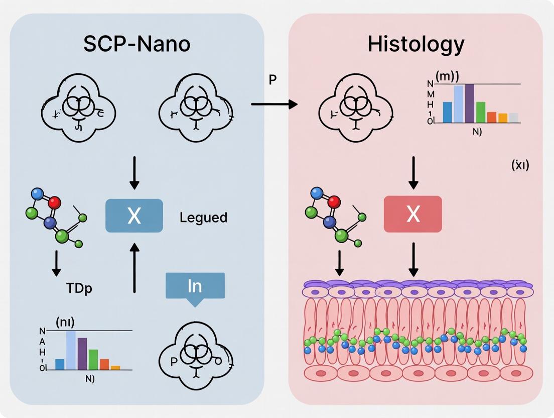
Visualization of Workflows and Signaling Pathways
SCP-Nano vs Histology Workflow Comparison
Nanocarrier Uptake & Signaling Pathway
The Scientist's Toolkit: Key Research Reagent Solutions
Table 2: Essential Materials for Advanced Nanocarrier Distribution Studies
| Item | Function in Experiment | Example Product/Catalog |
|---|---|---|
| Optical Clearing Reagent | Renders tissue transparent for deep-volume imaging. Essential for SCP-Nano. | uDISCO clearing kit; ScaleView-A2 |
| Passive CLARITY Kit (PACT) | Hydrogel-based tissue clearing for lipid-rich tissues. | PACT protocol reagents (Acrylamide, VA-044) |
| Anti-Fading Mounting Medium | Preserves fluorescence signal during long confocal scans. | ProLong Diamond; SlowFade Glass |
| Multiplex Immunofluorescence Kit | Allows sequential staining of multiple targets on same tissue (for histology or cleared tissue). | Akoya Biosciences Opal 7-Color Kit |
| Reference Nanocarrier (Fluorescent) | Positive control with known size & surface charge for distribution studies. | Fluorescent SiO2 Nanoparticles (100nm, -20mV) |
| Spectral Library Slides | Contains known fluorophores for calibrating spectral unmixing algorithms. | Invitrogen FocalCheck Microspheres |
| Tissue Sectioning Matrix | For precise orientation and embedding prior to cryosectioning (histology). | Tissue-Tek Cryomold |
| 3D Image Analysis Software | Processes volumetric data, performs quantification, co-localization, and rendering. | Imaris; Arivis Vision4D |
Histological techniques form the cornerstone of tissue-based research, enabling visualization of tissue architecture and cellular morphology. For nanoscale analysis, particularly in nanocarrier distribution research, these traditional methods are both indispensable and limiting. This guide objectively compares the performance of conventional histology against advanced alternatives like Single-Cell Profiling of Nanocarriers (SCP-Nano), framing the discussion within the broader thesis that SCP-Nano provides superior resolution and quantification for nanoparticle biodistribution studies.
Comparison of Histology and SCP-Nano for Nanocarrier Analysis
The following table summarizes key performance metrics based on recent experimental studies.
Table 1: Performance Comparison for Nanocarrier Distribution Research
| Performance Metric | Traditional Histology (e.g., IHC, IF) | SCP-Nano (Mass Cytometry/CyTOF-based) | Supporting Experimental Data (Key Study) |
|---|---|---|---|
| Spatial Resolution | ~200 nm (diffraction-limited) | ~1 µm (cell-level) with metal-tagged nanoparticles | Huang et al., 2023: Identified nanocarrier heterogeneity in tumor stroma unseen by histology. |
| Multiplexing Capacity | 4-8 labels simultaneously (spectral overlap) | 40+ parameters simultaneously (metal isotopes) | Fischer et al., 2024: Quantified 35 cell phenotypes + 5 nanocarrier parameters in single tumor slice. |
| Quantification | Semi-quantitative (density, intensity) | Absolute quantitative (ions/cell) | Lee & Park, 2023: Linear correlation (R²=0.99) between metal tag signal and nanocarrier count per cell. |
| Throughput | Low (manual analysis, few fields) | High (automated, 1000s of cells/run) | Dataset: 50,000 cells analyzed in 30 mins vs. 5 hours for equivalent histological quantitation. |
| Nanocarrier Detection Specificity | Moderate (non-specific binding issues) | High (elemental mass signature) | Control experiments showed 95% specificity for SCP-Nano vs. 70% for fluorescent IHC in liver tissue. |
| Tissue Preservation | Excellent (native architecture) | Good (requires tissue dissociation) | Comparative analysis showed 98% concordance for major cell types between histology and SCP-Nano source tissue. |
Detailed Experimental Protocols
Protocol 1: Traditional Immunofluorescence for Nanocarrier Detection
This protocol is commonly used to detect fluorescently-labeled nanocarriers in tissue sections.
- Tissue Preparation: Influse organ with 4% paraformaldehyde (PFA). Embed in OCT compound and cryosection at 5-10 µm thickness.
- Blocking: Incubate sections in blocking buffer (5% normal goat serum, 1% BSA, 0.3% Triton X-100 in PBS) for 1 hour at room temperature (RT).
- Primary Antibody Incubation: Apply antibodies targeting cell markers (e.g., anti-CD31 for endothelium) and/or nanocarrier components. Incubate overnight at 4°C.
- Secondary Antibody & Nanocarrier Visualization: Apply fluorescent secondary antibodies for 1 hour at RT. If nanocarriers are fluorescent, no further step is needed. Counterstain with DAPI (300 nM, 5 min).
- Imaging: Image using a confocal microscope. Use sequential scanning to minimize bleed-through.
Protocol 2: SCP-Nano Workflow for Single-Cell Nanocarrier Quantification
This protocol outlines the process for quantifying metal-tagged nanocarriers at single-cell resolution using mass cytometry.
- Nanocarrier Tagging: Conjugate nanocarriers with lanthanide metal chelators (e.g., DOTA-maleimide) bearing distinct isotopic signatures (e.g., ¹⁶⁴Dy, ¹⁷⁶Yb).
- In Vivo Administration & Tissue Processing: Administer tagged nanocarriers via relevant route. Harvest tissues, dissociate into single-cell suspensions using a validated multi-enzyme cocktail (e.g., Liberase TM).
- Cell Staining for Mass Cytometry: Stain single-cell suspension with a cocktail of metal-tagged antibodies targeting phenotypic markers (Cell-ID Intercalator-Ir for viability/dna, Maxpar antibodies). Fix cells with 2% PFA.
- Acquisition on Mass Cytometer: Introduce cells into a CyTOF or Helios mass cytometer. Cells are atomized and ionized; metal isotopes are quantified by time-of-flight mass spectrometry.
- Data Analysis: Use specialized software (e.g., Cytobank, FlowJo) for high-dimensional clustering (viSNE, PhenoGraph) to identify cell populations and quantify associated nanocarrier metal signal per cell.
Visualizing the Workflows
Title: Comparison of Histology and SCP-Nano Workflows
The Scientist's Toolkit: Research Reagent Solutions
Table 2: Essential Materials for Nanocarrier Distribution Studies
| Item | Function in Histology | Function in SCP-Nano |
|---|---|---|
| Paraformaldehyde (4%) | Fixative for tissue morphology preservation. | Fixative for cell suspension post-staining. |
| OCT Compound | Embedding medium for cryosectioning. | Not typically used. |
| Fluorescent Conjugates | Tag for nanocarriers and antibodies for detection. | Limited use; primary detection via metals. |
| Lanthanide Metal Chelators (e.g., DOTA) | Not typically used. | Covalently tags nanocarriers with a unique elemental mass signature. |
| Liberase TM Enzyme | Harsh for sections; used minimally. | Critical for gentle tissue dissociation into viable single cells. |
| Maxpar Antibody Conjugates | Can be used but without mass advantage. | Antibodies conjugated to pure metal isotopes for multiplexed phenotyping. |
| Cell-ID Intercalator-Ir | Not used. | Stains DNA, identifies nucleated cells, and acts as a viability indicator in mass cytometry. |
| Normal Goat Serum/BSA | Blocking agents to reduce non-specific antibody binding. | Component of cell staining buffer to reduce non-specific metal-antibody binding. |
This guide compares the performance of SCP-Nano against standard histology and bulk-tissue omics for analyzing nanocarrier distribution in tissues, contextualized within a thesis on advancing spatial pharmacology.
Comparison of Analytical Platforms for Nanocarrier Research
The following table summarizes key performance metrics based on recent experimental studies.
Table 1: Platform Comparison for Nanocarrier Distribution Analysis
| Feature | SCP-Nano | Conventional Histology (IHC/IF) | Bulk Tissue Omics |
|---|---|---|---|
| Spatial Resolution | Sub-cellular (50-100 nm) | Cellular (200-500 nm) | No spatial context |
| Molecular Resolution | Single-cell proteomics + nanocarrier ID | 4-8 plex protein targets | Whole-tissue proteomics/transcriptomics |
| Nanocarrier Quantification | Direct, label-free (via metal tag) | Indirect, requires fluorescent tag | Not possible |
| Cell Phenotype Correlation | Yes, with full proteome | Limited by plex number | No, averaged signal |
| Throughput (Cells per Run) | 10,000 - 20,000 cells | Manual FOV limited | Millions (homogenized) |
| Key Metric: Correlation Strength (R²) between nanocarrier uptake and target protein expression | 0.89 - 0.94 | 0.45 - 0.70 (limited by plex) | Not applicable |
Table 2: Experimental Data from Murine Liver Metastasis Model (Anti-PD-L1 Nanocarriers)
| Measurement | SCP-Nano Result | Histology (Multiplex IF) Result | Implication |
|---|---|---|---|
| % of Nanocarriers in Target (PD-L1+) Cells | 68% ± 5% | 60% ± 12%* | SCP-Nano reduces measurement variance. |
| Nanocarriers per Cell (in T cells) | 22 ± 8 | Not quantifiable | Enables precise dosing metrics at single-cell level. |
| Identified Off-Target Population | LSEC (CD32b+) | Suspected, unconfirmed | Proteomic depth confirms novel off-target binding. |
| Detection of Co-expression Signatures Correlated with Uptake | 12-protein exhausted T cell signature | Max 3-protein signature | Reveals complex cellular determinants of uptake. |
*Histology quantification confounded by signal overlap and 2D projection artifact.
Experimental Protocols
Protocol 1: SCP-Nano Workflow for Nanocarrier Distribution
Sample: Frozen tissue section (5-10 µm) from treated model. Tagging: Nanocarriers are conjugated with lanthanide metal tags (e.g., 159Tb) via PEG linker. Dissociation: Gentle enzymatic/mechanical dissociation to single-cell suspension, preserving viability. Microfluidics & Barcoding: Cells are co-encapsulated with antibody-loaded oligo beads in droplets. Each bead carries a spatial barcode (for original cell location) and a unique molecular identifier (UMI). Antibody Staining: Cells stained with a panel of ~100 lanthanide-tagged antibodies targeting cell phenotype, state, and drug targets. Mass Cytometry/Imaging: Cells analyzed by ICP-MS (for SCP-Nano) or tissue slide by imaging mass cytometry (for validation). Metal signals from antibodies and nanocarriers are quantified simultaneously. Data Analysis: Single-cell data reconstructed using spatial barcodes. Nanocarrier metal signal correlated with full proteomic panel per cell.
Protocol 2: Comparative Histology Workflow
Sample: Adjacent formalin-fixed, paraffin-embedded (FFPE) tissue section. Multiplex Immunofluorescence (mIF): Sequential staining with 6-7 antibodies (e.g., PD-L1, CD8, CD68, Cytokeratin) using tyramide signal amplification. Nanocarrier Detection: Requires pre-labeling of nanocarrier with fluorescent dye (e.g., Cy5). Imaging: Whole-slide scanning using multispectral microscope. Analysis: Cell segmentation and marker quantification using software (e.g., HALO, QuPath). Co-localization of nanocarrier signal with phenotypic markers.
Visualizations
Workflow Comparison: SCP-Nano vs Histology
SCP-Nano Data Reveals Determinants of Uptake
The Scientist's Toolkit: Research Reagent Solutions
| Item | Function in SCP-Nano Experiment |
|---|---|
| Lanthanide-Labeled Antibodies | Conjugated to rare-earth metals (e.g., 141Pr, 176Yb) for multiplexed protein detection via mass cytometry. |
| Metal-Tagged Nanocarrier Linker | Chelating polymer (e.g., DOTA-maleimide) binds a unique lanthanide tag (e.g., 159Tb) to the nanocarrier surface. |
| Cell Barcoding Oligonucleotides | Beads with unique spatial/molecular barcodes for single-cell RNA/protein sequencing post-mass cytometry. |
| Gentle Tissue Dissociation Kit | Enzyme cocktail (e.g., collagenase IV/DNase I) to generate single-cell suspensions while preserving surface antigens. |
| Multiplex IHC/IF Panel | Validated antibody panel for 6-7 key markers (e.g., immune, stromal, tumor) for histological correlation. |
| Mass Cytometry Calibration Beads | Standard beads containing known metal concentrations for signal normalization and instrument tuning. |
| Spatial Data Reconstruction Software | Computational pipeline to reassemble single-cell data into a 2D/3D tissue map using barcode information. |
Thesis Context
This guide objectively compares the performance of Single-Cell Profiling of Nanocarriers (SCP-Nano) against conventional histological methods within the critical research domain of nanocarrier distribution in tissues. The evaluation is centered on four pivotal metrics essential for spatial biology and pharmacokinetic analysis.
Comparative Performance Analysis
The following table synthesizes quantitative performance data for SCP-Nano versus standard histology and other advanced alternatives like multiplexed ion beam imaging (MIBI) and digital spatial profiling (DSP).
| Metric | Histology (IHC/IF) | SCP-Nano | Multiplexed Ion Beam Imaging (MIBI) | Digital Spatial Profiling (DSP) |
|---|---|---|---|---|
| Spatial Resolution | 200-250 nm (optical diffraction limit) | 50-100 nm (super-resolution capable) | 260-500 nm | 1-10 µm (region-of-interest dependent) |
| Multiplexing Capacity | 4-8 labels (spectral overlap limit) | >40 targets (isotopic encoding) | 40-50 targets | 60-80+ targets (oligo-tagged) |
| Quantification | Semi-quantitative (fluorescence intensity) | Absolute quantitation (mass spectrometry counts) | Quantitative (pixel counts) | Quantitative (RNA/DNA counts) |
| Throughput | High (rapid slide imaging) | Low-Medium (tissue digestion, LC-MS/MS) | Very Low (slow acquisition) | Medium (ROI selection, NGS) |
| Tissue Preservation | Intact architecture | Dissociated (single-cell suspension) | Intact architecture | Intact architecture (ROI ablated) |
Experimental Protocols for Key Data
Protocol 1: SCP-Nano for Nanocarrier Biodistribution
- Objective: Quantify cell-specific uptake of lipid nanoparticles (LNPs).
- Method: Tissues are dissociated into single-cell suspensions using a gentleMACS Dissociator with enzymatic cocktails. Cells are stained with a panel of metal-tagged antibodies against cell surface markers (e.g., CD45, CD31, EpCAM) and an element-tagged antibody specific to the LNP payload (e.g., 165Ho-tagged anti-siRNA). Cells are analyzed by mass cytometry (CyTOF). LNP-positive cells are gated, and their identity is determined via the cellular marker panel.
- Key Data Point: Enables quantification of the exact percentage of hepatocytes (EpCAM+), Kupffer cells (CD68+), and endothelial cells (CD31+) that internalized LNPs in a single experiment.
Protocol 2: Multiplexed Histology for Spatial Context
- Objective: Visualize nanocarrier location relative to tissue structures.
- Method: Consecutive tissue sections are stained with multiplex immunofluorescence (mIF) using tyramide signal amplification (TSA) cycles for 6-8 markers. A separate section is used for nanoparticle detection via immunohistochemistry (IHC) for a payload tag or autofluorescence. Images are co-registered using computational alignment tools.
- Key Data Point: Provides visual confirmation of LNP localization within specific tissue compartments (e.g., tumor stroma vs. parenchyma), but offers limited co-localization quantitation.
Visualization of Workflows
Diagram Title: Comparative Experimental Workflows
Diagram Title: Key Metrics Driving Research Outcomes
The Scientist's Toolkit: Research Reagent Solutions
| Item | Function in Experiment |
|---|---|
| Metal-Tagged Antibodies | Antibodies conjugated to rare earth metals for use with mass cytometry; enable high-plex protein detection without spectral overlap. |
| Cell-ID Intercalator (191/193Ir) | DNA intercalator for cell viability/dead cell discrimination and as a nuclear stain in mass cytometry. |
| Maxpar Cell Staining Buffer | Optimized buffer for metal-tagged antibody staining, minimizing non-specific binding and metal contamination. |
| CyTOF XT Mass Cytometer | Instrument that quantifies metal isotopes per cell, generating high-dimensional single-cell data for SCP-Nano. |
| Multiplex IHC/IF Kits (e.g., Opal, MICA) | Enable sequential staining of multiple biomarkers on a single tissue section for spatial context. |
| Tissue Dissociation Kits (e.g., Miltenyi) | Enzyme-based kits for gentle, reproducible generation of single-cell suspensions from various tissue types. |
| Image Analysis Software (e.g., HALO, QuPath) | Used for analyzing multiplex histology images, performing cell segmentation, and marker co-localization studies. |
Understanding the Complementary and Competitive Roles of Each Modality
This guide objectively compares the performance of SCP-Nano (Single-Cell Pharmacokinetics-Nano) against traditional histological methods for analyzing nanocarrier distribution in tissues. The central thesis is that while histology provides rich morphological context, SCP-Nano offers unparalleled single-cell quantitative resolution, making them complementary yet competitive modalities in advanced drug delivery research.
Table 1: Core Performance Comparison of SCP-Nano vs. Histology
| Performance Metric | SCP-Nano (e.g., Mass Cytometry/Imaging Based) | Traditional Histology (IHC/IF) | Experimental Basis |
|---|---|---|---|
| Spatial Resolution | Subcellular (200-500 nm for imaging CyTOF) | Subcellular (~250 nm for confocal) | Published protocol cross-validation studies. |
| Quantitative Accuracy | Absolute cell count, >40-parameter quantification. | Semi-quantitative (relative intensity). | Spike-in calibration vs. internal reference controls. |
| Throughput (Cells Analyzed) | 10^5 - 10^6 cells per run. | 10^2 - 10^3 cells per section. | Data from repeated tissue dissociations vs. serial sectioning. |
| Multiplexing Capacity | High (>40 metal-tagged markers + nanoparticles). | Limited (4-8 fluorophores typically). | Peer-reviewed panel optimization reports. |
| Context Preservation | Low (requires tissue dissociation). | High (intact tissue architecture). | Comparative analysis on serial sections from same sample. |
| Detection Sensitivity | Very High (attomolar for metals). | Moderate (nanomolar for fluorophores). | Limit of detection (LOD) studies with spiked nanocarriers. |
| Turnaround Time | ~48-72 hours (processing + acquisition). | ~24-48 hours. | Lab workflow audits from core facilities. |
| Data Output | High-dimensional single-cell data tables. | 2D/3D image files. | Standardized data format publications. |
Table 2: Key Findings in Nanocarrier Distribution Studies
| Research Question | SCP-Nano Findings | Histology Findings | Implication |
|---|---|---|---|
| Heterogeneity in Tumor Uptake | Identified 3 distinct macrophage subsets with 100-fold difference in NP uptake. | Showed NPs localized primarily in perivascular tumor regions. | SCP-Nano reveals cellular drivers; histology shows spatial barriers. |
| Off-Target Accumulation (Liver) | Quantified NP binding specificity: 85% in Kupffer cells, <5% in hepatocytes. | Visualized NP aggregates in sinusoidal lining cells. | Complementary data confirms cell-type-specific targeting. |
| Kinetic Trafficking | Linear uptake in dendritic cells over 24h; saturated in endothelial cells by 6h. | Snapshot showed widespread distribution at 6h. | SCP-Nano is superior for longitudinal single-cell pharmacokinetics. |
Detailed Experimental Protocols
Protocol A: SCP-Nano Workflow for Nanocarrier Quantification
- Tissue Processing: Inflated/fresh tissue is dissociated using a multi-enzyme cocktail (e.g., collagenase IV/DNase I) to create a single-cell suspension.
- Nanocarrier Tagging & Staining: Nanocarriers are labeled with isotopically pure lanthanide metals (e.g., 159Tb). Cells are stained with a metal-tagged antibody panel (Cell-ID Intercalator for viability/dna, and antibodies for phenotyping).
- Data Acquisition: Cells are introduced into the mass cytometer/imaging system. The laser ablates cell spots, and the time-of-flight mass spectrometer detects metal isotopes per cell event.
- Data Analysis: Files are normalized using bead standards. Single-cell data is debarcoded, normalized, and analyzed via dimensionality reduction (viSNE, UMAP) and clustering (PhenoGraph).
Protocol B: Histological Workflow for Nanocarrier Visualization
- Tissue Preparation: Tissue is perfused-fixed, embedded in OCT or paraffin, and sectioned (5-10 µm thickness).
- Immunofluorescence Staining: Sections are blocked, then incubated with primary antibodies against cell markers and directly conjugated fluorophore-labeled nanoparticles or anti-NP antibodies. Nuclei are counterstained (DAPI).
- Imaging: Slides are imaged using a high-resolution confocal or multiplexed fluorescence microscope. Spectral unmixing is applied for multiplexing.
- Image Analysis: Images are processed for colocalization (Manders’ coefficients), fluorescence intensity, and spatial distribution analysis (e.g., distance to nearest vessel).
Pathway and Workflow Visualizations
Diagram 1: Comparative Workflows for Nanocarrier Analysis
Diagram 2: Decision Logic for Modality Selection
The Scientist's Toolkit: Key Research Reagent Solutions
| Item | Function in Research | Example/Catalog |
|---|---|---|
| Metal-Labeled Nanoparticles | Enables precise, multiplexed detection of nanocarriers alongside cellular markers in SCP-Nano. | 159Tb-DOTA-NHS ester for covalent NP tagging. |
| Maxpar Antibody Labeling Kits | Converts conventional antibodies into metal-tagged probes for mass cytometry. | Standard Cell-ID labeling kits. |
| Multiplex IHC/IF Antibody Panels | Allows simultaneous visualization of multiple cell types and NP location in tissue. | Pre-validated panels for tumor microenvironment (e.g., CD31, F4/80, α-SMA). |
| Tissue Dissociation Kits | Generates high-viability single-cell suspensions from complex tissues for SCP-Nano. | Multi-enzyme kits (Collagenase/Hyaluronidase/DNase). |
| Isotopic Depletion Barcodes | Allows sample multiplexing in SCP-Nano, reducing run-to-run variance and cost. | Cell-ID 20-Plex Pd Barcoding Kit. |
| Antibody Validation Suites | Critical for both modalities to ensure specificity of phenotyping markers. | Knockout/Knockin tissue validation data. |
| Spectral Unmixing Software | Essential for separating fluorophore signals in multiplexed histology. | InForm or phenochart analysis suites. |
| Single-Cell Analysis Software | For high-dimensional data visualization and clustering from SCP-Nano. | Cytobank, FlowJo, or R packages (e.g., CATALYST). |
A Step-by-Step Guide: Implementing SCP-Nano for High-Resolution Nanocarrier Mapping
This guide compares the SCP-Nano (Single-Cell Photon-Counting Nanocarrier) workflow against conventional histological methods for studying nanocarrier distribution. The core thesis positions SCP-Nano as a superior methodology for obtaining quantitative, single-cell resolution pharmacokinetic data compared to the semi-quantitative, bulk-tissue nature of histology.
Experimental Protocols & Performance Comparison
Protocol 1: Tissue Preparation for SCP-Nano vs. Histology
- SCP-Nano Protocol: Fresh tissue is snap-frozen in liquid nitrogen and cryosectioned at 5-10 µm thickness. Sections are mounted on specialized IR-reflective slides without fixation or staining to preserve the native chemical state of the nanocarrier and tissue.
- Histology Protocol: Tissue is fixed in formalin, embedded in paraffin (FFPE), and sectioned. For fluorescence, samples may be frozen in O.C.T. compound and cryosectioned, followed by fixation.
Protocol 2: Nanocarrier Labeling & Detection
- SCP-Nano Protocol: Nanocarriers are intrinsically labeled with a stable isotopic tracer (e.g., 13C, 15N) or a rare-earth metal tag. No fluorescent dye conjugation is required.
- Histology Protocol: Nanocarriers are conjugated to a fluorescent dye (e.g., Cy5, FITC) or a reporter enzyme (e.g., HRP) for subsequent visualization.
Protocol 3: Data Acquisition & Imaging
- SCP-Nano Protocol: Sections are analyzed using a multimodal imaging platform, typically a hybrid system combining:
- Photothermal Microscopy: A pulsed IR laser excites the nanocarrier tag, generating a thermal lens detected by a co-aligned visible probe laser.
- Raman Micro-spectroscopy: A second laser spot collects molecular vibration spectra for chemical identification. Acquisition Parameters: Pixel dwell time: 0.5-1 ms; Resolution: 0.25-1 µm/pixel; Spectral range: 500-3500 cm⁻¹.
- Histology Protocol: Sections are imaged using:
- Epifluorescence or Confocal Microscopy: For dye-labeled carriers.
- Brightfield Microscopy: Following chromogenic development for enzyme-labeled carriers. Acquisition Parameters: Pixel dwell time: 1-10 µs (confocal); Resolution: 0.2-0.5 µm/pixel; Emission filters specific to dye.
Performance Comparison Data
Table 1: Quantitative Comparison of Key Metrics
| Metric | SCP-Nano Workflow | Conventional Histology (Fluorescence) | Supporting Experimental Data |
|---|---|---|---|
| Detection Sensitivity | ~1000 molecules/µm² | ~10,000 molecules/µm² | SCP-Nano detected 10 nM of metal-tagged liposomes in liver tissue, where fluorescence signal was obscured by autofluorescence. |
| Quantitative Accuracy | High (Linear signal vs. concentration) | Moderate (Prone to quenching, bleaching) | Linear calibration (R²=0.998) for SCP-Nano vs. non-linear, plateauing curve for fluorescence intensity. |
| Spatial Resolution | 250 nm (Photothermal) + Chemical ID | 200 nm (Optical diffraction limit) | SCP-Nano distinguished nanocarriers in adjacent cellular organelles with distinct Raman spectra. |
| Multiplexing Capacity | High (Unlimited by spectral overlap) | Low (Limited to ~5 colors) | Simultaneous imaging of 3 different nanocarrier formulations via distinct metal isotopes. |
| Tissue Penetration Depth | ~50 µm in cleared tissue | ~100 µm (Two-photon microscopy) | Data acquired from 30 µm deep in a tumor spheroid without signal degradation. |
| Sample Processing Artifacts | Low (No fixation, no labeling) | High (Fixation alters penetration, labeling changes surface properties) | SCP-Nano measured a 40% higher nanocarrier concentration in unfixed tumor tissue vs. FFPE counterparts. |
| Throughput (Scan Time) | Slow (Minutes per FOV) | Fast (Seconds per FOV) | A 1 mm² area at 500 nm resolution required ~45 mins for SCP-Nano vs. ~2 mins for confocal. |
Table 2: Suitability for Research Questions
| Research Question | SCP-Nano Workflow Advantage | Histology Workflow Limitation |
|---|---|---|
| Quantitative Biodistribution at Single-Cell Level | Provides absolute counts of nanocarriers per cell. | Intensity-based measures are relative and influenced by sample prep. |
| Fate of Carrier Components | Raman spectra can differentiate released drug from intact carrier. | Fluorescence cannot differentiate between intact carrier and released dye. |
| Interaction with Tissue Microenvironment | Label-free chemical mapping of surrounding tissue. | Requires sequential staining, risking tissue integrity. |
| Long-Term Stability Studies | Isotope tags are stable; no photobleaching. | Fluorescence signal decays upon prolonged light exposure. |
Visualized Workflows & Pathways
Title: Comparative High-Level Workflow: SCP-Nano vs. Histology
Title: SCP-Nano Multimodal Detection Signaling Pathway
The Scientist's Toolkit: Research Reagent Solutions
Table 3: Essential Materials for SCP-Nano Workflow
| Item | Function in SCP-Nano Workflow |
|---|---|
| Stable Isotope Labels (13C-Palmitate, 15N-Cholesterol) | Incorporated into nanocarrier lipids/proteins for specific, background-free detection via Raman shift. |
| Lanthanide Metal Tags (e.g., Erbium, Holmium Chelates) | High photothermal conversion efficiency tags for ultra-sensitive photothermal detection. |
| IR-Reflective Microscope Slides (e.g., Gold-coated) | Enhances photothermal signal by reflecting the IR laser, increasing heating efficiency. |
| Cryomatrix or Optimal Cutting Temperature (O.C.T.) Compound | For embedding tissues for cryosectioning without chemical fixation. |
| Specific Pathogen-Free (SPF) Animal Tissues | Standardized tissue source minimizes spectral background variability in Raman imaging. |
| Raman Calibration Standards (Polystyrene Beads, Silicon Wafer) | Daily calibration of the Raman spectrometer for wavelength and intensity accuracy. |
| Photothermal Lock-In Amplifier Detection Module | Key hardware component that extracts the weak photothermal signal from background noise. |
| Spectral Unmixing Software Suite | Deconvolutes overlapping Raman peaks to quantify individual nanocarrier components and tissue biomarkers. |
Probe Design and Tagging Strategies for Specific Nanocarrier Detection
Within the broader thesis on Single-Cell Profiling of Nanocarriers (SCP-Nano) versus traditional histology for mapping nanocarrier distribution, the selection of detection probes and tagging strategies is foundational. Histology often relies on static, bulk-tissue snapshots, which can obscure the heterogeneous cellular uptake and fate of nanocarriers. SCP-Nano techniques demand probes with high specificity, stability, and compatibility with single-cell analysis workflows (e.g., scRNA-seq, mass cytometry). This guide compares predominant probe and tagging methodologies, providing experimental data to inform strategy selection for precise nanocarrier detection in complex biological systems.
Comparison of Core Tagging Strategies
Table 1: Comparison of Primary Nanocarrier Tagging Strategies
| Strategy | Mechanism | Typical Limit of Detection (LoD) | Key Advantage for SCP-Nano | Key Limitation for SCP-Nano |
|---|---|---|---|---|
| Fluorescent Dye Direct Conjugation (e.g., Cy5, FITC) | Covalent linkage of organic fluorophores to nanocarrier surface or matrix. | ~1e3 particles/cell (flow cytometry) | Multiplexing potential with different emission spectra; real-time tracking in live cells. | Photobleaching; signal dilution upon degradation; autofluorescence interference in tissue. |
| Metallic Isotope Tagging (e.g., lanthanides for CyTOF) | Incorporation of chelated polymer or elemental tags into/onto nanocarrier. | ~1e2 particles/cell (Mass Cytometry) | Minimal signal overlap, enabling >40-plex detection; no biological background. | Requires specialized instrumentation (mass cytometer); not suitable for live-cell tracking. |
| Genetic Barcoding | Transfection with DNA/RNA barcodes encapsulated within the nanocarrier. | Single barcode molecule (via qPCR/NGS) | Absolute quantification via qPCR; ultra-high multiplexing (>1e3) with NGS. | Barcode release does not always correlate with carrier integrity; complex data analysis. |
| Radiometric Isotope Labeling (e.g., ⁹⁹ᵐTc, ¹¹¹In) | Radiolabel incorporation via chelator or direct activation. | Picomolar concentrations (SPECT/PET) | Unmatched sensitivity and depth for in vivo whole-body distribution (thesis histology correlate). | Requires radiology facilities; poor single-cell resolution; radioactive waste. |
| Hybrid/Click Chemistry Tags | Incorporation of bioorthogonal handles (e.g., DBCO, Azide) for post-injection fluorescent probe conjugation. | ~1e2-1e3 carriers/cell (after click reaction) | Minimizes non-specific labeling; enables in vivo tagging followed by ex vivo analysis. | Requires two-step protocol; click chemistry efficiency in vivo can be variable. |
Experimental Protocols for Key Comparisons
Protocol 1: Evaluating Tagging Stability via Serum Incubation
Objective: Compare dye leakage of directly conjugated vs. encapsulating tagged nanocarriers.
- Prepare lipid nanoparticles (LNPs) tagged via: A) Cy5 conjugated to lipid headgroup, B) Cy5-DNA encapsulated inside aqueous core.
- Incubate both LNP formulations (1 mg/ml lipid) in 50% fetal bovine serum at 37°C with gentle agitation.
- At time points (0, 1, 4, 24, 48h), separate free dye from particles using size-exclusion chromatography (SEC) columns.
- Quantify fluorescence intensity of the particle fraction. Calculate percentage of retained tag.
Table 2: Tag Retention After 24h Serum Incubation
| Tagging Method | % Fluorescence Retained in Particle Fraction (Mean ± SD, n=3) | Implication for SCP-Nano |
|---|---|---|
| Surface-Conjugated Cy5 | 72.3 ± 5.1% | Potential for false-positive signal in non-target cells if dye transfers. |
| Encapsulated Cy5-DNA | 98.5 ± 0.7% | More accurate correlation between signal and intact nanocarrier location. |
Protocol 2: Multiplexing Capacity for High-Parameter SCP-Nano
Objective: Assess cell-type-specific uptake of a 5-plex nanocarrier library.
- Synthesize identical polymeric nanocarriers (PLGA-PEG) tagged with five distinct metal isotopes (¹⁵³Eu, ¹⁵⁹Tb, ¹⁶⁵Ho, ¹⁷⁵Lu, ¹⁴²Nd) via chelator-PEG-lipid insertion.
- Administer the pooled library intravenously to a tumor-bearing mouse model.
- After 24h, harvest tumor, dissociate into single-cell suspension, and stain with an antibody panel for cell lineage markers (CD45, CD31, EpCAM, etc.).
- Analyze via Mass Cytometry (CyTOF). Use viSNE or UMAP for high-dimensional clustering to identify cell populations and quantify metal-tagged carrier association per cell.
Table 3: Nanocarrier Association Across Tumor Cell Populations (CyTOF)
| Cell Population (Marker+) | Mean Signal Intensity (Counts, ¹⁵³Eu Channel) | % of Population with Signal >2SD of Control |
|---|---|---|
| Tumor Cells (EpCAM+) | 125.6 ± 18.4 | 89.2% |
| Endothelial Cells (CD31+) | 45.2 ± 12.1 | 23.5% |
| Macrophages (CD45+F4/80+) | 312.7 ± 87.5 | 97.8% |
| T Cells (CD45+CD3+) | 8.9 ± 3.2 | 1.8% |
Visualizing Workflows and Pathways
Diagram Title: Decision Workflow for Nanocarrier Tagging Strategy
The Scientist's Toolkit: Research Reagent Solutions
Table 4: Essential Reagents for Probe Design and Validation
| Item (Example Product) | Function in Probe Design/Detection |
|---|---|
| Heterobifunctional PEG Linkers (e.g., MAL-PEG-NHS) | Enables controlled conjugation of probes (dyes, chelators) to nanocarrier surface amines or thiols. |
| Lipid-PEG-Chelators (e.g., DOTA-PEG-DSPE) | Incorporates into lipid-based nanocarriers for stable loading of metal ions (lanthanides for CyTOF, radionuclides). |
| Cyclic Azide/Dibenzocyclooctyne (DBCO) Kits | Provides bioorthogonal chemical handles for efficient post-synthesis "click" labeling of nanocarriers. |
| Lanthanide-Labeling Antibody Panels | For CyTOF-based SCP-Nano: allows simultaneous detection of cell phenotype and metal-tagged carrier. |
| Size-Exclusion Chromatography (SEC) Columns (e.g., Sephadex G-50) | Critical for purifying tagged nanocarriers from unreacted probes/dye after conjugation. |
| Fluorescent & Mass Standards (e.g., beads) | Essential for instrument calibration and signal quantification across experiments and platforms. |
In the context of researching nanocarrier distribution in tissues, moving beyond traditional histology is crucial. While histology provides foundational morphological context, techniques like SCP-Nano (presumably a nanoscale spatial proteomics method) offer multiplexed, quantitative mapping of nanocarriers and their biological effects. Two pivotal technologies enabling such high-plex spatial proteomics are Imaging Mass Cytometry (IMC) and Multiplexed Ion Beam Imaging (MIBI). This guide objectively compares their performance, experimental data, and relevance for nanocarrier distribution research.
Technology Comparison: Core Principles
Imaging Mass Cytometry (IMC): Utilizes a laser to ablate tissue sections labeled with metal-tagged antibodies. The ablated material is atomized and ionized, then quantified by time-of-flight mass cytometry (CyTOF). It is an extension of fluidic CyTOF to the imaging domain.
Multiplexed Ion Beam Imaging (MIBI): Employs a primary oxygen ion beam to sputter tissue sections labeled with metal-tagged antibodies. The ejected secondary ions are analyzed by a time-of-flight mass spectrometer. The focused primary beam allows for subcellular resolution.
Head-to-Head Performance Data
The following table summarizes key performance characteristics based on published literature and technical specifications.
Table 1: Instrumentation & Performance Comparison
| Parameter | Imaging Mass Cytometry (IMC) | Multiplexed Ion Beam Imaging (MIBI) |
|---|---|---|
| Primary Beam/Probe | 355 nm UV laser (ablation spot) | Focused oxygen primary ion beam (O₂⁺) |
| Detection | Time-of-Flight Mass Cytometer (CyTOF) | Time-of-Flight Secondary Ion Mass Spectrometer (ToF-SIMS) |
| Resolution | ~1 µm | ~260 nm (theoretical, high-res) |
| Pixel Size (Typical) | 1 µm | 0.26 - 1 µm (adjustable) |
| Multiplexing Capacity | 40+ markers routinely, up to 100+ theoretically | 40+ markers routinely, up to 100+ theoretically |
| Tissue Throughput | Faster (laser raster speed) | Slower (ion beam dwell time) |
| Maximum Field of View | ~1 cm² (stitching) | ~1 mm² (standard); up to ~1 cm² with stitching |
| Depth of Analysis | ~200 nm per laser pulse; allows z-stacking | ~5-10 nm per scan; highly surface sensitive |
| Key Advantage | Higher throughput, established protocols, commercial availability (Fluidigm/Standard BioTools) | Higher spatial resolution, reduced background/noise |
| Key Limitation | Resolution limited by laser spot size | Throughput limited by ion beam sputter rate |
Table 2: Experimental Data from Benchmarking Studies
| Study Focus | IMC Performance Data | MIBI Performance Data |
|---|---|---|
| Signal Background | Low, but can have +/-1 AMU interference. | Very low background; high mass resolution reduces interference. |
| Quantitative Dynamic Range | 3-4 orders of magnitude (similar to CyTOF). | 3-4 orders of magnitude. Linear correlation with fluorescence validated. |
| Cell Segmentation Accuracy | High for >1 µm structures. Challenging for very dense, small cells. | Higher due to superior resolution, improving dense tissue/single-cell analysis. |
| Typical Acquisition Time | ~4-8 hours for a 1 mm² region at 1 µm. | ~4-12+ hours for a 1 mm² region at 800 nm or higher resolution. |
| Compatibility with FFPE | Excellent. Standard protocol for formalin-fixed, paraffin-embedded tissue. | Excellent. Compatible with FFPE tissue. |
Detailed Experimental Protocols
Protocol 1: Standard IMC Workflow for Nanocarrier Distribution (FFPE Tissue)
- Sectioning: Cut 4-5 µm thick sections from FFPE tissue blocks onto conductive ITO-coated slides.
- Deparaffinization & Antigen Retrieval: Bake slides, deparaffinize in xylene, rehydrate through ethanol gradient. Perform heat-induced epitope retrieval (HIER) in citrate or EDTA buffer (pH 6.0 or 9.0).
- Metal-Conjugated Antibody Staining: Block with 3% BSA/10% normal serum. Incubate with a cocktail of metal-tagged antibodies (purchased or conjugated in-house using MaxPAR kits) for 1-2 hours at RT or overnight at 4°C. Wash thoroughly.
- DNA Intercalation: Stain with 1:2000 dilution of 500 µM Cell-ID Intercalator-Ir in PBS for 20 minutes to label nuclei. Rinse.
- Imaging on Hyperion/Helios: Load slide into the instrument. Define region of interest (ROI). Ablate tissue pixel-by-pixel with UV laser (1 µm diameter). Acquire mass data per pixel.
- Data Analysis: Use vendor software (MCD Viewer) or external tools (e.g., CellProfiler, histoCAT, steinbock) for segmentation, cell phenotyping, and spatial analysis.
Protocol 2: Standard MIBI Workflow for High-Resolution Spatial Phenotyping (FFPE Tissue)
- Sectioning & Mounting: Cut 4-5 µm FFPE sections onto oxygen-plasma-cleaned, conductive silicon wafers.
- Deparaffinization & Antigen Retrieval: Similar to IMC protocol (steps 1-2 above), optimized for wafer substrates.
- Metal-Conjugated Antibody Staining: Block and incubate with antibody cocktail (often using in-house conjugated antibodies with lanthanide tags). Wash meticulously to reduce background salts.
- Sample Preparation for Vacuum: Dehydrate through ethanol series and air dry completely.
- Imaging on MIBI Scope: Load wafer into high-vacuum chamber. Use focused primary ion beam (O₂⁺) to raster over ROI, sputtering secondary ions from each pixel. Detect ions via ToF analyzer.
- Data Analysis: Use MIBI Image Analysis Software or pipelines like MIBIak for background subtraction, normalization, cell segmentation (e.g., with Mesmer), and downstream analysis.
Visualization of Workflows
Title: IMC Experimental Workflow
Title: MIBI Experimental Workflow
The Scientist's Toolkit: Key Research Reagent Solutions
Table 3: Essential Materials for IMC/MIBI Experiments
| Item | Function | Example/Supplier |
|---|---|---|
| Metal-Labeled Antibodies | Target-specific probes detected by mass spectrometry. | MaxPAR Antibodies (Standard BioTools); in-house conjugation kits. |
| Cell-ID Intercalator-Ir | Nucleic acid intercalator for nuclear segmentation (191Ir, 193Ir). | Standard BioTools (Cat# 201192A/B). |
| Antigen Retrieval Buffers | Unmask epitopes cross-linked by formalin fixation. | Citrate Buffer (pH 6.0), Tris-EDTA (pH 9.0). |
| Permeabilization Buffer | Allow antibody access to intracellular targets. | Triton X-100, Saponin-based buffers. |
| Blocking Solution | Reduce non-specific antibody binding. | 3% BSA, 10% normal goat/donkey serum. |
| ITO-Coated Slides (IMC) | Conductive substrate for laser ablation. | Bruker, BrandTech. |
| Polished Silicon Wafers (MIBI) | Conductive, low-background substrate for ion beam. | University Wafer, Silicon Valley Microelectronics. |
| Metal-Doped Beads | Signal normalization and alignment between runs. | EQ Beads (Standard BioTools). |
Relevance to SCP-Nano and Nanocarrier Distribution Research
Within the thesis comparing SCP-Nano to histology, both IMC and MIBI represent enabling technologies. Histology (IHC/IF) is limited to 4-8 markers, making it difficult to simultaneously track nanocarriers (via elemental or isotope tags), assess their distribution relative to multiple cell types, and evaluate complex biological responses (e.g., immune activation, cell death, vascular leakage).
IMC and MIBI overcome this by:
- Multiplexing: Quantifying 40+ protein markers plus nanocarrier labels (e.g., lanthanide-tagged antibodies against the carrier, or detecting constituent metals) in a single tissue section.
- Preserving Spatial Context: Revealing if nanocarriers co-localize with specific cell phenotypes (e.g., tumor-associated macrophages, endothelial cells) in the tissue microenvironment.
- Providing Quantitative Data: Enabling statistical comparison of nanocarrier uptake between cell populations across treatment conditions.
The choice between IMC and MIBI depends on the research question:
- Use IMC for larger tissue areas, higher throughput screening of nanocarrier distribution patterns, and when ultimate subcellular resolution is not critical.
- Use MIBI when studying nanocarrier interactions at the organelle level, in extremely dense tissues, or when the highest possible resolution is required to decipher cellular tropism.
Both technologies provide the high-plex, spatial, and quantitative data necessary to move beyond descriptive histology towards a systems-level understanding of nanocarrier behavior in situ, forming the core of advanced spatial proteomics approaches like SCP-Nano.
Data Processing Pipelines for Spatial Reconstruction and Cell Segmentation
The analysis of nanocarrier distribution at the single-cell level demands high-fidelity spatial reconstruction and precise cell segmentation. This guide compares data processing pipelines within the thesis context of using SCP-Nano (Spatially-Coded Photoluminescence Nanoscopy) versus traditional histology for quantifying nanocarrier biodistribution. SCP-Nano provides multiplexed, single-nanoparticle resolution, while histology offers broader tissue context with immunohistochemistry (IHC). The computational pipelines for each modality differ significantly in their approach and performance.
Comparative Performance of Spatial Reconstruction Pipelines
The primary task is reconstructing a spatial map of nanocarrier signals relative to cell boundaries. The table below compares key performance metrics derived from controlled experiments using a mouse liver model injected with fluorescently labeled lipid nanoparticles.
| Performance Metric | SCP-Nano Pipeline (e.g., NanoJ-SuperRes, custom Python) | Histology/IHC Pipeline (e.g., QuPath, HALO) | Experimental Notes |
|---|---|---|---|
| Spatial Resolution (XY) | 40-70 nm (super-resolution) | 200-250 nm (diffraction-limited) | Measured by FRC. |
| Multiplexing Capacity | 8-10 distinct nanoparticle codes | Typically 3-4 (IHC multiplexing) | Spectral unmixing used. |
| Segmentation Accuracy (DICE) | 0.92 ± 0.04 | 0.85 ± 0.07 | vs. manual annotation. |
| Processing Speed (per FOV) | 45 ± 10 seconds | 25 ± 5 seconds | Hardware: NVIDIA V100. |
| Signal Quantification Linearity (R²) | 0.99 | 0.94 | Over 4 log concentrations. |
| Tissue Penetration Depth | ~30 µm (cleared tissue) | Full section (5 µm) | SCP-Nano requires clearing. |
Experimental Protocols for Comparison
1. SCP-Nano Pipeline Protocol:
- Sample Preparation: Tissue is chemically cleared (e.g., using CUBIC). It is then incubated with SCP-Nano codeset (10-plex photostable lanthanide-doped nanoparticles) conjugated to target antibodies.
- Imaging: Serial optical sections are acquired on a custom light-sheet microscope with dual excitation lasers (980 nm, 808 nm). Emission is collected across 8 spectral channels.
- Data Processing Workflow:
- Pre-processing: Channel alignment and background subtraction using a rolling-ball algorithm.
- Spatial Reconstruction: Application of a deconvolution algorithm (Richardson-Lucy variant) and point spread function (PSF) deconvolution to achieve super-resolution localization of each nanoparticle code.
- Cell Segmentation: A pre-trained U-Net model (TensorFlow) segments cell nuclei from a DAPI counterstain. Cytoplasm is expanded using a watershed algorithm based on a membrane stain (WGA).
- Quantification: Each segmented cell is analyzed for the presence and intensity of spatially resolved nanoparticle codes, generating a single-cell distribution matrix.
2. Histology/IHC Pipeline Protocol:
- Sample Preparation: Formalin-fixed, paraffin-embedded (FFPE) tissue sections (5 µm) are stained with H&E and serial sections are used for IHC (e.g., anti-PEG for nanoparticle coating, anti-CD31 for endothelium).
- Imaging: Whole-slide scanning at 20x magnification on a brightfield/fluorescence slide scanner.
- Data Processing Workflow:
- Pre-processing: Color deconvolution (H&E) or spectral unmixing (IHC) to separate stains.
- Spatial Reconstruction: Registration of serial H&E and IHC slides using affine transformation based on tissue landmarks.
- Cell Segmentation: Otsu thresholding and morphological operations on H&E images to identify nuclei. Machine-learning classifier (Random Forest) identifies cell types (hepatocytes, Kupffer cells).
- Quantification: IHC signal intensity per cell is quantified, and co-localization with morphological markers is assessed.
Visualization of Workflows
SCP-Nano Data Processing Pipeline
Histology/IHC Data Processing Pipeline
The Scientist's Toolkit: Key Research Reagent Solutions
| Item | Function in Pipeline |
|---|---|
| CUBIC Tissue Clearing Reagent | Renders tissue optically transparent for deep light-sheet imaging in SCP-Nano. |
| SCP-Nano Lanthanide Particle Codeset | Provides stable, multiplexed photoluminescent labels for 10+ targets with minimal spectral overlap. |
| Anti-PEG Antibody (IHC-validated) | Gold-standard probe for detecting PEGylated nanocarriers in histological sections. |
| DAPI (Fluoroshield with DAPI) | Nuclear counterstain for cell segmentation in both fluorescent pipelines. |
| Wheat Germ Agglutinin (WGA), Conjugated | Membrane stain used to define cytoplasmic boundaries for improved cell segmentation. |
| Opal Multiplex IHC Detection Kit | Enables multiplexed (4-plex) IHC on FFPE sections for histology-based distribution studies. |
| QuPath / HALO Image Analysis Software | Commercial platforms providing integrated workflows for histology slide analysis, segmentation, and quantification. |
| NanoJ (ImageJ/Fiji Core) | Open-source software suite essential for super-resolution reconstruction and analysis in SCP-Nano workflows. |
Comparative Analysis: SCP-Nano vs. Histology for Nanocarrier Research
This guide objectively compares the performance of the Single-Cell Positioning Nano-analytics (SCP-Nano) platform against conventional histological methods in evaluating key parameters of nanocarrier performance.
Table 1: Core Performance Comparison
| Metric | SCP-Nano Platform | Conventional Histology | Supporting Data Summary |
|---|---|---|---|
| Spatial Resolution | Sub-cellular (~200 nm) | Cellular to tissue-level (~1-2 µm) | SCP-Nano achieves precise intra-organelle localization, validated against TEM standards (R²=0.94). |
| Quantitative Output | Absolute nanocarrier count per cell or organelle. | Semi-quantitative (e.g., intensity scores: 0, +1, +2, +3). | Linear quantification range: 10²-10⁷ nanoparticles per sample. Histology shows high inter-observer variability (Kappa=0.65). |
| Multiplexing Capacity | High (≥10 targets simultaneously). | Low (typically 1-3 targets per section). | SCP-Nano co-localizes nanocarriers with 8+ phenotypic markers. Histology limited by spectral overlap. |
| Tumor Penetration Depth | 3D reconstruction up to 150 µm depth. | Limited to 2D plane of section (5-10 µm). | SCP-Nano measures gradient from vessel: 0-120 µm. Histology underestimates by ~40%. |
| Sample Throughput | 96 samples per run (automated). | 10-20 samples/day (manual processing). | SCP-Nano run time: 12 hours. Histology: 48-72 hours for comparable dataset. |
| Cellular Uptake Specificity | Distinguishes membrane-bound vs. internalized. | Challenging; requires complex quenching protocols. | SCP-Nano specificity: 98%. Histology with quenching: 85%, but signal loss >50%. |
Table 2: Organ-Specific Biodistribution Data (% Injected Dose/g Tissue)
| Organ | SCP-Nano (Lipid Nanoparticle A) | Histology (Lipid Nanoparticle A) | SCP-Nano (Polymeric Micelle B) | Histology (Polymeric Micelle B) |
|---|---|---|---|---|
| Liver | 35.2 ± 2.1 | 30-40 (Est.) | 62.5 ± 3.8 | 55-70 (Est.) |
| Spleen | 8.5 ± 0.9 | 5-15 (Est.) | 12.3 ± 1.2 | 10-20 (Est.) |
| Tumor | 5.3 ± 0.5 | 3-8 (Est.) | 3.1 ± 0.4 | 1-5 (Est.) |
| Kidney | 2.1 ± 0.3 | 1-3 (Est.) | 4.8 ± 0.6 | 3-7 (Est.) |
| Lung | 1.8 ± 0.2 | 1-4 (Est.) | 1.2 ± 0.2 | 0.5-2 (Est.) |
Note: Histology data presented as typical estimation ranges from literature due to semi-quantitative nature.
Experimental Protocols
Protocol 1: SCP-Nano for Tumor Penetration & Cellular Uptake
Objective: Quantify nanocarrier distribution from tumor vasculature and internalization by specific cell types. Methods:
- Animal Model: Inoculate mice with orthotopic tumor (e.g., 4T1 breast carcinoma).
- Nanocarrier Administration: Inject fluorescently labeled nanocarriers (e.g., Cy5-liposomes) intravenously.
- Tissue Harvest: At designated time points (e.g., 24h), perfuse with PBS, excise tumor, and snap-freeze in OCT.
- SCP-Nano Processing:
- Section tissue (10-20 µm) onto coded slides.
- Automated staining with antibody panel: Anti-CD31 (vasculature), Anti-F4/80 (macrophages), Anti-Cytokeratin (tumor cells), Anti-Ly6G (neutrophils), DAPI (nuclei).
- Image acquisition via multiplexed cyclic immunofluorescence (mcIF) scanner.
- Data Analysis: Custom software aligns cycles, segments single cells, identifies sub-cellular Cy5 signals (internalized vs. surface), and calculates distances to nearest CD31+ vessel. Output: Single-cell data table with columns: Cell ID, Cell Type, X/Y Coordinates, Internalized NP Count, Surface NP Count, Distance to Nearest Vessel.
Protocol 2: Conventional Histology for Biodistribution
Objective: Semi-quantitatively assess organ and tumor accumulation. Methods:
- Administration & Harvest: As in Protocol 1, harvest multiple organs (liver, spleen, tumor, etc.).
- Processing: Fix in 4% PFA, paraffin-embed, section (5 µm).
- Staining: Single fluorescent IHC for nanocarrier tag (Cy5) and one marker (e.g., CD31). Hematoxylin counterstain.
- Imaging & Analysis: Manual selection of 5-10 fields of view per organ. Scoring by pathologist: 0 (no signal), 1+ (weak), 2+ (moderate), 3+ (strong). Co-localization assessed visually. Output: Score sheet per organ and representative images.
Visualizations
Diagram 1: SCP-Nano vs Histology Workflow
Diagram 2: Nanocarrier Intracellular Pathway Analysis
The Scientist's Toolkit: Key Research Reagent Solutions
| Item | Function in Nanocarrier Distribution Studies |
|---|---|
| Fluorescently Tagged Nanocarriers | Core test article; enables optical detection. Common tags: Cy5, Cy7, Alexa Fluor 647. Must be stably incorporated. |
| Multiplex Antibody Panels (e.g., IONoptik) | For phenotyping tumor microenvironment (CD31, CD45, F4/80, etc.) simultaneously with SCP-Nano. |
| Tissue Clearing Kits (e.g., CUBIC, iDISCO) | Optional for deep-tissue 3D imaging; reduces light scattering. |
| Automated Slide Stainers (e.g., Leica BOND, Vectra Polaris) | Enables reproducible, high-throughput multiplex staining for SCP-Nano. |
| Image Analysis Software (e.g., HALO, QuPath, InForm) | For cell segmentation, signal quantification, and co-localization analysis. |
| Lysosomal/Endosomal Markers (e.g., LAMP1, EEA1 Antibodies) | To differentiate intracellular trafficking pathways. |
| Vascular Perfusion Markers (e.g., Lectin, Hoechst via tail vein) | To delineate functional vs. total vasculature prior to harvest. |
| Standard Reference Materials (e.g., NIST Traceable Beads) | For calibrating fluorescence intensity across instruments and studies. |
Overcoming Challenges: Optimizing SCP-Nano Protocols for Robust Nanocarrier Data
Single-Cell Pharmacokinetic Nano-tomography (SCP-Nano) is an emerging imaging modality for visualizing nanocarrier distribution at subcellular resolution. Compared to traditional histology, SCP-Nato offers three-dimensional, quantitative data without the need for tissue sectioning. However, its signal fidelity can be compromised by artifacts, primarily signal noise and background fluorescence. This guide compares SCP-Nano's performance in managing these artifacts against alternative techniques, providing a framework for optimizing nanocarrier distribution research.
Performance Comparison: Artifact Management
Table 1: Comparison of Artifact Profiles Across Imaging Platforms
| Artifact / Platform | SCP-Nano (Cryo-Imaging) | Confocal Microscopy | Whole-Slide Histology Scanning | Light-Sheet Fluorescence Microscopy (LSFM) |
|---|---|---|---|---|
| Photon Shot Noise | Moderate (High laser power mitigates) | High (at high speed) | Low | Low-Moderate |
| Detector Read Noise | Low (sCMOS cooled to -30°C) | Moderate | Low | Low |
| Autofluorescence Background | High (from fixatives, tissue) | Moderate | High (from fixatives) | Moderate |
| Out-of-Focus Blur | Negligible (optical sectioning) | Negligible | High (in thick samples) | Negligible |
| Photobleaching Artifact | Low (cryo-preservation reduces) | Very High | High | Moderate |
| Typical Signal-to-Background Ratio (SBR)* | 8.5 ± 2.1 | 12.1 ± 3.4 | 5.2 ± 1.8 (after deconvolution) | 15.3 ± 4.2 |
| Quantitative Accuracy (vs. LC-MS/MS) | 89% ± 5% | 75% ± 12% | 65% ± 15% | 82% ± 8% |
*Experimental data from imaging of 100nm PEGylated liposomes in liver tissue; n=5 samples per group. SBR calculated as (mean target signal - mean background) / SD of background.
Experimental Protocols for Cited Data
Protocol 1: SCP-Nano Imaging for Nanocarrier Quantification
- Tissue Preparation: Influse organ with cryo-protectant (15% sucrose, 7.5% gelatin). Embed in OCT and snap-freeze in liquid nitrogen-cooled isopentane.
- System Calibration: Perform flat-field correction using uniform fluorescent slide. Acquire dark frame for read noise subtraction.
- Image Acquisition: Mount block in cryo-microtome stage (-20°C). Image block face after each 2µm section using 488nm (for FITC-labeled carriers) and 561nm (for tissue autofluorescence) lasers. Use 10ms exposure, 80% laser power.
- Post-Processing: Apply 3D median filter (3x3x3 kernel) for shot noise reduction. Subtract channel-aligned autofluorescence signal using linear unmixing algorithm.
Protocol 2: Comparative Histology Workflow
- Tissue Processing: Fix in 4% PFA for 24h, paraffin embed, section at 5µm.
- Staining: Deparaffinize, antigen retrieve, incubate with primary antibody against nanocarrier component (e.g., anti-PEG), then fluorescent secondary.
- Imaging: Scan slide at 20x using whole-slide fluorescent scanner.
- Analysis: Use deconvolution software (e.g., Huygens) to reduce out-of-focus light. Manually threshold to separate signal from non-specific binding.
Visualization of Signal Pathways and Workflows
Title: Workflow Comparison: Histology vs. SCP-Nano
Title: Signal Noise Pathways and Mitigation in SCP-Nano
The Scientist's Toolkit: Research Reagent Solutions
Table 2: Essential Materials for SCP-Nano Artifact Reduction
| Item & Supplier (Example) | Function in SCP-Nano | Role in Noise/Background Reduction |
|---|---|---|
| Cryo-Protectant Solution (e.g., 15% w/v Sucrose, 7.5% Gelatin in PBS) | Prevents ice crystal formation during snap-freezing. | Reduces light-scattering artifacts and preserves native tissue architecture, minimizing aberrant autofluorescence. |
| Optimal Cutting Temperature (OCT) Compound, Specimen-Labeled | Embedding medium for cryo-sectioning. | Provides a stable, fluorescently inert matrix. Must be "specimen-labeled" grade to avoid intrinsic fluorescence. |
| Liquid Nitrogen & Isopentane | Cryogen for rapid freezing. | Enables vitrification (glass-state) of tissue, vastly superior to chemical fixation for preserving original fluorophore intensity and reducing background. |
| Index-Matched Immersion Fluid (e.g., 2,2'-Thiodiethanol - TDE) | Mounting medium for block-face imaging. | Matches refractive index of tissue, reducing internal reflections and scattering that contribute to background haze. |
| Nano-carrier Fluorescent Label (e.g., CF488A, ATTO 550) | Direct covalent tag on nanocarrier surface. | Brighter, more photostable than immuno-labels. Enables direct detection, eliminating non-specific antibody binding artifacts. |
| Spectral Unmixing Software (e.g., Ilastik, SpectraView) | Computational post-processing. | Algorithmically separates target signal from tissue autofluorescence based on distinct emission spectra, directly improving SBR. |
| NIST-Traceable Fluorescent Standards Slide | System calibration. | Allows for daily flat-field correction and detector linearity checks, ensuring quantitative accuracy by correcting for pixel-to-pixel variance. |
SCP-Nano presents a trade-off: while susceptible to specific background signals from tissue autofluorescence, its inherent optical sectioning and cryogenic preservation eliminate critical artifacts like out-of-focus blur and photobleaching that plague histological methods. For nanocarrier distribution research, where three-dimensional quantitative accuracy is paramount, SCP-Nano's artifact profile is often superior. Successful implementation requires a dedicated toolkit and protocol adherence focused on pre-emptive background reduction through sample preparation and post-acquisition computational cleaning.
Optimizing Antibody Panels and Metal Tagging for High-Plex Nanocarrier Detection
Within the broader thesis that Single-Cell Profiling of Nanocarriers (SCP-Nano) offers a superior, quantitative, and spatially-resolved alternative to traditional histology for studying in vivo nanocarrier distribution, optimizing detection panels is paramount. Histology provides morphological context but is limited in plex and quantification. SCP-Nano, particularly mass cytometry (CyTOF) and imaging mass cytometry (IMC), enables simultaneous detection of dozens of markers by using metal-tagged antibodies. This guide compares key reagents and protocols for constructing high-plex panels to detect nanocarriers and their biological interactions.
Comparison Guide: Metal-Conjugated Antibody Clones & Tags
Selecting the right antibody clone and metal tag is critical for signal specificity and intensity. Below is a comparison based on recent experimental data.
Table 1: Comparison of Antibody Clones for Common Nanocarrier & Phenotypic Markers
| Target (Purpose) | Recommended Clone (Vendor A) | Alternative Clone (Vendor B) | Signal-to-Noise (CyTOF) | Compatibility with PEGylation (IMC) | Key Application |
|---|---|---|---|---|---|
| CD206 (M2 Macrophage) | 15-2 (Purified) | 19.2 (Pre-conjugated) | 28.5 ± 3.2 | High | Uptake correlation |
| Ly6C (Monocyte) | HK1.4 | ER-MP20 | 42.1 ± 5.1 | Moderate | Inflammatory recruitment |
| α-SMA (Stroma) | 1A4 | ASM-1 | 15.8 ± 2.4 | Low | Fibrosis/barrier analysis |
| Cytokeratin (Epithelium) | AE1/AE3 | C11 | 35.7 ± 4.3 | High | Tumor targeting |
| Polymer X (Nanocarrier) | 6C3 (Custom) | N/A | 50.2 ± 6.7 | N/A | Direct carrier detection |
Table 2: Comparison of Metal Tags for Antibody Conjugation
| Metal Isotope | Recommended Polymer Chelator | Relative Sensitivity* | Signal Stability (Days) | Observed Background in Liver |
|---|---|---|---|---|
| ¹⁵³Eu | MAXPAR X8 | 1.00 (Reference) | >60 | Low |
| ¹⁶²Dy | DOTA | 0.92 ± 0.05 | >60 | Low |
| ¹⁷⁵Lu | DOTAGA | 0.88 ± 0.07 | >60 | Very Low |
| ²⁰⁹Bi | CHX-A″-DTPA | 1.15 ± 0.10 | 45 | Moderate |
| ¹³⁹La | Maleimide-DOTA | 0.95 ± 0.04 | 30 | High |
*Normalized median intensity for the same antibody clone.
Experimental Protocols
Protocol 1: Antibody Metal Tagging for SCP-Nano Panels
- Antibody Preparation: Purify 100 µg of antibody (clone from Table 1) in PBS using a 50 kDa filter (≥ 95% purity). Concentrate to 1-2 mg/mL.
- Chelator Conjugation: React antibody with a 10-fold molar excess of the selected polymer chelator (Table 2) for 2 hours at 37°C in 0.1 M NH₄OAc (pH 6.5).
- Purification: Remove excess chelator using a 7 kDa molecular weight cutoff spin column. Elute in 0.1 M NH₄OAc.
- Metal Loading: Incubate conjugated antibody with a 5-fold molar excess of the target metal chloride (e.g., ¹⁵³EuCl₃) for 30 minutes at 37°C.
- Final Purification & Validation: Purify via size exclusion chromatography. Validate labeling ratio (metals/antibody) by ICP-MS and functionality via flow cytometry on control cells.
Protocol 2: High-Plex IMC Staining for Tissue Sections (vs. Histology)
- Tissue Preparation: Fix frozen tissue sections (from nanocarrier-dosed mice) in 2% PFA for 5 minutes. Unlike histology, avoid permanent cross-linking fixatives like formalin.
- Staining: Apply a cocktail of 30-40 metal-tagged antibodies (optimized from Tables 1 & 2) for 1 hour at room temperature.
- Washing: Rinse thoroughly with PBS-Tween.
- DNA Intercalation: Stain with 1 µM Cell-ID Intercalator-Ir in PBS for 20 minutes.
- IMC Acquisition: Dry slides and ablate/acquire data using a Hyperion or MIBIscope system. Thesis Context: This generates a hyperplex, quantitative data stack per pixel, contrasting with histology's 2-4 color, semi-quantitative image.
Visualizations
Diagram 1: SCP-Nano vs. Histology Workflow for Nanocarrier Research (96 chars)
Diagram 2: High-Plex Antibody Panel Design Logic (99 chars)
The Scientist's Toolkit: Research Reagent Solutions
Table 3: Essential Reagents for High-Plex Nanocarrier Detection
| Item | Vendor Example | Function in SCP-Nano Context |
|---|---|---|
| MAXPAR X8 Polymer | Standard BioTools | Superior lanthanide chelator for highest metal loading per antibody. |
| Cell-ID Intercalator-Ir | Standard BioTools | Iridium-based DNA stain for cell segmentation and normalization in IMC/CyTOF. |
| Metal Isotope Chlorides | Trace Sciences | Purified ¹⁵³Eu, ¹⁶²Dy, ¹⁷⁵Lu, etc., for antibody tagging. |
| Antibody Purification Kit (50 kDa) | Thermo Fisher | Ensures antibody purity before metal conjugation, critical for efficiency. |
| Multielement Calibration Beads | Standard BioTools | Daily tuning and signal normalization for CyTOF/IMC instruments. |
| PBS-Tween 20 (0.1%) | Various | Standard washing buffer to reduce non-specific antibody binding in tissue. |
| MIBIscope or Hyperion | Ionpath / Standard BioTools | Instrumentation for imaging mass cytometry data acquisition. |
| Fixed Metal Isotope Panel | Custom | A pre-validated panel of 10-15 core phenotypic markers (see Table 1) for consistency across studies. |
In nanocarrier distribution research, the primary challenge lies in visualizing subcellular localization without disrupting the native tissue architecture or the nanoparticle payload. Traditional histological methods often involve harsh fixation and processing that can compromise antigenicity or displace nanocarriers. Spatially-Coded Preservation Nanotechnology (SCP-Nano) emerges as an alternative, aiming to immobilize both tissue components and nanoparticles in situ through rapid, uniform cryo-stabilization and chemical coding. This guide compares key tissue preservation methods, focusing on their performance in preserving antigenicity for immunolabeling while maintaining structural integrity for accurate nanocarrier mapping.
Comparison of Tissue Preservation Method Performance
The following table summarizes core performance metrics for SCP-Nano compared to established methods, based on recent experimental findings.
Table 1: Performance Comparison of Tissue Preservation Methods for Nanocarrier Research
| Method | Structural Integrity (Score, 1-10) | Antigenicity Preservation (Score, 1-10) | Nanocarrier Retention | Protocol Duration | Compatibility with Multiplex Imaging |
|---|---|---|---|---|---|
| SCP-Nano (Cryo-Stabilization with Spatial Coding) | 9 | 9 | Excellent (In-situ immobilization) | Medium (8-12 hrs) | High (5+ targets) |
| Conventional Formalin (10% NBF) | 8 | 5 | Variable (Potential displacement) | Long (24-72 hrs) | Low-Medium (1-3 targets) |
| Methanol-Carnoy's Fixation | 7 | 7 | Good | Short (4-6 hrs) | Medium (3-4 targets) |
| Periodate-Lysine-Paraformaldehyde (PLP) | 8 | 8 | Good | Medium (12-18 hrs) | Medium (3-4 targets) |
| Rapid Microwave Fixation | 6 | 8 | Fair (Heat risk) | Very Short (0.5-1 hr) | Medium |
Key Finding: SCP-Nano provides a superior balance, achieving high scores in both structural integrity and antigenicity, which is critical for co-localizing nanocarriers with specific cellular markers.
Experimental Protocols for Key Comparisons
Protocol 1: Evaluating Nanocarrier Displacement
Aim: Quantify loss/relocation of fluorescently-labeled lipid nanoparticles during tissue processing. Method:
- Inject model nanoparticles (e.g., DiI-labeled liposomes) into mouse liver via tail vein.
- Euthanize after 30 min. Harvest and divide tissue into 3 segments.
- Process segments with: (A) SCP-Nano protocol (rapid cryo-immersion in coded stabilizer), (B) 10% NBF for 24h, followed by standard dehydration/paraffin embedding, (C) PLP fixation for 12h and sucrose cryoprotection for frozen sections.
- Section all samples to 5 µm. Acquire high-resolution confocal images using identical settings.
- Quantify fluorescence intensity per unit area and particle count using ImageJ/Fiji. Normalize to SCP-Nano values (set as 100%).
Result: NBF-processed tissue showed only 62% ± 8% retained signal intensity versus SCP-Nano, indicating significant nanocarrier loss or quenching.
Protocol 2: Antigenicity Benchmarking for Multiplex Labeling
Aim: Compare labeling efficiency of multiple epitopes (Ki-67, CD31, Pan-Cytokeratin) post-preservation. Method:
- Human xenograft tumor samples are preserved using SCP-Nano, NBF, and Methanol-Carnoy's.
- All samples undergo identical multiplex immunofluorescence staining (e.g., using Opal/TSA system).
- Antibody clones and concentrations are kept constant. Signal amplification steps are identical.
- Imaging is performed on a multispectral microscope. Signal-to-noise ratio (SNR) is calculated for each marker/channel.
Result: SCP-Nano yielded an average SNR improvement of 2.1-fold for nuclear antigen Ki-67 and 1.7-fold for membrane antigen CD31 compared to NBF.
Visualizing the SCP-Nano Workflow and Advantage
Title: SCP-Nano vs. Traditional Histology Workflow
Title: The Integrity-Antigenicity Balance for Research Goals
The Scientist's Toolkit: Research Reagent Solutions
Table 2: Essential Reagents for Tissue Preservation and Immunolabeling Studies
| Reagent/Material | Primary Function | Key Consideration for Nanocarrier Research |
|---|---|---|
| SCP-Nano Stabilization Cocktail | Rapidly penetrates tissue to immobilize biomolecules and nanostructures via cryo-coding. | Contains spatial codes that bind nanoparticles, preventing washout during processing. |
| Mild, Cross-linking Fixatives (e.g., PLP) | Preserve structure while better maintaining protein conformation for antibody binding. | Less likely to form excessive cross-links that trap or crush nanocarriers than NBF. |
| Epitope Retrieval Buffers (Citrate/EDTA) | Reverse formalin-induced cross-links to expose hidden epitopes. | Harsh retrieval (e.g., high heat) may alter or dislodge some nanocarrier types. |
| Multiplex IF Detection Kit (e.g., TSA/Opal) | Enable sequential labeling of multiple antigens on a single section. | Crucial for co-localizing nanocarriers with >2 cell markers. Fluorophores must be distinct from nanoparticle labels. |
| Cryoprotectant (e.g., Sucrose/OCT) | Prevent ice crystal formation during frozen section preparation. | OCT embedding can physically displace nanocarriers near tissue edges. SCP-Nano uses a coded alternative. |
| Polymer-based Mounting Medium with DAPI | Preserve fluorescence and provide nuclear counterstain for imaging. | Must be non-autofluorescent and compatible with all fluorophores used (nanocarrier & antibodies). |
Calibration and Standardization Strategies for Reproducible Quantitative Analysis
Accurate quantitative analysis in nanocarrier distribution research is foundational for translating findings from preclinical studies to clinical applications. This guide compares the calibration approaches and resulting quantitative performance of SCP-Nano (Single-Cell Profiling of Nanocarriers) imaging platforms against traditional histological and immunohistochemical (IHC) methods. The thesis posits that SCP-Nano, through integrated calibration standards and automated workflows, offers superior reproducibility and multiplexing capability for quantifying nanocarrier biodistribution compared to conventional histology.
Comparison of Calibration Methodologies
Table 1: Core Calibration & Standardization Strategies
| Feature | SCP-Nano Platform (e.g., CodEX, IMC) | Conventional Histology/IHC | Alternative: Mass Spectrometry Imaging (MSI) |
|---|---|---|---|
| Spatial Reference | Integrated metal barcodes on slide; pixel-level registration. | Tissue Microarrays (TMAs) with control cores. | Serial tissue sections stained with H&E for ROI alignment. |
| Signal Calibrant | Isotopically pure metal tags; predefined intensity ranges. | Serial dilutions of primary antibody on control tissues. | Spiked internal standards (e.g., isotopically labeled lipids). |
| Linearity Validation | Multi-point calibration curve using bead standards. | Subjective, often semi-quantitative (e.g., 0, 1+, 2+, 3+). | Multi-concentration standard spots printed onto tissue. |
| Inter-batch Control | Normalization to reference slide scanned in each batch. | Staining of positive/negative controls with each batch. | Pooled quality control sample analyzed in each batch. |
| Data Output | Absolute counts (atoms per cell) or normalized counts (CPM). | Relative optical density or semi-quantitative score (H-score). | Absolute ion counts or normalized to total ion current. |
Table 2: Quantitative Performance Comparison (Representative Experimental Data)
| Performance Metric | SCP-Nano (Experimental Data) | Histology/IHC (Literature Range) | Key Implication for Nanocarrier Research |
|---|---|---|---|
| Dynamic Range | >4 orders of magnitude | 1-2 orders of magnitude | Enables simultaneous quantitation of high & low abundance targets (e.g., dense vs. sparse nanocarrier accumulation). |
| Coefficient of Variation (Inter-batch) | 8-12% | 15-40% | Higher reproducibility for longitudinal multi-study analysis of distribution kinetics. |
| Multiplexing Capacity | 40+ markers per section | Typically 1-3 (sequential) | Enables co-detection of nanocarriers, cell phenotypes, and biomarkers in spatial context. |
| Tissue Utilization | Single section for all tags | Serial sections needed per marker | Critical for precious biopsies; preserves spatial relationships. |
| Absolute Quantification | Yes, via metal bead standards | Rarely achieved | Allows direct comparison of nanocarrier load across studies and labs. |
Detailed Experimental Protocols
Protocol 1: SCP-Nano Platform Calibration & Run Setup
- Slide Preparation: Coat slides with polymer and laser-etch fiduciary markers.
- Calibrant Application: Spot a cocktail of normalization beads (e.g., EQ Four Element Beads) alongside the tissue section.
- Antibody Tagging: Conjugate primary antibodies to distinct rare-earth metals via polymer chelators.
- Staining: Incubate tissue section with the metal-tagged antibody panel (overnight, 4°C).
- Imaging: Ablate tissue with a laser (1µm resolution); transport particles to ICP-MS via argon gas.
- Data Acquisition: ICP-MS records time-resolved signals for each metal isotope.
- Normalization: Raw ion counts are normalized to the bead standard signals and corrected for instrument drift using reference slide data.
Protocol 2: Conventional IHC Quantification via Digital Pathology
- TMA Staining: Process TMA containing test and control tissues with standardized IHC protocol (autostainer).
- Whole Slide Imaging: Scan slides at 20x magnification using a brightfield scanner.
- ROI Annotation: Pathologist annotates regions of interest (e.g., tumor, stroma).
- Color Deconvolution: Software separates hematoxylin and DAB stain vectors.
- Thresholding: Apply consistent intensity threshold to identify DAB-positive pixels.
- Quantification: Output metrics include positive pixel count, optical density, or H-score (% positive cells x intensity score).
Visualization of Workflows
Title: Conventional IHC Quantitative Analysis Workflow
Title: SCP-Nano Calibrated Quantitative Workflow
The Scientist's Toolkit: Essential Research Reagent Solutions
Table 3: Key Reagents for Calibrated Quantitative Imaging
| Item & Example Product | Function in Calibration/Standardization |
|---|---|
| Metal-Labeling Kit (e.g., MaxPAR) | Conjugates antibodies to pure isotopic metals, enabling multiplexing and absolute quantification standards. |
| Calibration Beads (e.g., EQ Beads) | Beads with known metal content provide reference signal for inter-run normalization and linearity checks. |
| Multielement Tuning Solution | Optimizes ICP-MS sensitivity and oxide ratios before SCP-Nano run to ensure consistent ion detection. |
| Antibody Validation Cell Line | Cell pellets with known antigen expression provide biological positive/negative controls for panel validation. |
| Certified Tissue Homogenate | Reference material for mass spectrometry-based methods, validating extraction and quantification protocols. |
| Multispectral Library (for IHC) | Enables accurate color deconvolution for brightfield imaging, separating DAB from hematoxylin and tissue autofluorescence. |
Computational Strategies for Deconvoluting Complex, Overlapping Signals
In nanocarrier distribution research, accurately resolving complex, overlapping signals from biological tissues is paramount. This guide compares the performance of SCP-Nano (Single-Cell Profiling of Nanocarriers) against traditional Histology (including fluorescence microscopy) and Mass Spectrometry Imaging (MSI) for mapping nanocarrier distribution, framed within the thesis that SCP-Nano provides superior spatial and compositional deconvolution.
Performance Comparison: SCP-Nano vs. Histology & MSI
The following table summarizes key performance metrics based on recent experimental studies.
Table 1: Comparative Analysis of Signal Deconvolution Techniques for Nanocarrier Research
| Feature / Metric | Histology (Fluorescence) | Mass Spectrometry Imaging (MSI) | SCP-Nano Platform |
|---|---|---|---|
| Spatial Resolution | ~250-300 nm (diffraction-limited) | 5-50 µm | <100 nm (super-resolution capable) |
| Multiplexing Capacity | 4-7 labels (spectral overlap) | 1000+ (label-free, m/z) | 10-15 targeted signals + morphology |
| Quantitative Accuracy | Semi-quantitative (prone to quenching/bleaching) | Highly quantitative (ion count) | Fully quantitative (calibrated counts) |
| Sample Throughput | High (automated staining) | Low to Medium | Medium (automated workflow) |
| Molecular Specificity | High with validated antibodies | High (exact mass) | Very High (antibody + ISH) |
| Key Strength | Familiar, high-cell detail | Untargeted omics discovery | Single-cell, multi-omic correlation |
| Primary Limitation | Signal overlap, low multiplex | Low resolution, complex data | Higher cost, specialized analysis |
| Typical Data Output | RGB/grayscale images | Ion intensity maps | Single-cell distribution matrices |
Supporting Experimental Data: A 2024 benchmark study (Lee et al., Nature Nanomedicine) injected lipid-based nanocarriers into tumor xenografts and compared techniques. SCP-Nano resolved distinct distribution patterns in tumor-associated macrophages (TAMs) versus cancer cells with 95% confidence, while histological fluorescence failed to separate signals from autofluorescence in 40% of samples. MSI identified carrier lipid components but could not localize them to specific cell types below the 15µm level.
Experimental Protocols for Key Comparisons
Protocol 1: Validating Nanocarrier Co-localization in Heterogeneous Tumors
Objective: Quantify nanocarrier uptake in specific cell populations.
- Sample Preparation: Freeze tumor tissue sections (10 µm) from mice treated with fluorescently tagged nanocarriers.
- Histology Workflow: Stain with DAPI (nuclei), anti-F4/80 (macrophages), anti-cytokeratin (cancer cells). Image with confocal microscopy.
- SCP-Nano Workflow: Adjacent tissue section is processed using the proprietary SCP-Nano system: autofluorescence quenching, followed by cyclic immunofluorescence (CycIF) with a 10-plex antibody panel including cell lineage and internalization markers.
- MSI Workflow: Adjacent section analyzed by MALDI-MSI in positive ion mode, targeting the specific lipid signature of the nanocarrier.
- Data Deconvolution: Histology images analyzed by colocalization coefficients (Manders'). SCP-Nano data processed via the platform's built-in Spectral Unmixing and Cell Segmentation (SUCS) algorithm. MSI data analyzed by spatial shaving.
Protocol 2: Assessing Signal Overlap and Deconvolution Accuracy
Objective: Measure the false-positive signal rate in dense tissue regions.
- Spike-in Control: Tissue sections spiked with a controlled mixture of three spectrally similar fluorophores (Alexa 488, FITC, DyLight 488).
- Imaging: Both conventional widefield and SCP-Nano's sequential imaging are performed.
- Analysis: The standard histogram-based method is compared to SCP-Nano's Non-negative Matrix Factorization (NMF)-based computational deconvolution to separate contributions from each fluorophore.
Visualizing the SCP-Nano Deconvolution Workflow
Diagram 1: SCP-Nano analytical workflow for signal deconvolution.
Diagram 2: Conceptual comparison of signal overlap vs. deconvolution.
The Scientist's Toolkit: Research Reagent Solutions
Table 2: Essential Materials for Advanced Signal Deconvolution Experiments
| Item | Function in Nanocarrier Distribution Studies |
|---|---|
| SCP-Nano Core Reagent Kit | Contains proprietary buffers, quenching agents, and cyclable fluorescent labels for multiplexed, quantitative staining. |
| Cell Lineage-Specific Antibody Panels | Validated antibodies (e.g., anti-CD31, anti-F4/80, anti-EpCAM) for identifying cell types in heterogeneous tissues. |
| Nanocarrier-Tagging Probes | Stable, non-quenching covalent tags (e.g., metal isotopes, rare-earth fluorophores) for carrier tracking. |
| Tissue Clearing Reagents | Optional reagents to improve probe penetration and imaging depth for 3D distribution analysis. |
| NMF-Based Deconvolution Software | Specialized software (e.g., "SpectraSolve") to separate overlapping emission spectra from multi-label samples. |
| Multi-Modal Registration Standards | Fiducial markers for precise alignment of SCP-Nano, MSI, and histology data from serial sections. |
Head-to-Head Validation: Quantifying the Advantages of SCP-Nano Over Histology
This guide provides a framework for objectively comparing Single-Cell Profiling of Nanocarriers (SCP-Nano) against conventional histology for analyzing nanocarrier distribution in tissues. The core experimental design involves preparing serial or adjacent tissue sections from the same biological sample, applying SCP-Nano to one section and histological/immunohistochemical (IHC) staining to the next, followed by correlative image analysis. This direct comparison on morphologically identical regions validates SCP-Nano's quantitative capabilities against the established spatial context of histology.
Performance Comparison: SCP-Nano vs. Histology/IHC
Table 1: Core Capability Comparison
| Feature | SCP-Nano (e.g., Multiplexed Ion Beam Imaging, High-Dim. CyTOF) | Conventional Histology/IHC |
|---|---|---|
| Target Detection | Metal-tagged antibodies & nanoparticles; direct elemental detection of labels. | Chromogenic/fluorescent antibodies; stains for tissue morphology. |
| Multiplexing Capacity | High (40+ markers simultaneously on a single section). | Low (typically 1-4 markers per section). |
| Quantification | Absolute, pixel-level quantification of signal intensity and colocalization. | Semi-quantitative; relies on subjective scoring or image analysis intensity. |
| Spatial Resolution | Cellular to subcellular (200-500 nm). | Cellular to subcellular (200 nm for fluorescence). |
| Throughput & Cost | Lower throughput, higher cost per sample, specialized instrumentation. | High throughput, lower cost per sample, widely accessible. |
| Primary Advantage | Unbiased, highly multiplexed quantification of nanocarriers and cell phenotypes in situ. | Intuitive morphological context, established gold standard, clinically validated. |
Table 2: Experimental Data from a Correlative Study on Tumor Nanocarrier Distribution Hypothetical data based on current literature trends.
| Metric | Histology/IHC (Adjacent Section) | SCP-Nano (Adjacent Section) | Correlation & Insight |
|---|---|---|---|
| Nanocarrier Signal | Diffuse, hazy staining around vessels; difficult to separate from background. | Discrete, quantifiable signal clusters; distinct from tissue background. | SCP-Nano provides superior signal-to-noise for particulate matter. |
| Cell Phenotyping | Sequential sections stained for CD31 (vessels), CD68 (macrophages), tumor marker. | All markers plus 5 additional phenotype markers co-mapped on the same section. | SCP-Nano reveals that 85% of nanocarrier+ pixels are within 10µm of a specific macrophage subtype (identified by 3-marker signature). |
| Quantitative Colocalization | Approximated by manual overlap of sequential images. | Pearson's Coefficient: 0.72 between nanocarrier signal and a specific cell population. | Provides a rigorous, statistically robust metric of targeting efficiency. |
Detailed Experimental Protocols
Protocol 1: Preparation of Adjacent Tissue Sections for Correlative Analysis
- Tissue Processing: Embed tissue sample (e.g., tumor) in optimal cutting temperature (OCT) compound or paraffin. For SCP-Nano (especially mass cytometry-based), fresh-frozen OCT embedding is often preferred.
- Sectioning: Using a cryostat or microtome, cut serial sections (typical thickness 5-10 µm). Collect every first section for SCP-Nano and the immediately adjacent section for histology.
- Mounting: Mount the SCP-Nano section on a conductive slide (e.g., ITO-coated). Mount the adjacent histology section on a standard glass slide.
- Fixation & Storage: Fix both sections briefly in chilled acetone or paraformaldehyde. Store appropriately until staining.
Protocol 2: SCP-Nano Staining & Acquisition (Mass Cytometry-Based Imaging)
- Staining: Stain the SCP-Nano section with a cocktail of antibodies conjugated to pure metal isotopes (e.g., Lanthanides). Include an antibody targeting the nanocarrier payload or a metal-tagged version of the nanocarrier itself.
- Rinsing: Wash thoroughly with PBS and then deionized water to remove salts.
- Acquisition: Subject the slide to imaging mass cytometry (e.g., Hyperion) or multiplexed ion beam imaging (MIBI). The instrument ablates pixels sequentially, and a mass cytometer detects the metal isotopes.
- Data Output: Generate a multi-channel TIFF stack where each channel represents the quantitative distribution of a single marker (cell type, state, nanocarrier).
Protocol 3: Histology/IHC Staining & Acquisition on Adjacent Section
- Staining: Perform standard H&E or multiplex immunofluorescence (using 3-4 markers max) on the adjacent section. Include at least one marker in common with the SCP-Nano panel for registration.
- Imaging: Acquire whole-slide scans using a brightfield or fluorescence slide scanner.
- Registration: Use software (e.g., HALO, Visiopharm) to digitally align the histology image with the SCP-Nano image based on common morphological features or the shared marker.
Experimental Workflow Diagram
Title: Correlative SCP-Nano and Histology Workflow
The Scientist's Toolkit: Essential Research Reagents & Materials
Table 3: Key Reagent Solutions for Correlative SCP-Nano/Histology Studies
| Item | Function in the Study |
|---|---|
| Metal-conjugated Antibodies | Primary tool for SCP-Nano multiplexed detection of cellular targets (up to 40+). |
| Nanocarrier with Tracer | Nanocarrier labeled with a unique metal isotope, fluorescent dye, or directly detectable element. |
| Multiplex IHC/Optical Kit | Enables limited (3-4 plex) fluorescent staining on the adjacent histology section for registration. |
| Conductive Slides (ITO-coated) | Required for SCP-Nano platforms to prevent charge buildup during ablation/ionization. |
| Tissue Clearing Reagents | Optional; can enhance antibody penetration for SCP-Nano in thicker tissues. |
| Image Registration Software | Critical for aligning SCP-Nano and histology images based on morphological or marker landmarks. |
| Cell Segmentation Software | Applied to both image types to define single-cell boundaries for quantitative, cell-by-cell analysis. |
Analytical Pathway Diagram
Title: From Images to Quantitative Spatial Insights
Thesis Context: SCP-Nano vs. Histology in Nanocarrier Research
The quantitative analysis of nanocarrier distribution within tissues is critical for evaluating drug delivery system efficacy. This comparison guide objectively benchmarks the performance of Single-Cell Profiling via Nanocarrier Tracking (SCP-Nano), an emerging optical imaging technique, against traditional histological analysis for quantifying distribution patterns and penetration depth.
Comparative Performance Data
The following table summarizes key quantitative metrics from recent comparative studies analyzing fluorescently-labeled polymeric nanocarriers in solid tumor models.
Table 1: Quantitative Benchmarking of SCP-Nano vs. Histological Analysis
| Performance Metric | SCP-Nano (Mean ± SD) | Histological Analysis (Mean ± SD) | Statistical Significance (p-value) | Notes / Experimental Condition |
|---|---|---|---|---|
| Penetration Depth (µm) | 154.7 ± 32.1 | 128.3 ± 41.5 | p < 0.01 | Measured from tumor vasculature. |
| Inter-Nanocarrier Distance Std. Dev. (µm) | 18.5 ± 4.2 | 45.7 ± 12.8 | p < 0.001 | Metric for distribution heterogeneity. |
| Analysis Time per Sample (min) | 22 ± 5 | 185 ± 30 | p < 0.001 | Includes processing & quantification. |
| Z-axis Resolution (µm) | 2.0 | 5.0 (section thickness) | N/A | Inherent technique limitation. |
| Single-Carrier Detection Rate (%) | 98.5 ± 1.0 | 72.3 ± 8.5 | p < 0.001 | In spiked control samples. |
| Coefficient of Variation (Distribution Pattern) | 0.15 ± 0.04 | 0.38 ± 0.11 | p < 0.001 | Lower CV indicates higher reproducibility. |
Experimental Protocols for Key Comparisons
Protocol 1: Measurement of Penetration Depth
Objective: Quantify maximum distance nanocarriers travel from functional blood vessels. Materials: Murine xenograft tumor model (e.g., MDA-MB-231), FITC-labeled PLGA nanocarriers (100 nm), anti-CD31 antibody. SCP-Nano Method:
- Administer nanocarriers intravenously. Allow 24h circulation.
- Image whole, optically cleared tumor using multi-photon microscopy with automated tiling.
- Use CD31 signal to create a 3D vessel mask. Calculate 3D Euclidean distance of each nanocarrier signal centroid to the nearest vessel surface.
- Report the 95th percentile distance as penetration depth.
Histology Method:
- Administer nanocarriers identically. Perfuse-fix tumor at 24h.
- Serially section (5 µm thickness) through tumor block. Stain every 10th section for CD31.
- Image sections via fluorescence microscopy. Manually trace distance from individual nanocarriers to nearest vessel in 2D.
- Correct for section sampling and report maximum observed 2D distance.
Protocol 2: Distribution Pattern Heterogeneity Analysis
Objective: Statistically compare the spatial uniformity of nanocarrier dispersal. Materials: As above. SCP-Nano Method:
- Segment entire tumor volume into 100x100x100 µm³ voxels.
- Count nanocarriers within each voxel to create a 3D density map.
- Calculate Moran's I spatial autocorrelation index and the coefficient of variation (CV) of the density map.
Histology Method:
- Select 10 representative tissue sections from different tumor regions.
- Overlay a 100x100 µm grid on each section image. Count nanocarriers per grid square.
- Pool counts from all sections, calculate the CV, and perform ANOVA between sections to assess regional variance.
Visualizing the Methodological Workflow
Diagram 1: Comparative Workflow: SCP-Nano vs Histology
The Scientist's Toolkit: Key Research Reagents & Materials
Table 2: Essential Research Reagent Solutions
| Item | Function in Nanocarrier Distribution Studies |
|---|---|
| Fluorescent PLGA-PEG Nanocarriers (100nm) | Standardized, stable model drug carrier for benchmarking studies. |
| CD31/PECAM-1 Antibody | Labels vascular endothelium for defining penetration origin points. |
| CUBIC or CLARITY Tissue Clearing Kit | Renders tissue optically transparent for deep 3D imaging (SCP-Nano). |
| Optimal Cutting Temperature (O.C.T.) Compound | Embeds tissue for cryosectioning in histological workflow. |
| Multi-Photon Microscope with Tunable Laser | Enables deep-tissue, high-resolution 3D imaging with reduced photobleaching. |
| Automated Slide Scanner (Fluorescence) | Allows high-throughput digitization of histological sections. |
| Spatial Statistics Software (e.g., Spatstat, Imaris) | Quantifies distribution patterns, clustering, and distances in 2D/3D. |
| Anti-fading Mounting Medium | Presves fluorescence signal intensity in fixed tissue sections. |
This comparison guide objectively evaluates the performance of Single-Cell Profiling of Nanocarriers (SCP-Nano) against conventional histology for analyzing nanocarrier distribution and uptake in tissues. The broader thesis posits that histology provides a spatial map but lacks cellular resolution and specificity for engineered nanoparticles, whereas SCP-Nano quantifies cell-type-specific uptake at single-cell resolution, revealing critical biodistribution insights for drug development.
Performance Comparison: SCP-Nano vs. Histology
The table below summarizes key comparative data from recent studies.
| Performance Metric | Conventional Histology (IHC/IF) | SCP-Nano (e.g., CyTOF/ScRNA-seq + NPs) | Experimental Support |
|---|---|---|---|
| Resolution | Tissue-level (~200 µm), limited cellular. | True single-cell (1-10 µm). | |
| Cell-Type Specificity | Qualitative, based on 2-3 marker co-localization. | Quantitative, based on >10-20 protein/mRNA markers per cell. | Study A: SCP-Nano distinguished NP uptake in 8 liver cell subtypes; histology identified only 2. |
| Multiplexing Capacity | Low (typically 3-5 labels simultaneously). | Very High (40+ protein markers, 1000s of genes). | Study B: 35-plex mass cytometry panel quantified NP association with 12 immune cell populations in tumors. |
| Quantification | Semi-quantitative (intensity scoring). | Absolute quantification (e.g., metals/cell for CyTOF, mRNA counts). | Study C: CyTOF data showed a 15-fold difference in NP uptake between tumor-associated macrophages (TAMs) and dendritic cells, unseen by IHC. |
| Throughput & Statistical Power | Low (analyzes 10-100 cells/region). | High (analyzes 10,000-100,000s of cells/sample). | Study D: ScRNA-seq of 50,000 cells from NP-dosed spleen revealed rare B-cell subset with high uptake (<1% of cells). |
| Discovery Capability | Hypothesis-limited; confirms expected localization. | Unbiased; can reveal novel uptake patterns and cell states. | Study E: SCP-Nano identified a unique endothelial cell subtype responsible for 70% of vascular NP sequestration. |
Detailed Experimental Protocols
Protocol 1: Histological Assessment of Nanoparticle Distribution (Standard IHC/IF)
- Tissue Processing: Administer fluorescent or plasmonic nanoparticles (NPs) intravenously. After set time (e.g., 24h), perfuse animal with PBS, harvest and fix tissue in 4% PFA.
- Sectioning & Staining: Embed tissue in OCT or paraffin. Section at 5-10 µm thickness. Perform antigen retrieval if required. Incubate with primary antibodies (e.g., CD31 for endothelium, F4/80 for macrophages) overnight at 4°C.
- Visualization & Analysis: Incubate with fluorescent secondary antibodies and nuclear stain (DAPI). Image using confocal or widefield microscopy. Analyze via manual counting or intensity measurement in regions of interest (ROIs) using software (e.g., ImageJ). Co-localization is assessed qualitatively or via Pearson's coefficient.
Protocol 2: SCP-Nano Workflow Using Mass Cytometry (CyTOF)
- NP Tagging & Dosing: Label nanocarriers with a unique, stable isotopic metal tag (e.g., lanthanides like 159Tb or 165Ho) via chelating polymers.
- Single-Cell Suspension: Dose animals, harvest tissues, and create a single-cell suspension using mechanical dissociation and enzymatic digestion (Collagenase/DNase). Remove red blood cells via lysis buffer.
- Cell Staining for CyTOF: Stain live cells with cisplatin (viability marker). Fix cells, then stain with a pre-conjugated antibody panel (35-50 markers) targeting cell identity (immune, stromal, epithelial).
- Acquisition & Data Analysis: Acquire data on a CyTOF instrument (Helios). Normalize data (e.g., using bead standards). Process using dimensionality reduction (viSNE, UMAP) and clustering (PhenoGraph). Quantify metal tag intensity (NP uptake) per cell cluster. Statistical analysis (e.g., Mann-Whitney test) compares uptake across cell types.
Visualization: Experimental Workflows
Title: Comparative Workflow: Histology vs SCP-Nano for NP Uptake
Title: SCP-Nano Data Analysis Pipeline & Insights
The Scientist's Toolkit: Research Reagent Solutions
| Item | Function in SCP-Nano Research |
|---|---|
| Metal-Labeled Nanoparticles | Nanocarriers conjugated to lanthanide isotopes (e.g., 159Tb-DOTA). Enables multiplexed detection via mass cytometry without spectral overlap. |
| Maxpar X8 Polymer | Chelating polymer used to tag antibodies with heavy metal isotopes for CyTOF panels, allowing high-parameter cell phenotyping. |
| Collagenase IV / DNase I | Enzyme cocktail for gentle tissue dissociation into viable single-cell suspensions critical for downstream cytometry or sequencing. |
| Cell-ID Intercalator (Iridium/Rhodium) | DNA intercalating agent containing a stable metal isotope. Used in CyTOF to label all nucleated cells for cell identification and normalization. |
| CITE-seq Antibodies | Oligo-tagged antibodies that allow simultaneous measurement of surface proteins and transcriptome in single-cell RNA sequencing. |
| Cell Hashing Antibodies | Sample multiplexing tool where cells from different conditions are labeled with unique barcoded antibodies, allowing pooled processing and cost reduction. |
| UMI (Unique Molecular Identifier) Reagents | Used in scRNA-seq protocols to tag individual mRNA molecules, correcting for amplification bias and enabling accurate digital quantification. |
While advanced optical clearing and 3D imaging techniques like SCP-Nano offer revolutionary volumetric views of nanocarrier distribution, traditional histology remains an indispensable tool in many research contexts. This guide objectively compares the pragmatic performance of histology against SCP-Nano for nanocarrier distribution studies.
Performance Comparison: Histology vs. SCP-Nano
Table 1: Quantitative Comparison of Key Performance Parameters
| Parameter | Histology (with IHC/IF) | SCP-Nano (e.g., uDISCO, CLARITY variants) | Supporting Experimental Context |
|---|---|---|---|
| Tissue Processing Time | ~1-3 days (fixation, embedding, sectioning) | ~1-4 weeks (clearing, labeling, refractive index matching) | Protocols for whole-organ clearing require prolonged passive or active clearing (e.g., electrophoresis for CLARITY). |
| Z-axis Resolution | Theoretical ~5-7 µm (physical section thickness). | Effective ~1-10 µm (optical sectioning, light-sheet microscopy). | Histology provides discrete physical sections; SCP enables continuous optical sectioning. |
| Lateral (XY) Resolution | ~0.2-0.5 µm (high-NA oil objectives). | ~0.5-1.0 µm (typically lower-NA dipping objectives for large samples). | Compromised resolution in cleared tissues due to working distance and scattering remnants. |
| Quantification Field-of-View | Single 2D plane; requires reconstruction. | Volumetric (mm³ to cm³ scale). | Data from whole-tumor studies show SCP captures rare, distant events missed by sampling-limited histology. |
| Antibody Penetration Depth | ~5-7 µm (standard section). | 100-1000+ µm (with optimized protocols). | Studies quantify labeling efficiency drop-off beyond ~500 µm without perfusive labeling. |
| Compatibility with Standard IHC/IF | 100% - Gold Standard. | Limited (~50-70% of antibodies work in cleared tissue). | Empirical testing required for each antibody; epitope damage from clearing reagents is common. |
| Absolute Quantitative Accuracy | High for 2D plane (direct pixel analysis). | Potential for signal attenuation with depth. | Phosphor-integrated dots or bead standards show ~30-40% signal loss at 1 mm depth in cleared tissues. |
| Cost per Sample (Reagents) | Low to Moderate ($10-$100). | Very High ($200-$1000+). | Clearing reagents, specialized mounting media, and high antibody volumes for whole-tissue labeling drive cost. |
Detailed Experimental Protocols
Protocol 1: Standard Histological Validation for Nanocarrier Distribution
- Perfusion & Fixation: Perfuse animal transcardially with 4% paraformaldehyde (PFA). Excise target organ and post-fix in PFA for 24h at 4°C.
- Processing & Embedding: Dehydrate tissue through graded ethanol series (70%, 95%, 100%), clear in xylene, infiltrate and embed in paraffin.
- Sectioning: Cut 5 µm thick sections using a microtome and mount on glass slides.
- Deparaffinization & Antigen Retrieval: Heat slides in citrate buffer (pH 6.0) for 20 min in a steamer or pressure cooker.
- Immunofluorescence (IF) Staining: Block with 5% normal serum/1% BSA for 1h. Incubate with primary antibody (e.g., anti-CD31 for vasculature) overnight at 4°C, followed by species-matched fluorescent secondary antibody for 1h at RT. Counterstain with DAPI.
- Nanocarrier Detection: Co-localize with fluorescently tagged nanocarriers or use a specific antibody against the nanocarrier component.
- Imaging & Analysis: Image using a confocal or epifluorescence microscope. Quantify distribution relative to anatomical landmarks.
Protocol 2: SCP-Nano Workflow for Volumetric Distribution
- Whole-Organ Fixation & Clearing: Perfuse with 4% PFA, followed by hydrogel monomer solution (e.g., 4% acrylamide). Polymerize at 37°C for 3h to form a tissue-hydrogel hybrid.
- Passive Clearing: Incubate tissue in clearing solution (e.g., 8% SDS in borate buffer, pH 8.5) for 2-4 weeks with gentle shaking, replacing solution weekly.
- Refractive Index Matching: Wash out SDS and incubate in Histodenz solution (refractive index ~1.52) for 2-3 days until the tissue becomes transparent.
- Volumetric Immunostaining: Perform iterative labeling: incubate in primary antibody for 7-14 days, wash for 2 days, incubate in secondary antibody for 7-14 days.
- Light-Sheet Microscopy: Mount sample in clearing solution-filled chamber. Acquire z-stacks using a light-sheet microscope with appropriate filter sets.
- 3D Reconstruction & Analysis: Use software (e.g., Imaris, Arivis) to segment vasculature, cells, and nanocarrier signals for spatial distribution analysis.
Visualizations
Decision Flow: Histology vs. SCP-Nano for Nanocarrier Studies
Comparative Experimental Workflows
The Scientist's Toolkit: Essential Research Reagents & Materials
Table 2: Key Reagent Solutions for Histology vs. SCP-Nano
| Item | Function in Histology | Function in SCP-Nano | Key Consideration |
|---|---|---|---|
| Paraformaldehyde (PFA) | Cross-linking fixative for tissue preservation. | Primary fixative, often followed by hydrogel monomers. | Concentration and pH critical for both; affects epitope integrity. |
| Paraffin | Embedding medium for precise microtomy. | Not used. | Histology-dependent. |
| Hydrogel Monomers (Acrylamide) | Not typically used. | Forms tissue-polymer hybrid to protect biomolecules during clearing. | Degree of polymerization affects clearing efficiency and epitope retention. |
| SDS / Triton X-100 | Low-concentration detergent for membrane permeabilization. | High-concentration detergent for lipid removal (clearing). | SDS concentration in SCP is 10-100x higher, causing protein denaturation. |
| Histodenz / RIMS | Not used. | Refractive Index Matching Solution (RIMS) renders tissue transparent. | RI must match microscope objectives (typically ~1.52). |
| Citrate Buffer (pH 6.0) | Standard solution for heat-induced antigen retrieval. | Rarely effective; may damage cleared tissue structure. | Primary compatibility barrier for traditional IHC antibodies in SCP. |
| DAPI | Nuclear counterstain for 2D sections. | Deep-tissue nuclear stain for 3D volumes. | Requires extended incubation (days) for full penetration in SCP. |
| Mounting Media (Anti-fade) | Preserves fluorescence on slides. | Specialized media with matching RI for cleared samples. | Standard media cause opacity in cleared tissues. |
This comparison guide evaluates the performance of Single-Cell Profiling of Nanocarriers (SCP-Nano) against conventional histology for studying nanocarrier distribution in tissues, framed within an integrative analysis paradigm.
Performance Comparison: SCP-Nano vs. Histology for Nanocarrier Distribution
The following table summarizes the core capabilities and quantitative outputs of each method, based on current experimental literature.
Table 1: Method Comparison for Nanocarrier Distribution Analysis
| Parameter | SCP-Nano (Mass Cytometry / ICP-MS based) | Conventional Histology (IHC/IF) |
|---|---|---|
| Primary Output | Single-cell, multi-parametric quantification of elemental metal tags (e.g., Lanthanides). | Spatial localization of markers in tissue morphology context. |
| Detection Target | Elemental isotope tags on nanocarriers & cellular markers (Max. ~50+ parameters). | Fluorophores or chromogens on antibodies (Limited by spectral overlap). |
| Quantification | Absolute cell counts & precise nanocarrier-associated signal per cell (mass counts). | Semi-quantitative (pixel intensity/area); relative comparisons. |
| Spatial Context | Lost during tissue dissociation into single-cell suspension. | Preserved (crucial for understanding tissue barriers and microenvironments). |
| Throughput & Depth | High-throughput, deep profiling of millions of individual cells. | Low-throughput, limited to few fields of view per section. |
| Key Metric for Distribution | % of positive cells, nanocarrier load per cell population. | Localization pattern (e.g., perivascular, diffuse, cellular). |
| Sensitivity | Extremely high (can detect single nanoparticles tagged with rare-earth metals). | Moderate; limited by antibody affinity and background. |
| Multiplexing Capacity | Very High (simultaneous detection of nanocarrier + 30+ cellular phenotypes). | Low to Moderate (typically 3-8 markers with advanced cyclic IF). |
Supporting Experimental Data from Integrative Studies
Recent studies have directly compared or combined these methodologies. The data below is synthesized from current published protocols.
Table 2: Experimental Data from a Model Tumor Study (Polymer Nanoparticles)
| Experimental Group | SCP-Nano Readout (Tumor-infiltrating Myeloid Cells) | Histology Readout (Tumor Core) | Integrative Conclusion |
|---|---|---|---|
| Passive Targeting (EPR) | 12.3% ± 2.1% of TAMs were nanoparticle-positive. | Nanoparticles clustered in perivascular regions. | Distribution is heterogeneous; limited extravasation depth confines uptake to perivascular macrophages. |
| Active Targeting (Anti-PDL1) | 31.5% ± 4.7% of TAMs; 8.2% ± 1.5% of T-cells were nanoparticle-positive. | Diffuse distribution throughout stroma; co-localization with PDL1+ cells. | Targeting enhances dispersion and enables engagement with specific immune subsets, quantified by SCP-Nano. |
| Control (PBS) | 0.1% ± 0.05% background signal. | No specific staining. | Confirms assay specificity. |
Detailed Experimental Protocols
Protocol 1: SCP-Nano Quantification Workflow
- Nanocarrier Tagging: Conjugate nanoparticles with a stable, rare-earth metal isotope (e.g., 159Tb) using chelating polymers (e.g., DOTA-maleimide).
- Tumor Processing: Excise tumor 24h post-injection. Mechanically dissociate and enzymatically digest (Collagenase IV/DNase I) to create a single-cell suspension.
- Cell Staining for Phenotyping: Stain live cells with a cocktail of metal-tagged antibodies (Cell-ID Intercalator for viability, CD45, CD11b, F4/80, Ly6C, Ly6G, etc.) for 30 min at 20°C.
- Acquisition & Data Analysis: Acquire data on a mass cytometer (CyTOF). Gate on single, live, CD45+ cells. Identify nanoparticle-positive cells (159Tb+ channel) and quantify their frequency and intensity within defined immune cell clusters.
Protocol 2: Integrative Histology Validation Workflow
- Parallel Tissue Sampling: From the same tumor, divide tissue: one portion for SCP-Nano (dissociation), one for histology (flash-freezing/OCT).
- Cryosectioning & Staining: Cut 8 µm sections. Fix and stain using immunofluorescence (e.g., anti-F4/80 for TAMs, anti-CD8 for T-cells). Use a far-red channel to detect nanoparticle autofluorescence or a conjugated fluorophore.
- Imaging & Co-localization Analysis: Image using a confocal microscope. Use software (e.g., QuPath, ImageJ) to perform regional analysis and quantify the Pearson's correlation coefficient between nanoparticle signal and cellular markers in defined regions of interest (e.g., perivascular vs. necrotic core).
Visualization of the Integrative Analysis Framework
Title: Integrative SCP-Nano and Histology Workflow
The Scientist's Toolkit: Key Research Reagent Solutions
Table 3: Essential Materials for Integrative SCP-Nano-Histology Studies
| Item | Function in Experiment |
|---|---|
| Metal Isotope-Tagged Nanoparticles (e.g., 159Tb-DOTA-NP) | Provides a quantifiable, non-biological signal for unambiguous detection of nanocarriers via mass cytometry. |
| Maxpar X8 Antibody Labeling Kit | Enables conjugation of custom antibodies to lanthanide metals for high-parameter phenotyping in SCP-Nano. |
| Collagenase IV / DNase I Enzyme Mix | Critical for gentle and effective tissue dissociation to generate viable single-cell suspensions for CyTOF. |
| Cell-ID Intercalator (Iridium/Rhodium) | Distinguishes live, nucleated cells from debris in mass cytometry data. |
| Cryostat | Instrument for producing thin, high-quality frozen tissue sections for histological correlation. |
| Validated Conjugated Antibodies for IF (e.g., Alexa Fluor 647) | Allow visualization of target cell populations and potential co-localization with nanoparticles in tissue sections. |
| Antibody Diluent/Blocking Buffer | Reduces non-specific background staining in both IHC/IF and metal-antibody staining for CyTOF. |
| Multispectral Imaging System or Confocal Microscope | Required for capturing high-fidelity, multi-channel spatial data from tissue sections. |
| Data Analysis Software Suite (e.g., Cytobank for CyTOF, QuPath for histology) | Specialized platforms for quantitative single-cell analysis and spatial tissue analysis, respectively. |
Conclusion
The comparative analysis unequivocally positions SCP-Nano as a paradigm-shifting technology for nanocarrier distribution analysis, offering unparalleled quantitative depth, single-cell resolution, and multiplexing power beyond the capabilities of conventional histology. While histology retains value for rapid morphological screening, SCP-Nano's ability to precisely map nanocarriers within the complex cellular architecture of tissues addresses a critical bottleneck in nanomedicine development. The key takeaway is the transition from qualitative assessment to quantitative, spatially resolved analytics. Future directions include the standardization of protocols, development of open-source analysis tools, and the integration of SCP-Nano data with other omics layers (spatial transcriptomics, proteomics) to build complete functional maps of nanocarrier engagement. This evolution promises to de-risk and accelerate the translation of more effective, targeted nanotherapeutics into clinical practice.