3D Nanoscale Shape Analysis: Advanced AFM Techniques for Nanoparticle Characterization in Drug Development
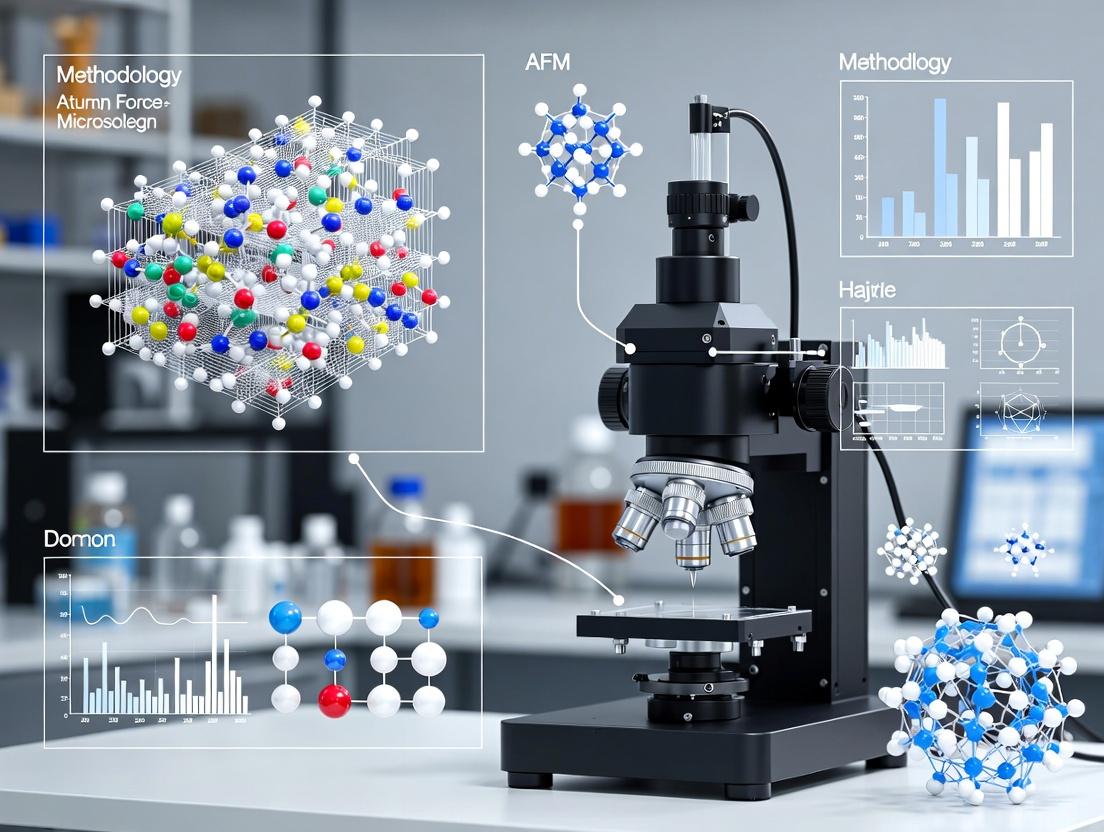
This article provides a comprehensive guide to Atomic Force Microscopy (AFM) for the three-dimensional shape characterization of nanoparticles, critical for drug delivery and nanomedicine.
3D Nanoscale Shape Analysis: Advanced AFM Techniques for Nanoparticle Characterization in Drug Development
Abstract
This article provides a comprehensive guide to Atomic Force Microscopy (AFM) for the three-dimensional shape characterization of nanoparticles, critical for drug delivery and nanomedicine. It begins with foundational principles, explaining why 3D morphology beyond simple size is essential for understanding nanoparticle behavior, biodistribution, and therapeutic efficacy. We then detail advanced AFM methodologies, including peak force tapping and high-resolution imaging modes, for accurate topographical mapping. The guide addresses common challenges like tip convolution and sample preparation, offering optimization strategies for reliable data. Finally, it validates AFM against complementary techniques like SEM and TEM, positioning it as an indispensable tool for researchers and scientists in pharmaceutical development who require quantitative, nanoscale 3D shape analysis.
Why 3D Shape Matters: The Critical Role of Nanoparticle Morphology in Biomedical Applications
The physicochemical properties of nanoparticles (NPs)—size, surface charge, and chemistry—have long been the focus of nanomedicine design. However, a critical parameter often oversimplified in 2D projections is 3D shape. Recent research, accelerated by advanced characterization tools like Atomic Force Microscopy (AFM), reveals that 3D shape (e.g., rods, disks, spheres, stars) profoundly impacts biological fate. This document details application notes and protocols for investigating shape-dependent biological behaviors, framed within a thesis utilizing AFM for precise 3D nanoscale shape characterization.
Table 1: Influence of Nanoparticle 3D Shape on Key Biological Parameters
| Nanoparticle Shape (Model) | Aspect Ratio (AR)* | Cellular Uptake Efficiency (vs. Sphere) | Circulation Half-life (t₁/₂) | Tumor Targeting Index (vs. Sphere) | Key Reference System |
|---|---|---|---|---|---|
| Sphere (Isotropic) | ~1.0 | 1.0 (Reference) | Moderate (e.g., 8-12 h) | 1.0 (Reference) | PEGylated Liposomes, Polymeric NPs |
| Rod / Filament | 2.0 - 5.0 | Increased (1.5 - 4x) | Significantly Prolonged (up to 2-5x) | Enhanced (2-3x) | Gold Nanorods, Filomicelles |
| Disk / Platelet | Low height, high width | Variable (Cell-type dependent) | Moderate to Prolonged | High Margination to Vasculature | Silicon Disks, Nanoplates |
| Star / Branched | N/A | Greatly Enhanced (3 - 10x) | Reduced (due to rapid clearance) | High for Avid Capture | Gold Nanostars |
| Cube / Polyhedron | ~1.0 - 1.5 | Similar or Slightly Enhanced | Similar to Sphere | Moderate | Metal-Organic Frameworks |
*AR = Length/Width or similar major/minor axis ratio. Data synthesized from recent literature (2022-2024).
Experimental Protocols
Protocol 3.1: AFM-Based 3D Shape Characterization of Synthesized Nanoparticles
Objective: To obtain high-resolution 3D topography and quantitative shape descriptors (height, aspect ratio, surface roughness) of nanoparticles. Materials: See "Scientist's Toolkit" (Section 6). Procedure:
- Substrate Preparation: Dilute nanoparticle suspension in appropriate solvent (e.g., DI water, ethanol) to ~0.5-1 µg/mL. Deposit 10-20 µL onto freshly cleaved mica substrate. Allow adsorption for 2-5 minutes. Rinse gently with DI water and dry under a gentle nitrogen stream.
- AFM Imaging: Mount substrate on AFM sample stage. Use tapping mode in air or PeakForce Tapping in liquid. Employ a sharp silicon probe (k ~ 40 N/m, f₀ ~ 300 kHz).
- Scan Parameters: Set scan size to capture 10-20 individual particles. Use a scan rate of 0.5-1.0 Hz with 512-1024 samples/line.
- 3D Shape Analysis: Use AFM software to:
- Measure particle height (H) and lateral diameter (D). Note: True lateral size is convolved with tip geometry.
- Calculate Aspect Ratio (3D): Use height vs. mean lateral dimension, or from 3D reconstruction.
- Determine Surface Roughness (Rq) of individual particles.
- Export 3D topography data for further analysis (e.g., sphericity index calculation).
Protocol 3.2: Assessing Shape-Dependent Cellular Uptake via Flow Cytometry
Objective: To quantify the internalization of fluorescently labeled, shape-variant nanoparticles by target cells. Materials: Shape-variant NPs (fluorescent label, e.g., Cy5), cell culture, flow cytometer, cold PBS/NaN₃ solution. Procedure:
- Cell Seeding: Seed cells in 24-well plates at 1x10⁵ cells/well and culture overnight.
- NP Exposure: Apply fluorescent NPs (e.g., 50 µg/mL) to cells in serum-free media. Incubate at 37°C for a set time (e.g., 2h). Include 4°C controls for binding-only correction.
- Quenching & Harvest: Remove media. Wash cells 3x with cold PBS. For membrane-bound fluorescence quenching, add trypan blue (0.4% in PBS) for 1 min, then wash. Harvest cells with trypsin/EDTA.
- Analysis: Resuspend cells in cold PBS/NaN₃. Analyze fluorescence intensity of 10,000 events per sample via flow cytometry. Report geometric mean fluorescence intensity (gMFI) normalized to spherical NP control.
Protocol 3.3: In Vivo Circulation Kinetics and Biodistribution
Objective: To evaluate the effect of NP shape on blood residence time and organ accumulation. Materials: Radiolabeled (¹¹¹In, ⁶⁴Cu) or NIRF-labeled (DIR, ICG) shape-variant NPs, IVIS or SPECT/CT imaging system, animal model. Procedure:
- NP Administration: Inject NPs (n=5/group) intravenously via tail vein at a standardized dose (e.g., 1 mg/kg, 100 µCi).
- Blood Circulation: Collect blood samples (5-10 µL) from tail nick at serial time points (5 min, 30 min, 2h, 8h, 24h). Measure radioactivity/NIRF signal. Fit data to a two-compartment model to calculate half-life (t₁/₂β).
- Biodistribution: At terminal time points (e.g., 24h, 48h), euthanize animals. Harvest major organs (liver, spleen, kidneys, heart, lungs, tumor). Weigh tissues and quantify signal. Express as % Injected Dose per Gram (%ID/g).
Signaling Pathways and Experimental Workflows
Diagram 1: Cellular Uptake Mechanisms by Shape
Diagram 2: AFM Shape Char. to Bio. Testing Workflow
Research Reagent Solutions & Essential Materials
Table 2: Key Research Reagents and Materials for Shape-Dependent Studies
| Item | Function/Application | Example Product/Catalog |
|---|---|---|
| AFM Probes (Tapping Mode) | High-resolution imaging of NP 3D topography without lateral force damage. | Bruker RTESPA-150, Olympus AC240TS |
| Freshly Cleaved Mica Substrate | Atomically flat, negatively charged surface for NP adsorption for AFM. | Ted Pella, Grade V1 Mica Discs |
| PEG-Thiol (SH-PEG-COOH) | Provides stealth coating and functional handles for conjugation on metal NPs. | Creative PEGWorks, MW: 5000 Da |
| Cell Culture-Tested, Fluorescent NPs | Shape-variant nanoparticles (rods, spheres) with encapsulated dye for uptake studies. | nanoComposix, Cy5-labeled Au Nanorods/Spheres |
| Trypan Blue Solution (0.4%) | Quenches extracellular fluorescence to differentiate internalized vs. surface-bound NPs. | Thermo Fisher, T10282 |
| Near-Infrared Fluorescent Dye (DIR) | Lipophilic carbocyanine for in vivo labeling and tracking of NPs via IVIS. | Thermo Fisher, D12731 |
| MicroBCA Protein Assay Kit | Quantifies protein corona formed on NPs after incubation in plasma/serum. | Thermo Fisher, 23235 |
| Matrigel Matrix | For establishing 3D cell culture models to study NP penetration in tumor spheroids. | Corning, 356231 |
Principles of Operation
Atomic Force Microscopy (AFM) generates topographical images of surfaces at the nanoscale by scanning a sharp tip attached to a flexible cantilever across a sample. The core principle is the detection of forces between the tip and the sample surface. A laser beam is reflected off the back of the cantilever onto a position-sensitive photodetector (PSPD). As the tip interacts with surface features, the cantilever deflects, causing a change in the laser spot position on the detector. This signal is used to construct a three-dimensional topographical map.
Primary Imaging Modes
Two primary modes are used for topographical imaging:
- Contact Mode: The tip is in constant physical contact with the sample surface. Topography is measured by maintaining a constant cantilever deflection (constant force) via a feedback loop that adjusts the scanner height.
- Dynamic (Intermittent Contact/Tapping) Mode: The cantilever is oscillated at or near its resonance frequency. Tip-sample interactions (van der Waals, capillary forces) cause changes in the oscillation's amplitude, phase, or frequency. This change is used by the feedback loop to maintain a constant oscillation amplitude while scanning, thereby tracking the topography.
Table 1: Comparison of Primary AFM Topographical Imaging Modes
| Parameter | Contact Mode | Dynamic (Tapping) Mode |
|---|---|---|
| Tip-Sample Interaction | Constant, repulsive contact | Intermittent, oscillating contact |
| Forces Exerted | Higher (lateral, normal) | Significantly lower (primarily normal) |
| Typical Resolution | Atomic lattice on hard samples | High, exceptional on soft samples |
| Sample Damage Risk | High for soft, adhesive, or loosely bound samples | Low to moderate |
| Best For | Very hard, rigid surfaces (e.g., mica, HOPG, silicon wafer) | Soft, fragile, or adhesive samples (e.g., polymers, cells, nanoparticles in matrix) |
| Key Measured Signal | Cantilever deflection | Oscillation amplitude/phase/frequency shift |
Experimental Protocol for 3D Nanoparticle Shape Characterization
This protocol details the use of intermittent contact mode AFM for characterizing the 3D shape of nanoparticles (NPs) deposited on a solid substrate, a critical step in drug delivery system research.
Materials & Sample Preparation
- Nanoparticle Suspension: e.g., polymeric NPs (PLGA), liposomes, or inorganic NPs.
- Substrate: Freshly cleaved mica (for most NPs) or silicon wafer (for hydrophobic NPs). Mica provides an atomically flat, negatively charged surface.
- Cationic Solution: 10-100 mM MgCl₂ or poly-L-lysine solution (0.1% w/v) for facilitating NP adhesion to mica.
- Ultrapure Water (e.g., Milli-Q) and analytical grade solvents.
- AFM Probe: Silicon cantilever with a sharp tip (typical resonance frequency: 150-400 kHz, spring constant: 5-40 N/m, tip radius <10 nm).
Protocol Steps
Step 1: Substrate Preparation
- Cleave the top layer of a mica disk (≈1 cm²) using adhesive tape to expose a fresh, atomically flat surface.
- (Optional but recommended for charged NPs) Apply 20-50 µL of the cationic solution (e.g., 50 mM MgCl₂) onto the mica surface for 1-2 minutes. Rinse gently with 1 mL of ultrapure water and dry under a gentle stream of inert gas (N₂ or Ar).
Step 2: Nanoparticle Deposition
- Dilute the NP suspension in an appropriate buffer or water to a concentration that prevents aggregation and ensures isolated particles for analysis (typically 1-10 µg/mL). Optimize via series dilution.
- Apply 20-50 µL of the diluted NP suspension onto the prepared mica substrate.
- Allow adsorption for a defined time (e.g., 5-15 minutes) in a covered Petri dish to prevent evaporation.
- Rinse the surface gently with 1-2 mL of ultrapure water to remove loosely bound NPs and salts. Dry thoroughly under a gentle stream of inert gas.
Step 3: AFM Instrument Setup & Imaging
- Mount the prepared sample onto the AFM sample stage securely.
- Mount an appropriate cantilever (e.g., RTESPA-150 from Bruker) into the probe holder.
- Align the laser spot onto the end of the cantilever and center the reflected beam on the PSPD.
- Tune the cantilever to find its resonance frequency and set the drive amplitude.
- Engage the tip in intermittent contact mode. Set initial scan parameters: Scan size: 1-5 µm, Scan rate: 0.5-1.0 Hz, Points/Lines: 512 x 512.
- Optimize the feedback parameters (Integral and Proportional gains, setpoint ratio) to achieve stable imaging with minimal tip-sample interaction force.
- Acquire images at multiple locations on the sample to ensure statistical relevance.
Step 4: Data Analysis for 3D Shape Parameters
- Apply a first-order flattening or plane-fit to all images to correct for sample tilt.
- Use particle analysis software (e.g., Gwyddion, Nanoscope Analysis) to identify and segment individual nanoparticles.
- For each nanoparticle, extract quantitative 3D parameters:
- Height (H): Maximum z-value from the substrate baseline.
- Diameter (D): Full-width at half-maximum (FWHM) or equivalent circle diameter from cross-section analysis at half-height.
- Aspect Ratio (H/D): Indicator of sphericity (1=sphere).
- Volume: Calculated by integration of pixels above the baseline.
- Surface Roughness (Rq): Within a single nanoparticle.
Table 2: Example Quantitative AFM Data for Nanoparticle Shape Characterization
| NP Type (Theoretical) | Measured Height (nm) Mean ± SD | Measured Diameter (nm) Mean ± SD | Aspect Ratio (H/D) | Calculated Volume (x10³ nm³) | Surface Roughness, Rq (nm) |
|---|---|---|---|---|---|
| Spherical PLGA NPs | 102.3 ± 8.7 | 105.1 ± 9.2 | 0.97 ± 0.05 | 588 ± 150 | 0.5 ± 0.2 |
| Liposomes | 18.5 ± 2.1 | 89.3 ± 12.4 | 0.21 ± 0.03 | 72 ± 25 | 0.3 ± 0.1 |
| Gold Nanorods | 48.2 ± 5.6 | 32.1 ± 4.8 | 1.50 ± 0.15 | 39 ± 12 | 1.1 ± 0.3 |
The Scientist's Toolkit: Essential Research Reagents & Materials
Table 3: Key Research Reagent Solutions for AFM of Nanoparticles
| Item | Function & Importance |
|---|---|
| Freshly Cleaved Mica | Provides an atomically flat, inert, and negatively charged substrate for NP deposition, crucial for accurate height measurement. |
| Divalent Cation Solution (MgCl₂) | Promotes electrostatic adhesion of negatively charged NPs to the mica surface, preventing displacement by the AFM tip. |
| Ultrapure Water (18.2 MΩ·cm) | Used for rinsing to remove salts and non-adhered material without introducing particulate contaminants that can interfere with imaging. |
| Silicon AFM Probes (Tapping Mode) | High-frequency, sharp-tipped cantilevers are essential for high-resolution imaging of nanoscale particles with minimal sample damage. |
| Standard Nanoparticle Reference (e.g., NIST-traceable gold NPs) | Used for AFM tip characterization (shape convolution assessment) and periodic verification of lateral scale calibration. |
Visualization of Key Concepts
Application Notes
Within the framework of Atomic Force Microscopy (AFM) methodology for 3D nanoparticle characterization, these four descriptors are foundational for correlating nanoparticle morphology with biological function and therapeutic efficacy. AFM provides the true 3D topographical data necessary for their precise calculation, surpassing 2D imaging techniques.
- Height: The vertical dimension (Z-axis) measured from the substrate to the highest point of the nanoparticle. It is critical for understanding cellular uptake mechanisms, as size thresholds exist for endocytic pathways. It also directly influences the surface area available for ligand conjugation in targeted drug delivery systems.
- Aspect Ratio: Defined as the ratio of the particle's major axis length to its minor axis length (or height, depending on orientation). It is a primary determinant of biodistribution and circulation time; high-aspect-ratio particles (e.g., rod-shaped) often exhibit prolonged circulation but may face different macrophage clearance rates compared to spherical particles.
- Surface Roughness: Typically quantified as the Root Mean Square (RMS) Roughness (Rq) or Average Roughness (Ra) over the nanoparticle's topographical surface. Increased roughness enhances protein adsorption (opsonization) and cellular adhesion, directly impacting immune recognition, targeting specificity, and degradation kinetics.
- Sphericity: A measure of how closely the shape of a nanoparticle approximates a perfect sphere. Calculated from volume and surface area, it influences packing density in solid drug formulations, suspension viscosity, and predictability of fluid dynamics in biological systems.
Table 1: Quantitative Impact of 3D Shape Descriptors on Drug Delivery Parameters
| Descriptor | Typical AFM-derived Range | Key Influence on Drug Delivery | Correlation with Cellular Uptake Efficiency |
|---|---|---|---|
| Height | 1 – 200 nm | Controls steric interaction with cell membrane proteins. | Optimal uptake often reported between 40-60 nm height. |
| Aspect Ratio | 1 (sphere) to >5 (rod) | Alters flow dynamics and margination in vasculature. | Low ratio (~1) favors clathrin-mediated endocytosis; high ratio may trigger alternative pathways. |
| RMS Roughness (Rq) | 0.1 – 5 nm | Modulates the conformation of adsorbed serum proteins. | Increased roughness (Rq > 2nm) often correlates with enhanced phagocytosis by immune cells. |
| Sphericity | 0.7 – 1.0 | Affects the uniformity of shear stress and ligand presentation. | High sphericity (>0.9) leads to more predictable and uniform internalization rates. |
Experimental Protocols
Protocol 1: AFM Imaging for Descriptor Extraction Objective: Acquire high-fidelity 3D topographical images of nanoparticles deposited on a flat substrate for shape descriptor quantification.
- Sample Preparation: Dilute nanoparticle suspension in appropriate solvent (e.g., Milli-Q water). Deposit 10 µL onto freshly cleaved mica substrate. Allow adsorption for 10 minutes, then gently rinse with water and dry under a mild nitrogen stream.
- AFM Imaging: Use tapping mode in air or liquid. Employ a sharp silicon probe (tip radius < 10 nm, spring constant ~40 N/m). Set scan size to encompass 10-20 particles, with resolution of at least 512x512 pixels.
- Image Processing: Apply a first-order flattening to correct for substrate tilt. Use non-destructive filtering (e.g., Gaussian low-pass) only to remove high-frequency electronic noise.
- Descriptor Analysis:
- Height: Use section analysis tool to measure particle height from substrate plane to apex.
- Aspect Ratio: Fit particle footprint with an ellipse; calculate ratio of major to minor axis lengths.
- Surface Roughness: Select a region of interest encompassing the top 80% of the particle's height. Calculate RMS Roughness (Rq) via software algorithms.
- Sphericity: Calculate from particle volume (V) and surface area (S) derived from AFM data: Ψ = (π^(1/3)(6V)^(2/3))/S.
Protocol 2: Correlating Roughness with Protein Adsorption Objective: Quantify the relationship between nanoparticle surface roughness and serum protein adsorption.
- Nanoparticle Incubation: Incubate characterized nanoparticles (from Protocol 1) in 100% fetal bovine serum (FBS) at 37°C for 1 hour.
- Washing: Centrifuge nanoparticles at 14,000 rpm for 15 minutes. Discard supernatant and resuspend pellet in phosphate-buffered saline (PBS). Repeat twice.
- AFM Re-imaging: Re-deposit washed nanoparticles on mica and image using AFM in tapping mode in liquid (PBS).
- Analysis: Measure the increase in particle height and change in surface roughness (Rq) compared to pre-incubation data. A significant increase in both indicates formation of a protein corona, the thickness and uniformity of which can be roughness-dependent.
Title: AFM Shape Descriptor Correlation Workflow
Title: Descriptor-Biological Outcome Relationship Map
The Scientist's Toolkit: Essential Research Reagents & Materials
Table 2: Key Reagents and Materials for AFM Nanoparticle Shape Characterization
| Item | Function in Research |
|---|---|
| Freshly Cleaved Mica Discs | Atomically flat, negatively charged substrate for nanoparticle adsorption, essential for high-resolution AFM imaging. |
| AFM Probes (Tapping Mode) | Silicon tips with known radius (<10 nm) and spring constant; the primary tool for interacting with and mapping nanoparticle topography. |
| Certified Nanoparticle Size Standards | Polystyrene or gold nanoparticles with traceable diameter, used for AFM lateral and vertical calibration. |
| Ultrapure Water (e.g., Milli-Q) | Solvent for nanoparticle dilution and rinsing to prevent salt crystallization and artifacts on the mica substrate. |
| Nitrogen Gas (High Purity, Dry) | Used for gentle, artifact-free drying of prepared AFM samples. |
| Fetal Bovine Serum (FBS) | Complex protein mixture used in in vitro studies to investigate protein corona formation on nanoparticles of defined shape. |
| Image Analysis Software (e.g., Gwyddion, Gatan) | Specialized software for processing AFM data, performing plane correction, and calculating 3D shape descriptors. |
The Limitations of 2D Electron Microscopy and the Case for AFM's Z-axis Fidelity.
Accurate three-dimensional shape characterization of nanoparticles (NPs) is critical in drug development, where parameters like aspect ratio, surface roughness, and ligand distribution directly influence biodistribution, cellular uptake, and efficacy. While Transmission and Scanning Electron Microscopy (TEM/SEM) are standard, they provide fundamentally 2D projections or surface topographs with limited z-axis fidelity. This document frames the limitations of 2D EM within the broader thesis that Atomic Force Microscopy (AFM) methodology is indispensable for true 3D nanoscale metrology, offering quantitative z-axis data essential for rigorous structure-activity relationship studies.
Quantitative Comparison of 2D EM vs. AFM for 3D Characterization
The following table summarizes key limitations and capabilities based on current literature and instrument specifications.
Table 1: Comparative Analysis of Techniques for Nanoparticle 3D Shape Characterization
| Parameter | Transmission Electron Microscopy (TEM) | Scanning Electron Microscopy (SEM) | Atomic Force Microscopy (AFM) |
|---|---|---|---|
| Primary Data | 2D Projection (Mass-Thickness Contrast) | 2D Surface Topography (Secondary Electron Signal) | 3D Surface Topography (Tip-Sample Interaction Force) |
| Z-axis Resolution | N/A (Projection) | ~1-10 nm (for in-lens detectors) | <0.1 nm (True Vertical Resolution) |
| Lateral Resolution | <0.1 nm | 0.5-4 nm | 0.5-5 nm (dependent on tip) |
| Sample Environment | High Vacuum | High Vacuum (typically) | Ambient, Liquid, Vacuum |
| Sample Preparation | Complex (grids, staining, drying) | Moderate (conductive coating often required) | Minimal (often requires immobilization only) |
| Quantitative Z-Data | Indirect, requires tomography (complex, dose-limited) | Indirect, tilt-dependent; poor for soft materials | Direct, quantitative height measurement |
| Key Limitation for 3D | Projection ambiguity; beam damage to soft NPs; tomography is resource-intensive. | Conductive coating alters dimensions; limited z-axis quantification on nanoscale features. | Tip convolution effects lateral dimensions; slower imaging speed. |
| Strength for Drug Dev. | Unparalleled lattice/atomic imaging. | Fast, large-area surveying. | True 3D shape, roughness, and mechanical properties in physiologically relevant conditions. |
Application Notes: Critical Z-axis Parameters in NP Characterization
- Aspect Ratio (AR): TEM-derived AR from 2D projections can be erroneous for non-symmetric particles lying in non-principal axes. AFM height data provides the true minor axis dimension.
- Surface Roughness (Rq/Ra): Critical for understanding protein corona formation. SEM cannot reliably quantify nanoscale roughness on NP surfaces. AFM directly measures Rq with sub-nanometer precision.
- Ligand Shell Thickness: Measuring the hydrodynamic shell via DLS is indirect. AFM can directly profile the height increase from a bare core to a ligand-coated core on a substrate.
- Aggregation State: While EM identifies aggregation, AFM differentiates between aggregation (particle-particle contact) and coalescence (fusion with shared z-height), crucial for stability studies.
Detailed Protocols
Protocol 4.1: AFM-based 3D Shape Characterization of Polymeric Nanoparticles
Objective: To obtain accurate 3D topography, height distribution, and surface roughness of PEG-PLGA nanoparticles in buffer. Materials: See "The Scientist's Toolkit" below. Workflow:
- Substrate Preparation: Cleave a fresh sheet of Muscovite Mica using adhesive tape. Treat with 10 µL of 0.01% poly-L-lysine (PLL) for 5 minutes, then rinse gently with ultra-pure water and dry under a gentle nitrogen stream. PLL provides a cationic surface for electrostatic immobilization.
- Sample Immobilization: Dilute the NP suspension in the desired buffer (e.g., 1x PBS) to ~0.01 mg/mL. Pipette 20 µL onto the PLL-treated mica. Allow to adsorb for 15 minutes in a humidity chamber.
- Gentle Rinsing: Tilt the mica substrate and rinse with 2 mL of the same buffer to remove loosely adhered particles and salts. Carefully dry the edges with a laboratory wipe.
- AFM Imaging:
- Mount the sample on the AFM stage.
- Engage a sharp, nitride-lever silicon tip (k ~ 0.7 N/m, f0 ~ 75 kHz) in air or directly inject buffer for liquid imaging.
- Use PeakForce Tapping or Quantitative Imaging (QI) Mode to minimize lateral forces. Set the peak force to < 100 pN to prevent particle deformation.
- Acquire images of at least 5 different 5 µm x 5 µm areas at 512 x 512 or 1024 x 1024 resolution.
- Data Analysis:
- Apply a first-order flattening to each image.
- Use particle analysis software to identify individual NPs. For each particle, extract: Max Height (H), Diameter at Base (D), and Root Mean Square Roughness (Rq) of the top surface.
- Calculate true Aspect Ratio as H / D. Generate population histograms for H and Rq.
Protocol 4.2: Correlative TEM-AFM for Method Validation
Objective: To highlight z-axis information loss in TEM by direct comparison with AFM on identical particles. Workflow:
- Prepare NPs on a findER or similar finder grid with numbered squares.
- First, perform AFM in liquid (as per Protocol 4.1) on selected grid squares. Map and record coordinates of specific particles of interest.
- Carefully rinse and dry the grid (critical point drying preferred).
- Image the exact same particles using TEM at an accelerating voltage of 80-100 kV to minimize damage.
- Measure the lateral diameter (DTEM) from TEM and the height (HAFM) and lateral diameter (DAFM, noting tip convolution) from AFM. Tabulate to demonstrate DTEM ≈ DAFM but HAFM provides the missing third dimension.
Visualizing the Methodological Argument
Title: Analytical Pathways for 3D Nanoparticle Characterization
Title: AFM 3D Nanometrology Protocol and Outputs
The Scientist's Toolkit: Essential Research Reagents & Materials
Table 2: Key Materials for AFM-based 3D Nanoparticle Characterization
| Item | Function & Rationale |
|---|---|
| Muscovite Mica (V1 Grade) | An atomically flat, easily cleavable substrate that provides a pristine surface for NP immobilization and height reference. |
| Poly-L-Lysine (PLL) Solution (0.01-0.1% w/v) | A cationic polymer coating for mica; electrostatically immobilizes anionic or neutral nanoparticles, preventing drift during scanning. |
| Sharp Silicon Nitride AFM Probes (e.g., Bruker ScanAsyst-Fluid+) | Tips optimized for PeakForce Tapping in liquid. Low spring constant (~0.7 N/m) minimizes deformation of soft nanomaterials. |
| AFM Calibration Grating (e.g., TGZ1, TGX1) | Grid with known pitch and step height for periodic verification of the scanner's x, y, and z dimensional accuracy. |
| Phosphate Buffered Saline (PBS), pH 7.4 | Standard physiologically relevant imaging buffer. Must be filtered (0.02 µm) before use to remove particulates. |
| FindER TEM Finder Grids | Grids with alphanumeric coordinates enabling reliable correlation between AFM and TEM imaging of the exact same particles. |
| Critical Point Dryer | For sample preparation in correlative studies; removes liquid without surface tension-induced collapse, preserving NP structure for vacuum-based EM. |
This application note, framed within a thesis on Atomic Force Microscopy (AFM) methodology for 3D nanoparticle shape characterization, provides a primer on establishing quantitative structure-function relationships (SFRs) for nanomedicines. For drug developers, linking the precise physical structure of nanoparticles (NPs)—including shape, size, surface topography, and mechanical properties—to biological function is critical for rational design. AFM offers unparalleled 3D topographic imaging and nanomechanical mapping under near-physiological conditions, making it indispensable for this correlative research.
Key Structural Parameters and Functional Correlations
Table 1: Nanoparticle Structural Parameters and Their Functional Impact
| Structural Parameter | Primary Measurement Method | Key Functional Correlations | Typical Target Range for Drug Delivery |
|---|---|---|---|
| 3D Shape / Aspect Ratio | AFM Topography Imaging | Cellular uptake, circulation time, biodistribution. Rods/filaments show prolonged circulation vs. spheres. | Aspect Ratio: 1 (Spheres) to 3-5 (Rods) |
| Size (Height/Width) | AFM Cross-Section Analysis | Renal clearance (<6 nm), RES uptake (>200 nm), EPR effect (10-200 nm). | 20-150 nm for EPR-based tumor targeting |
| Surface Roughness (Rq/Ra) | AFM Surface Analysis | Protein corona composition, cell adhesion, macrophage evasion. | Optimized roughness can reduce opsonization. |
| Elasticity / Young's Modulus | AFM Force Spectroscopy | Cell internalization mechanism, tissue penetration, clearance. | Softer particles (MPa range) may improve tumor penetration. |
| Surface Charge (ζ-Potential) | Dynamic Light Scattering (DLS) | Colloidal stability, protein adsorption, cellular interaction. | ±20-30 mV for stable suspensions in physiological media. |
Application Notes: AFM Protocols for Structural Characterization
Protocol 1: 3D Topography and Shape Analysis of Lipid Nanoparticles (LNPs)
Objective: To quantify the morphology, size, and uniformity of mRNA-LNPs. Materials: Freshly prepared LNP formulation, freshly cleaved mica substrate, AFM with tapping mode capability, PBS buffer (pH 7.4). Procedure:
- Sample Preparation: Dilute LNP sample in filtered PBS to ~0.05 mg/mL total lipid concentration. Apply 20 µL to a clean mica surface. Incubate for 5 minutes. Rinse gently with 1 mL of ultrapure water to remove unbound particles and salts. Dry under a gentle stream of nitrogen.
- AFM Imaging: Mount the sample. Use a silicon cantilever (k ~ 40 N/m, f₀ ~ 300 kHz). Engage in tapping mode in air. Scan an area of 5x5 µm² at a resolution of 512x512 pixels.
- Shape Analysis: Use AFM software to measure particle height (most reliable AFM size metric) and width at half-height. Calculate aspect ratio (width/height). Generate roughness (Rq) values for individual particles and the overall sample deposition.
Protocol 2: Nanomechanical Mapping of Polymeric NPs in Fluid
Objective: To correlate NP elasticity with in vitro endothelial cell uptake rates. Materials: PLGA NPs (varied polymer MW), AFM with fluid cell and quantitative nanomechanical mapping (QNM) mode, cantilevers with calibrated spring constant and known tip geometry, relevant cell culture medium. Procedure:
- Sample Preparation: Adsorb NPs onto poly-L-lysine coated glass coverslips. Immerse in cell culture medium.
- Force Volume/QNM Imaging: In fluid, perform a grid of force-distance curves (e.g., 64x64 over a 2x2 µm area). Fit the retraction curve using the Derjaguin–Muller–Toporov (DMT) model to extract the Young's Modulus for each NP.
- Data Correlation: Plot NP Young's Modulus (kPa to GPa) against percentage internalization efficiency determined from parallel flow cytometry experiments.
Research Reagent Solutions & Essential Materials
Table 2: Key Research Toolkit for AFM-Based NP Characterization
| Item | Function/Application | Key Considerations |
|---|---|---|
| Freshly Cleaved Mica Discs | Atomically flat substrate for NP adsorption and imaging. | Provides a clean, reproducible surface for high-resolution topography. |
| Silicon Cantilevers for Tapping Mode | For high-resolution imaging in air or fluid. | Choose appropriate resonance frequency and spring constant (e.g., 300 kHz, 40 N/m). |
| Calibrated Cantilevers for Force Spectroscopy | For nanomechanical property measurement. | Require precise spring constant and tip radius calibration (via thermal tune or reference sample). |
| PBS Buffer (Filtered, 0.1 µm) | For sample dilution and fluid imaging. | Filtering prevents contamination from particulates that interfere with imaging. |
| Poly-L-Lysine Solution | For enhancing adhesion of charged NPs to substrates. | Critical for imaging in liquid where weak adhesion can lead to particle displacement. |
| NP Reference Standards (e.g., Gold Nanospheres) | For lateral calibration and tip morphology characterization. | Essential for validating measurement accuracy and accounting for tip convolution effects. |
Visualizing the Structure-Function Correlation Workflow
Diagram 1: Workflow for Correlating NP Structure & Function
Diagram 2: Key NP Properties Influencing Biological Outcomes
Integrating detailed 3D structural characterization via AFM with biological performance assays is fundamental for advancing nanomedicine. The protocols and correlations outlined here provide a foundational framework for drug developers to move beyond empirical formulation and towards the rational design of nanoparticles with predictably enhanced therapeutic function. This approach directly supports the overarching thesis that high-fidelity 3D shape analysis is a critical enabling methodology in next-generation nanotherapeutics development.
Step-by-Step AFM Protocols: From Sample Prep to 3D Topography Mapping
Optimized Substrate Selection and Sample Immobilization Techniques
Within the broader thesis on Atomic Force Microscopy (AFM) methodology for 3D nanoparticle shape characterization, precise and reliable sample preparation is the foundational step. Accurate topographical imaging and dimensional analysis of nanoparticles (NPs)—critical for drug delivery system evaluation—are wholly dependent on the selection of an appropriate substrate and an effective immobilization protocol. Sub-optimal preparation leads to artifacts such as nanoparticle aggregation, diffusion, or deformation, compromising data integrity. These Application Notes detail current best practices for preparing metallic, polymeric, and lipid-based nanoparticles for high-resolution AFM analysis.
Quantitative Comparison of Common AFM Substrates
The choice of substrate is dictated by nanoparticle composition, required interaction strength, and background roughness.
Table 1: Key Properties of Common Substrates for Nanoparticle AFM
| Substrate Material | Typical RMS Roughness (nm) | Primary Immobilization Mechanism | Optimal For NP Type | Key Advantage | Key Limitation |
|---|---|---|---|---|---|
| Freshly Cleaved Mica | 0.05 - 0.1 | Electrostatic adsorption, Cation-mediated adhesion | Lipid nanoparticles, Exosomes, Proteins, Soft polymers | Atomically flat, negatively charged surface | Low adhesion for hydrophobic/neutral particles |
| Silicon (Si) | 0.1 - 0.3 | Hydrophobic interaction, Van der Waals, Functionalization | Metallic NPs (Au, Ag), Polymeric micelles | Easily functionalized, good for tapping mode | Higher intrinsic roughness than mica |
| Silicon with Thermal Oxide (SiO₂) | 0.2 - 0.5 | Electrostatic, Covalent functionalization (e.g., APTES) | Functionalized NPs, Drug-loaded nanocapsules | Tunable surface chemistry, widely used | Roughness increases with oxide thickness |
| Gold (Au) on Mica | 0.2 - 0.4 | Thiol-gold covalent bonding, Chemisorption | Thiol-coated NPs, DNA-functionalized particles | Excellent for specific, strong immobilization | Expensive, requires evaporation equipment |
| Amino-coated Glass | 0.3 - 0.8 | Electrostatic, Covalent coupling | Carboxylated or charged nanoparticles | Cost-effective, compatible with optical checks | Roughness can be high; batch variability |
Detailed Experimental Protocols
Protocol 1: Cation-Mediated Immobilization of Lipid Nanoparticles on Mica
This protocol is optimized for soft, negatively charged nanoparticles like liposomes or lipid NPs.
- Substrate Preparation: Cleave a sheet of muscovite mica (Grade V1) using adhesive tape to expose a fresh, atomically flat surface.
- Cation Solution Application: Immediately apply 20 µL of a divalent cation solution (e.g., 10 mM MgCl₂ or NiCl₂ in ultrapure water) onto the mica surface.
- Nanoparticle Deposition: After 1 minute, add 10 µL of the diluted nanoparticle suspension (typically 5-10 µg/mL in a suitable buffer) directly into the droplet. Mix gently by pipetting.
- Incubation: Allow the sample to incubate for 5-10 minutes at room temperature in a humidified chamber to prevent evaporation.
- Rinsing and Drying: Gently rinse the mica surface with 2-3 mL of ultrapure water (or filtered buffer) to remove unbound particles and salts. Carefully dry the edges with a laboratory wipe and dry under a gentle stream of filtered nitrogen or argon gas.
- AFM Mounting: Mount the prepared sample onto the AFM metal puck using a double-sided adhesive tab.
Protocol 2: Functionalized Substrate Preparation for Covalent Immobilization
This protocol creates an amine-reactive surface on silicon/silicon oxide for covalent attachment of carboxylated nanoparticles.
- Substrate Cleaning: Sonicate a silicon wafer piece in acetone for 5 minutes, followed by ethanol for 5 minutes. Rinse with ultrapure water and dry with N₂. Treat with oxygen plasma for 2-3 minutes to activate surface hydroxyl groups.
- Silanization: Immerse the clean substrate in a 2% (v/v) solution of (3-Aminopropyl)triethoxysilane (APTES) in anhydrous toluene for 1 hour at room temperature under anhydrous conditions.
- Rinsing: Rinse the substrate sequentially with toluene, ethanol, and ultrapure water to remove unbound silane.
- Curing: Cure the APTES-coated substrate at 110°C for 20 minutes.
- Crosslinker Application: Incubate the aminated substrate with a 2.5% glutaraldehyde solution in PBS for 30 minutes. Rinse thoroughly with PBS and water.
- Nanoparticle Immobilization: Apply the carboxylated nanoparticle solution (in a low-salt buffer, pH ~6.0) onto the aldehyde-activated surface for 1 hour. The carboxyl groups on NPs form Schiff base linkages with surface amines.
- Quenching and Rinsing: Quench the reaction by incubating with a 50 mM glycine solution for 5 minutes. Rinse with the imaging buffer.
Protocol 3: Adsorption of Polymeric Nanoparticles on Hydrophobic Silicon
Suitable for hydrophobic polymeric nanoparticles like PLGA or chitosan.
- Substrate Hydrophobization: Clean a silicon wafer as in Protocol 2, Step 1. Alternatively, use a highly doped, native oxide Si wafer which is inherently hydrophobic after cleaning.
- Sample Preparation: Dilute the hydrophobic nanoparticle suspension in a volatile organic solvent compatible with the NPs (e.g., chloroform, acetone) at a low concentration (~1 µg/mL). Note: Ensure solvent compatibility to avoid NP dissolution.
- Spin-Coating: Place a drop (50-100 µL) of the NP suspension onto the silicon substrate. Spin-coat at 2000-4000 rpm for 30-60 seconds to achieve a dispersed monolayer.
- Drying: Allow the substrate to air-dry in a fume hood to ensure complete solvent evaporation.
- AFM Imaging: Proceed directly to AFM imaging, typically in tapping mode to minimize lateral forces.
The Scientist's Toolkit: Research Reagent Solutions
Table 2: Essential Materials for Nanoparticle Immobilization
| Item | Function & Rationale |
|---|---|
| Muscovite Mica (V1 Grade) | Provides an atomically flat, negatively charged, and inert substrate for electrostatic immobilization. |
| APTES ((3-Aminopropyl)triethoxysilane) | Silane coupling agent used to introduce primary amine groups onto oxide surfaces for subsequent covalent chemistry. |
| MgCl₂ or NiCl₂ (High Purity) | Divalent cations that bridge negatively charged nanoparticles and the mica surface, enhancing adhesion. |
| Glutaraldehyde (25% Solution) | Homobifunctional crosslinker that reacts with amine groups to create aldehyde-terminated or crosslinked surfaces. |
| Filtered Ultrapure Water (0.02 µm filtered) | Used for rinsing to avoid contamination by particulates that can be mistaken for nanoparticles. |
| Oxygen Plasma Cleaner | Activates silicon/glass surfaces by generating reactive hydroxyl groups, essential for uniform silanization. |
| Anhydrous Toluene | Solvent for silanization reactions; must be anhydrous to prevent APTES self-polymerization. |
| Laboratory Parafilm | Used to create incubation chambers for small liquid volumes on substrate surfaces. |
Visualization of Experimental Workflows
Decision Workflow for Immobilization Protocol Selection (94 characters)
Workflow for Cation-Mediated Immobilization on Mica (61 characters)
Within the broader thesis on Atomic Force Microscopy (AFM) methodology for 3D nanoparticle shape characterization in drug delivery research, selecting the optimal imaging mode is a critical determinant of data fidelity and biological relevance. This document provides detailed Application Notes and Protocols for three primary AFM modes—PeakForce Tapping (PFT), Contact Mode (CM), and Non-Contact Mode (NC)—focusing on their application for characterizing soft, often functionalized, nanoparticles (e.g., polymeric NPs, liposomes, exosomes). The choice of mode directly influences resolution, sample integrity, and the accuracy of derived 3D morphological parameters essential for correlating structure with function in therapeutic development.
Mode Comparison: Principles and Quantitative Performance
The following table summarizes the core operating principles, key performance metrics, and primary applications of each mode, based on current literature and instrument specifications.
Table 1: Quantitative Comparison of AFM Imaging Modes for Nanoparticle Characterization
| Feature | PeakForce Tapping (PFT) | Contact Mode (CM) | Non-Contact Mode (NC) |
|---|---|---|---|
| Core Principle | Intermittent, controlled tip-sample contact at a user-defined peak force. | Tip in constant physical contact with the sample surface. | Tip oscillates near its resonance frequency above the sample without contact. |
| Typical Force Control | < 100 pN to ~1 nN (precisely set). | 0.5 nN to 10 nN (lateral forces significant). | Forces < 100 pN (van der Waals attraction dominant). |
| Lateral Shear Forces | Negligible. Minimizes sample deformation. | Very High. Can deform or sweep soft samples. | None. |
| Optimal Resolution | Sub-nanometer vertical; ~1 nm lateral on NPs. | Atomic step resolution on hard samples; poor on soft. | ~1 nm vertical; lateral limited by tip-sample distance. |
| Sample Damage Risk | Very Low when optimized. | Very High for soft, biological, or loosely adsorbed NPs. | Lowest. Ideal for delicate surfaces. |
| Ambient/Liquid Imaging | Excellent in both environments. | Challenging in liquid due to meniscus forces. | Primarily used in ambient/UHV; difficult in liquid. |
| Simultaneous Data Channels | Height, DMT Modulus, Adhesion, Deformation, Dissipation. | Height, Deflection (Error). | Height, Phase, Amplitude. |
| Best Suited For | Soft, adhesive nanoparticles (liposomes, polymersomes, exosomes), nanomechanical mapping. | Hard, flat, and rigid surfaces (crystalline materials, mica). | Atomic-scale topography of clean, rigid surfaces; moisture-sensitive samples. |
Detailed Experimental Protocols
Protocol 3.1: Sample Preparation for Nanoparticle AFM Imaging
- Objective: To immobilize nanoparticles without aggregation or deformation for high-resolution 3D shape analysis.
- Materials:
- Freshly cleaved muscovite mica (Grade V-1).
- Poly-L-lysine (PLL) solution (0.01% w/v in Milli-Q water) or APTES ((3-Aminopropyl)triethoxysilane) for functionalization.
- Nanoparticle suspension in appropriate buffer (e.g., PBS, HEPES).
- Milli-Q water.
- Nitrogen gas stream.
- Procedure:
- Substrate Preparation: Cleave mica to obtain a fresh, atomically flat surface.
- Functionalization (Optional but recommended for NPs in buffer): Apply 50 µL of PLL solution to the mica for 5 minutes. Rinse thoroughly with Milli-Q water and dry with gentle N₂. This creates a positively charged surface to electrostatically trap negatively charged NPs.
- Sample Deposition: Dilute the NP suspension to ~1-5 µg/mL in a low-salt buffer (e.g., 1-5 mM HEPES, pH 7.4). Pipette 20-50 µL onto the mica substrate. Incubate for 10-20 minutes in a humid chamber to prevent evaporation.
- Rinsing and Drying: Gently rinse the substrate with 2-3 mL of Milli-Q water to remove salts and unbound particles. Dry thoroughly with a gentle stream of nitrogen. Note: For liquid imaging, skip drying and proceed directly to mounting in the fluid cell.
Protocol 3.2: Imaging Polymer Nanoparticles Using PeakForce Tapping Mode
- Objective: To obtain high-resolution 3D topography and simultaneous nanomechanical properties of soft polymeric NPs.
- AFM Setup: Bruker Dimension FastScan or Icon system with a ScanAsyst-Air (or Fluid) probe.
- Parameters:
- Scan Rate: 0.5 - 1.0 Hz.
- Scan Points: 512 x 512.
- PeakForce Setpoint: Start at 100 pN, adjust incrementally upward until stable imaging is achieved (typically 200-500 pN).
- PeakForce Frequency: 1-2 kHz.
- Feedback Gains: Use auto-optimization feature if available.
- Procedure:
- Engage the probe in air (or liquid) using standard procedures.
- Select PeakForce Tapping mode and input the initial parameters.
- Engage on the sample surface. The system will automatically maintain the defined peak force.
- Optimize the setpoint to the lowest stable value that maintains tip-sample contact. This minimizes deformation.
- Acquire images of multiple areas to ensure reproducibility. Capture Height, DMT Modulus, and Adhesion channels simultaneously.
- Use particle analysis software (e.g., NanoScope Analysis) to extract diameter, height, and volume from the Height image.
Protocol 3.3: Imaging Hard Nanocrystals Using Contact Mode
- Objective: To achieve high-resolution topography of rigid, well-adsorbed nanoparticles (e.g., gold nanospheres, quantum dots).
- AFM Setup: Standard AFM with rigid cantilevers (e.g., Bruker RTESPA, k ~40 N/m).
- Parameters:
- Scan Rate: 1.0 - 2.0 Hz.
- Scan Points: 512 x 512.
- Deflection Setpoint: Maintain as low as possible (e.g., 0.5 - 1.0 V) to minimize applied force.
- Feedback Gains: Integral gain 0.3-0.5, Proportional gain 0.8-1.2.
- Procedure:
- Engage in deflection channel.
- Immediately reduce the setpoint after engagement to lower the normal force.
- Carefully increase gains until tracking is stable without oscillation.
- Scan and record Height and Deflection (Error) signal images.
- Critical Check: Continuously monitor the Deflection image. Any streaking or distortion indicates the tip is pushing the particles. Stop and switch to PFT.
Protocol 3.4: High-Resolution Imaging in Non-Contact Mode
- Objective: To image surface topography of nanoparticles with minimal interaction.
- AFM Setup: AFM capable of frequency modulation (FM) or amplitude modulation (AM) NC mode, in a controlled environment (low humidity/vacuum). Use stiff, high-frequency probes (e.g., PPP-NCHR, k ~42 N/m, f₀ ~330 kHz).
- Parameters (AM-NC):
- Oscillation Amplitude (A₀): 5-10 nm.
- Drive Frequency: Slightly below the resonant frequency.
- Amplitude Setpoint (A_sp): ~95% of A₀.
- Scan Rate: 0.3 - 0.6 Hz.
- Procedure:
- Tune the cantilever to find its resonance frequency and peak amplitude.
- Set the oscillation parameters and engage using the amplitude setpoint.
- The feedback loop maintains a constant oscillation amplitude by adjusting the tip-sample distance.
- Imaging in ambient air requires careful control of humidity to minimize the water meniscus.
Visualization of AFM Mode Selection Logic
Diagram 1: AFM Mode Selection Logic Flow for NPs
The Scientist's Toolkit: Key Research Reagent Solutions
Table 2: Essential Materials for AFM Nanoparticle Characterization
| Item | Function & Rationale |
|---|---|
| Muscovite Mica (V-1 Grade) | Provides an atomically flat, negatively charged substrate for sample deposition. Easily cleavable for a pristine surface. |
| Poly-L-Lysine (PLL) Solution | Creates a positively charged monolayer on mica to electrostatically trap anionic nanoparticles (e.g., most biological NPs), improving immobilization. |
| (3-Aminopropyl)triethoxysilane (APTES) | Silane coupling agent for functionalizing silicon or silica substrates with amine groups for covalent or electrostatic NP attachment. |
| HEPES Buffer (Low Salt, 1-5 mM) | Ideal deposition buffer. Maintains physiological pH while minimizing salt crystallization upon drying, which can obscure nanoparticles. |
| SCANASYST-AIR (or FLUID) Probes | Proprietary probes with optimized geometry and coating for PeakForce Tapping. Provide consistent, low-force performance and high-resolution. |
| RTESPA Probes | Stiff, sharp probes with conductive coating. Suitable for Contact Mode on hard samples and some electrical characterization modes. |
| PPP-NCHR Probes | High-frequency, sharp silicon probes for high-resolution Non-Contact and Tapping Mode imaging in air. |
| Nitrogen Gas (Dry, Filtered) | For drying samples after preparation without leaving contaminants or causing aggregation due to rapid evaporation. |
Application Notes
Within a thesis on Atomic Force Microscopy (AFM) methodology for 3D nanoparticle shape characterization, precise control of instrument parameters is paramount. These settings directly determine measurement accuracy, resolution, sample integrity, and the fidelity of 3D reconstructions for nanoparticles in pharmaceutical research. Optimizing setpoint, scan rate, and resolution is not a generic exercise but a sample-specific protocol critical for meaningful quantitative analysis.
Setpoint: This parameter defines the tip-sample interaction force during imaging. In ambient or fluid conditions, it is typically expressed as a percentage of the cantilever's free oscillation amplitude (in tapping/intermittent contact mode) or as a force setpoint in piconewtons (pN) in contact mode. An excessively high setpoint can deform soft nanoparticles (e.g., polymeric drug carriers, liposomes) and compromise shape accuracy. Conversely, a very low setpoint risks tip instability and loss of contact, leading to artifacts.
Scan Rate: Measured in Hz (lines per second), this controls the speed of the tip's lateral movement. A slower scan rate enhances signal-to-noise ratio, allowing for higher resolution and more accurate edge detection for nanoparticle shape analysis. However, it increases acquisition time and susceptibility to thermal drift. For nanoparticle mapping, a balance must be struck to capture features before drift distorts dimensions.
Resolution (Pixel Density): Defined by the number of data points per scan line (e.g., 512x512, 1024x1024). Higher pixel density captures finer topographic details essential for characterizing nanoparticle surface roughness, facets, and aspect ratios. The chosen resolution must be logically paired with the scan size and scan rate; a 1024x1024 scan over a large area at a high rate can induce scanner distortion.
Quantitative Parameter Interdependencies
Table 1: Impact of Parameter Variation on 3D Nanoparticle Characterization
| Parameter | Typical Range for Nanoparticles | Too Low Effect | Too High Effect | Optimal Goal for Shape Fidelity |
|---|---|---|---|---|
| Setpoint | 70-90% (amp. reduction), 50-200 pN (force) | Tip instability, false non-contact, noise. | Particle deformation, tip wear, flattened height. | Stable tracking with minimal indentation (<5% height error). |
| Scan Rate | 0.5 - 2.0 Hz (for 500nm scans) | Thermal drift dominates, long acquisition times. | Scanner lag, image distortion, missed features. | Maximize while maintaining feature accuracy (validated by line profiles). |
| Pixel Resolution | 512x512 to 1024x1024 | Loss of surface detail, poor edge definition. | Large file size, potential coupling with scan rate artifacts. | Nyquist criterion: Pixel size < (smallest feature of interest)/2. |
Experimental Protocols
Protocol 1: Iterative Optimization for Soft Nanoparticle Imaging (e.g., Liposomes)
Objective: Determine non-destructive parameters for accurate 3D shape and volume calculation. Materials: See "Scientist's Toolkit" below. Procedure:
- Sample Preparation: Deposit dilute liposome suspension on freshly cleaved mica. Allow adsorption (15 min), rinse gently with filtered DI water, and image in liquid (PBS buffer) if required.
- Initialization: Engage in tapping mode in fluid. Set a moderate scan rate (1 Hz) and resolution (256x256) for a large scan (5x5 µm) to locate particles.
- Setpoint Calibration:
- Start with a low setpoint (~85% of free amplitude). Engage and obtain a scan.
- Gradually decrease the setpoint (increase force) in 2% increments, rescanning the same nanoparticle cluster.
- Monitor the apparent particle height. The point just before a consistent decrease in measured height occurs indicates the onset of deformation. Set the operational setpoint 5-10% above this threshold.
- Resolution & Scan Rate Optimization:
- Zoom onto a single, representative nanoparticle.
- Fix the optimized setpoint. Acquire images at 512x512 pixels, sequentially reducing scan rate from 2.0 Hz to 0.5 Hz.
- Analyze the cross-sectional profiles for each image. The scan rate at which the profile and roughness parameters stabilize is optimal.
- With the optimal scan rate, acquire a final high-resolution image at 1024x1024 pixels for 3D shape analysis.
Protocol 2: High-Resolution Shape Characterization of Rigid Nanoparticles (e.g., Gold Nanorods)
Objective: Achieve atomic-scale edge resolution for facet and aspect ratio analysis. Materials: See "Scientist's Toolkit" below. Procedure:
- Sample Preparation: Sputter a 2-5 nm layer of Au/Pd onto a silicon wafer. Deposit nanoparticle solution, rinse, and dry under nitrogen.
- Initialization: Engage in tapping mode in air. Use a high-resonance-frequency, sharp tip (e.g., TESP).
- Setpoint Optimization for Edge Detection:
- Use a high setpoint (~80% of free amplitude) initially to ensure tracking.
- After locating particles, reduce the setpoint to the lowest stable value (often 88-92%). This minimizes lateral forces that can blur edges.
- Ultra-Slow Scan for Lattice/Shape Resolution:
- On a single nanorod, set scan rate to 0.2 - 0.5 Hz.
- Increase resolution to 1024x1024 for a scan size just encompassing the particle.
- Perform a dual-direction scan (trace and retrace). Align the fast-scan axis perpendicular to the rod's long axis to minimize time lag on edges.
- Compare trace and retrace images. If they diverge, further reduce the scan rate until they superimpose, indicating minimal scanner hysteresis.
Visualizations
Title: AFM Parameter Optimization Logic for Nanoparticle Imaging
Title: Workflow for AFM Nanoparticle Shape Characterization
The Scientist's Toolkit
Table 2: Essential Research Reagents & Materials for AFM Nanoparticle Characterization
| Item | Function in Experiments | Example/Criteria |
|---|---|---|
| AFM with Liquid Cell | Enables imaging in physiological buffer, critical for soft, hydrated nanoparticles (liposomes, micelles). | Bruker Dimension FastScan, Cypher ES. Must have acoustic/vibration isolation. |
| Sharp AFM Probes | Defines lateral resolution. High-frequency tips for fine details; soft levers for force-sensitive samples. | TESP (300 kHz) for high-res; SNL (0.35 N/m) for soft samples in liquid; Scanasyst-Fluid+. |
| Atomically Flat Substrate | Provides a clean, reproducible background for accurate height measurement. | Freshly cleaved muscovite mica (V1 grade). Functionalized silica wafers. |
| PBS or Relevant Buffer | Maintains nanoparticle stability and native conformation during fluid imaging. | Filtered through 0.02 µm membrane to remove particulates. |
| Calibration Grid | Verifies scanner accuracy in X, Y, and Z dimensions, essential for quantitative shape analysis. | TGZ1 (Ted Pella) or similar, with known pitch and step height. |
| Image Analysis Software | Extracts 3D shape parameters (height, volume, surface roughness, aspect ratio) from AFM data. | Gwyddion, NanoScope Analysis, MountainsSPIP, custom MATLAB/Python scripts. |
| Vibration Isolation Table | Minimizes environmental noise, crucial for high-resolution imaging at slow scan rates. | Active or passive isolation system integral to the AFM setup. |
Within the broader thesis on Atomic Force Microscopy (AFM) methodology for 3D nanoparticle shape characterization, imaging soft, nanoscale particles (e.g., liposomes, polymeric nanoparticles, protein aggregates) presents unique challenges. Their low mechanical stiffness and high adhesion propensity require specialized approaches to prevent deformation and obtain true topographical data. This protocol details best practices for reliable imaging.
The primary challenges in imaging soft nanoparticles are deformation during scanning and tip-particle adhesion. The following table summarizes key parameters and their quantitative impact.
Table 1: Key Parameters & Their Impact on Imaging Soft Nanoparticles
| Parameter | Typical Range for Hard Particles | Optimized Range for Soft Particles | Impact on Measurement |
|---|---|---|---|
| Setpoint Ratio | 0.7 - 0.9 | 0.85 - 0.95 (AC mode); >0.95 (Force-Distance) | Higher setpoint reduces applied force, minimizing deformation. |
| Scanning Force | 100 - 500 pN | < 50 pN | Directly correlates with sample indentation; lower force is critical. |
| Cantilever Spring Constant (k) | 10 - 40 N/m | 0.1 - 2 N/m | Softer cantilevers reduce applied force for a given deflection. |
| Drive Amplitude (AC mode) | 200 - 400 mV | 50 - 150 mV | Lower amplitude allows gentler tapping, reducing energy transfer. |
| Scan Rate | 1.0 - 2.0 Hz | 0.3 - 0.8 Hz | Slower scanning reduces lateral forces and improves tracking. |
| Image Resolution (pixels) | 256 x 256 | 512 x 512 or higher | Higher resolution captures finer details of delicate structures. |
| Sample Preparation Buffer | Often dry state | Liquid (PBS, HEPES, etc.) | Maintains hydration, reduces capillary forces, preserves native state. |
Detailed Experimental Protocols
Protocol 3.1: Substrate Preparation for Particle Immobilization
Objective: To immobilize soft nanoparticles with minimal flattening and without drying artifacts.
- Materials: Freshly cleaved mica substrate, (3-aminopropyl)triethoxysilane (APTES) or poly-L-lysine (PLL), appropriate buffer (e.g., 10 mM HEPES, pH 7.4), nanoparticle suspension.
- Procedure: a. Treat freshly cleaved mica with 20 µL of 0.01% w/v PLL solution for 5 minutes. b. Rinse gently but thoroughly with 2 mL of ultrapure water to remove unbound PLL. c. Blot edges with cleanroom wick; ensure surface remains moist. d. Immediately apply 20 µL of dilute nanoparticle suspension (≈ 1-5 µg/mL concentration) onto the treated mica. e. Allow adsorption for 15-20 minutes in a humidity chamber to prevent evaporation. f. Gently rinse with 2 mL of imaging buffer to remove loosely adhered particles. Do not let the substrate dry at any point.
Protocol 3.2: AC Mode (Tapping Mode) Imaging in Fluid
Objective: To acquire high-resolution topographical images with minimal vertical force.
- Materials: AFM with fluid cell, soft cantilever (k ≈ 0.1-0.7 N/m, f₀ ≈ 10-30 kHz in fluid), imaging buffer.
- Procedure: a. Mount the prepared substrate into the fluid cell. Inject 100-200 µL of imaging buffer to fully immerse the substrate. b. Install a soft, sharp tip (nominal radius < 10 nm). Engage the laser and adjust photodiode alignment in fluid. c. Tune the cantilever in fluid to find its resonant frequency and peak amplitude (A₀). Typically, A₀ will be 5-15 nm. d. Set the drive amplitude to achieve a free amplitude (A₀) of 5-10 nm. e. Engage with a high setpoint ratio (>0.9). If tracking is unstable, reduce the scan rate to 0.5 Hz and/or slightly lower the setpoint. f. Acquire images at 512 x 512 pixels. Use a slow scan rate (0.3-0.6 Hz). Always scan along the particle's long axis (fast-scan direction) to reduce shear forces. g. Perform an initial force calibration on a clean area of the substrate to estimate the maximum applied force (F_max ≈ k * A₀ * √(1-SetpointRatio²)).
Protocol 3.3: PeakForce Tapping Quantitative Nanomechanical Mapping (QNM)
Objective: To obtain simultaneous topographical and nanomechanical property maps with controlled, ultra-low force.
- Materials: AFM equipped with PeakForce Tapping mode, soft cantilever (k ≈ 0.1-0.4 N/m), calibrated for deflection sensitivity and spring constant.
- Procedure: a. Prepare substrate and load into fluid cell as in Protocol 3.2. b. Calibrate the cantilever’s deflection sensitivity on a hard, clean surface (e.g., sapphire) in the same buffer. c. Perform a thermal tune to determine the accurate spring constant. d. Set PeakForce parameters: PeakForce Setpoint to 50-150 pN, Frequency to 0.5-2 kHz, and PeakForce Amplitude to 10-20 nm. e. Engage and optimize the feedback to maintain a constant peak force. The setpoint should be the minimum value that provides stable tracking. f. Acquire data at 128-256 pixels per line to balance resolution and data acquisition time. The direct force control allows for reliable imaging of particles with Young's modulus as low as 1-100 MPa.
Experimental Workflow & Data Analysis
Diagram Title: AFM Workflow for Soft Nanoparticle 3D Shape Analysis
The Scientist's Toolkit: Research Reagent Solutions
Table 2: Essential Materials for Imaging Soft Nanoscale Particles
| Item | Function & Rationale |
|---|---|
| Freshly Cleaved Mica | Atomically flat, negatively charged substrate. Provides a clean, reproducible surface for particle adsorption. |
| Poly-L-Lysine (PLL), 0.01% w/v | Cationic polymer coating for mica. Electrostatically immobilizes negatively charged particles (e.g., most liposomes, EVs) without harsh covalent chemistry. |
| Muscovite Mica Discs (Grade V1) | Standard size for AFM fluid cells. Ensures compatibility and easy handling. |
| Soft Silicon Nitride Cantilevers (e.g., SNL, MLCT-Bio) | Low spring constant (0.1-0.7 N/m) minimizes indentation. Sharp tips (R < 10 nm) improve lateral resolution. |
| Ultra-Sharp Silicon Tips on Soft Levers (e.g., USC-F0.3-k0.3) | Very low spring constant (~0.3 N/m) with tip radius < 5 nm. Ideal for high-res imaging of delicate structures. |
| HEPES Buffered Saline (10 mM, pH 7.4) | Common physiological imaging buffer. Maintains sample hydration, ionic strength, and pH without phosphate crystallization. |
| Humidity Chamber | A simple Petri dish with moist filter paper. Prevents droplet evaporation during particle adsorption, crucial for maintaining native structure. |
| PeakForce Tapping Fluid Cantilevers (e.g., ScanAsyst-Fluid+) | Specifically designed for the mode, with calibrated spring constants and very soft levers (k ≈ 0.7 N/m) for force-controlled imaging. |
Within Atomic Force Microscopy (AFM) methodology for nanoparticle (NP) shape characterization, generating accurate 3D models from raw height data is paramount. This protocol details the critical software workflow to transform raw AFM scan files into quantitative, analyzable 3D reconstructions, enabling rigorous analysis of NP morphology—a key parameter in drug delivery system development.
Research Reagent Solutions (Software Toolkit)
| Software/Tool | Primary Function in AFM 3D Workflow |
|---|---|
| Gwyddion | Open-source software for initial AFM data import, leveling, line correction, and basic metric extraction. |
| SPIP (Image Metrology) | Commercial package for advanced image processing, particle analysis, and roughness calculations. |
| MountainsMap (Digital Surf) | Software for sophisticated 3D visualization, topographic parameter calculation, and ISO 25178-compliant analysis. |
| GMSL/Gwyddion SPM Library | Library for custom script-based batch processing of AFM images. |
| ParaView | Open-source, high-performance platform for rendering and animating complex 3D models from exported topography data. |
| MATLAB/Python (w/ SciKit-Image) | Custom scripting environments for developing tailored shape descriptor algorithms (e.g., sphericity, fractal dimension). |
| Fiji/ImageJ | For conversion of AFM data to standard image formats and application of generic image analysis macros. |
Protocol: From Raw AFM Scan to Analyzable 3D Model
Protocol 1: Primary Data Preprocessing & Leveling
Objective: Remove instrumental artifacts to obtain a true topographic representation.
- Data Import: Open raw scan file (.001, .spm, .tiff) in Gwyddion or SPIP.
- Row Alignment: Apply "Align rows" function to correct for scan line shifts.
- Plane Leveling: Execute 3rd-order polynomial fitting to remove sample tilt. Avoid over-flattening.
- Scar Removal: Use "Mask scars" or "Erase bad lines" to eliminate vertical spikes from tip crashes.
- Outlier Filter: Apply a median or mean filter (3x3 kernel) to suppress high-frequency noise.
- Data Export: Save the processed file as a universally readable format (e.g., ASCII .xyz, .txt) for downstream analysis.
Protocol 2: Nanoparticle Segmentation & Isolation
Objective: Isolate individual nanoparticles from the substrate for single-particle analysis.
- Substrate Identification: Use a thresholding tool (e.g., "Grain analysis" in MountainsMap) to define the substrate plane.
- Particle Detection: Apply an appropriate height threshold (e.g., >5 nm from substrate) to identify NP regions.
- Watershed Segmentation: Execute a watershed algorithm to separate touching or aggregated particles.
- Mask Application: Create a mask for each isolated nanoparticle.
- Data Extraction: For each mask, export a new data file containing only the height data of the single NP.
Protocol 3: 3D Model Reconstruction & Quantitative Shape Descriptor Extraction
Objective: Generate a 3D mesh model and calculate quantitative shape parameters.
- Mesh Generation: In ParaView or SPIP, import the isolated NP .xyz data. Use the "Delaunay 2D" or "Grid Reconstruct" filter to create a 3D triangulated mesh.
- Base Plane Subtraction: Subtract the substrate plane to set the NP base to zero height.
- Descriptor Calculation:
- Volume: Calculate from mesh above the base plane.
- Surface Area: Calculate from the 3D triangulated mesh.
- Height (H): Maximum z-value.
- Equivalent Projection Area Diameter (Deq): Diameter of a circle with the same base area.
- Sphericity (Ψ): Calculate using the formula: Ψ = (π^(1/3) * (6V)^(2/3)) / A, where V is volume and A is surface area.
- Aspect Ratio: Calculate as H / Deq.
- Data Compilation: Aggregate descriptors for all NPs in a population into a summary table.
Data Presentation: Quantitative Shape Analysis Output
Table 1: Exemplar Quantitative 3D Shape Descriptors for a Population of PLGA Nanoparticles Derived from AFM Analysis (n=50)
| Nanoparticle ID | Height (H) [nm] | Eq. Diameter (Deq) [nm] | Volume [nm³] | Surface Area [nm²] | Sphericity (Ψ) | Aspect Ratio (H/Deq) |
|---|---|---|---|---|---|---|
| NP_01 | 12.4 ± 0.3 | 58.7 ± 1.2 | 21,540 | 11,850 | 0.82 ± 0.04 | 0.21 |
| NP_02 | 14.1 ± 0.4 | 61.2 ± 1.5 | 25,890 | 13,220 | 0.79 ± 0.05 | 0.23 |
| ... | ... | ... | ... | ... | ... | ... |
| Mean ± SD | 13.2 ± 2.1 | 60.5 ± 5.7 | 23,450 ± 4,200 | 12,500 ± 2,100 | 0.80 ± 0.07 | 0.22 ± 0.03 |
Workflow & Relationship Diagrams
AFM 3D Nanoparticle Analysis Workflow
Shape Descriptor Derivation from 3D Mesh
Solving Common AFM Challenges: Tips for Accurate and Reproducible Nanoparticle Data
Within the broader thesis on advancing Atomic Force Microscopy (AFM) methodology for precise 3D nanoparticle shape characterization in drug delivery research, a central challenge is the distortion of true topography by the finite dimensions of the scanning probe. This artifact, known as tip convolution, leads to overestimation of lateral dimensions and loss of fine surface detail, critically compromising the accuracy of shape, volume, and surface roughness analyses. These Application Notes detail a dual-strategy framework combining a priori probe selection with post hoc computational deconvolution to minimize these artifacts, enabling more reliable nanometric characterization of therapeutic nanoparticles.
Key Concepts and Artifact Mechanisms
Tip convolution occurs when the tip geometry (radius, aspect ratio, shape) interacts with sample features of comparable or smaller size. The recorded image is a mathematical convolution of the tip shape and the true sample topography. The primary artifact is lateral broadening, where narrow features appear wider, and deep, narrow pits appear shallower or vanish. The severity depends on the Tip-to-Feature Size Ratio (TFSR).
Research Reagent Solutions & Essential Materials
| Item / Reagent | Function / Rationale |
|---|---|
| High-Resolution AFM Probes (e.g., ATEC-NC) | Ultralow (~1 nm) tip radius for minimal physical convolution. Carbon nanotube or super sharp silicon tips. |
| Tip Characterization Sample (e.g., TGT1) | Grating with sharp, known spikes (e.g., 10° cone angle) to empirically determine the Effective Tip Shape (ETS). |
| Reference Nanoparticles (NIST-traceable, e.g., 100nm Au) | Calibrated samples for validating deconvolution protocols and probe performance. |
| Adhesion-Reducing Coating (e.g., DDS) | Hydrophobic coating on tips to minimize meniscus forces in ambient scans, improving tracking. |
| Stiff Cantilevers (k > 40 N/m) | For tapping mode in liquids, minimizes phase lag and improves tracking of steep nanoparticle flanks. |
| Deconvolution Software (e.g., Gwyddion, SPIP, WSxM) | Implements algorithms like Blind Reconstruction or two-dimensional deconvolution for image restoration. |
Probe Selection Strategy: Minimizing Convolution at Source
The optimal probe balances sharpness, stiffness, and operational environment.
Quantitative Probe Performance Comparison
| Probe Type | Nominal Tip Radius (nm) | Aspect Ratio | Best For | Key Limitation |
|---|---|---|---|---|
| Standard Silicon (NCH-type) | 5-10 | Low (~3:1) | Large features (>50 nm), roughness. | Severe broadening on sub-20 nm particles. |
| Super Sharp Silicon (SSS) | 2-5 | Moderate (~5:1) | General high-res, 20-100 nm particles. | Fragile, wear increases radius rapidly. |
| Carbon Nanotube (CNT) | 1-3 (tube end) | Very High (>10:1) | High aspect ratio features, deep pores. | Flexible, can wobble; challenging attachment. |
| Quartz-Like Silicon (QLS) | 1-2 | High (~7:1) | Ultimate resolution on sub-10 nm features. | Extreme fragility and high cost. |
| Electroplated Wire Tips | 10-30 (custom) | Very High | Specialized deep trench measurements. | Poor consistency, non-standard shapes. |
Protocol 4.1: Empirical Tip Shape Characterization
- Sample: Scan a certified tip characterization sample (TGT1) in the primary imaging mode (e.g., tapping in air).
- Imaging: Acquire a 1×1 µm image at high resolution (512×512 pixels). Ensure spikes are cleanly imaged.
- Analysis: Use the software's "Tip Characterize" function. The image of a sharp spike is an inverted representation of the tip's shape.
- Extraction: Save the derived Effective Tip Shape (ETS) profile. This digital file is essential for later deconvolution.
Selection Decision Framework
For 3D nanoparticle characterization:
- Sub-10 nm particles (e.g., exosomes, viral vectors): Mandatory use of QLS or CNT probes.
- 10-50 nm particles (e.g., liposomes, polymeric NPs): SSS or QLS probes.
- >50 nm particles with fine surface detail: SSS probes provide optimal cost/benefit.
- Imaging in liquid (physiological buffer): Use stiff, sharp probes (e.g., ATEC-FM) with resonant frequency optimized for liquid.
Deconvolution Strategies: Computational Image Restoration
When probe minimization is insufficient, deconvolution algorithms mathematically "sharpen" the image.
Common Deconvolution Algorithms
| Algorithm | Principle | Input Required | Advantages | Disadvantages |
|---|---|---|---|---|
| 2D Linear Deconvolution | Assumes a constant tip shape. Applies inverse filter in Fourier space. | Measured or modeled tip shape. | Fast, simple, good for mild artifacts. | Noise amplification, assumes linearity. |
| Blind Reconstruction (BR) | Iteratively estimates both tip shape and sample surface. | Raw AFM image, initial tip estimate. | Can recover lost deep features; no perfect tip model needed. | Computationally heavy; risk of artifacts. |
| Morphological Reconstruction | Uses dilation/erosion operations with a tip-structuring element. | Measured tip shape. | Intuitive, based on physical model. | Can be less accurate for complex tips. |
Protocol 5.1: Iterative Blind Reconstruction using Open-Source Software (Gwyddion)
- Data Import: Load the raw AFM topography image (.spm, .nid, etc.).
- Pre-processing: Apply a basic plane leveling. Do not use aggressive low-pass filtering.
- Module Access: Navigate to
Process → Deconvolution → Blind Estimation. - Parameter Setting:
- Set Tip Parameter File to an initial guess (from Protocol 4.1 or a generic sharp tip model).
- Set Iteration Count to 50-200.
- Enable Dump Intermediate Results to monitor progress.
- Execution: Run the algorithm. The software will output the deconvoluted image and a refined tip estimate.
- Validation: Compare line profiles on a known, isolated nanoparticle. True width should reduce, and height may increase slightly.
Integrated Workflow for 3D Shape Characterization
AFM 3D Nanoparticle Shape Analysis Workflow
Validation and Best Practices
Protocol 6.1: Validation via Reference Nanospheres
- Image NIST-traceable polystyrene or silica nanospheres of known diameter (e.g., 30 nm) using the selected probe and protocol.
- Measure the Full Width at Half Maximum (FWHM) of 5-10 isolated particles in the raw image.
- Apply the chosen deconvolution method.
- Re-measure the FWHM. The deconvoluted FWHM should be closer to the known value than the raw measurement.
- Acceptance Criterion: Deconvoluted width within 10% of certified diameter. Height should remain constant.
Summary Table: Artifact Reduction Performance
| Strategy Combination | Measured Width Error* (50 nm sphere) | Ability to Resolve 5 nm Gap | Suitability for Soft Matter |
|---|---|---|---|
| Standard Tip, No Deconvolution | +80% to +120% | Poor | Good (low force) |
| Sharp Tip (SSS), No Deconvolution | +20% to +40% | Fair | Good |
| Standard Tip + Blind Reconstruction | +30% to +60% | Fair to Good | Risk of distortion |
| Sharp Tip (SSS) + 2D Deconvolution | +5% to +15% | Good | Good |
| Ultra-Sharp Tip (QLS) + BR | +0% to +8% | Excellent | Poor (risk of damage) |
*Error = (Measured - True)/True. Positive indicates broadening.
For robust 3D nanoparticle shape characterization within AFM methodology, a systematic approach to tip convolution is non-negotiable. The integrated protocol of selecting the sharpest feasible probe for the target feature size, empirically characterizing the tip shape, followed by appropriate computational deconvolution, significantly reduces artifacts. This enables the accurate measurement of lateral dimensions, volume, and shape descriptors that are critical for correlating nanoparticle structure with function in drug delivery systems.
Preventing Particle Movement and Deformation on the Substrate
Within the broader thesis on Atomic Force Microscopy (AFM) methodology for 3D nanoparticle shape characterization, a fundamental challenge is the accurate and stable immobilization of particles on a substrate. Movement or deformation during scanning corrupts topographical data, leading to erroneous dimensional analysis—a critical flaw in pharmaceutical research where nanoparticle shape influences drug delivery efficacy, cellular uptake, and biodistribution. This document provides application notes and protocols for robust particle fixation, enabling reliable AFM characterization.
Key Challenges & Mechanisms of Instability
Nanoparticles (e.g., polymeric NPs, liposomes, inorganic carriers) exhibit several behaviors that compromise AFM imaging:
- Lateral Movement: Driven by tip-particle adhesion forces exceeding particle-substrate adhesion.
- Rotation/Tilting: Caused by asymmetric tip forces.
- Deformation/Compression: Especially critical for soft, polymeric, or lipid-based nanoparticles under applied AFM tip force.
- Capillary Force Artifacts: In ambient conditions, water meniscus formation between tip and sample can drag particles.
Research Reagent Solutions & Essential Materials
The following table lists key reagents and materials for effective substrate preparation and particle immobilization.
Table 1: Essential Materials for Nanoparticle Immobilization in AFM Studies
| Item Name | Function & Rationale | Example Product/Chemical |
|---|---|---|
| Freshly Cleaved Mica (V-1 Grade) | Provides an atomically flat, negatively charged surface. Easy cleavage exposes a clean surface for adsorption. Ideal for electrostatic immobilization. | Muscovite Mica Sheets, SPI Supplies |
| (3-Aminopropyl)triethoxysilane (APTES) | Silane coupling agent. Functionalizes silica or mica substrates with primary amine groups (+ve charge at neutral pH) for electrostatic binding of negatively charged particles. | APTES, Sigma-Aldrich (≥98%) |
| Poly-L-Lysine (PLL) | Cationic polymer solution. Coats substrates to provide a uniform positive charge density for adsorbing diverse anionic nanoparticles. Minimizes patchy adhesion. | Poly-L-Lysine solution (0.1% w/v), MilliporeSigma |
| 1-Pyrenemethylamine (PMA) | Molecular linker. The pyrene moiety adsorbs strongly to hydrophobic surfaces (e.g., HOPG), while the amine group binds particles. Reduces lateral mobility. | 1-Pyrenemethylamine hydrochloride, TCI Chemicals |
| Highly Ordered Pyrolytic Graphite (HOPG) | Provides an atomically flat, hydrophobic, and conductive surface. Useful for immobilization via hydrophobic interactions or with PMA linkers. | HOPG, Grade ZYB, Bruker |
| Glutaraldehyde (0.1-2% Solution) | Crosslinking agent. Can covalently fix amine-containing particles to aminated substrates (e.g., APTES-mica), preventing displacement by tip forces. | Glutaraldehyde, 25% aqueous solution, Electron Microscopy Sciences |
| Ultrapure Water (Type I) | For substrate rinsing and sample preparation. Eliminates salts and contaminants that cause non-specific aggregation or poor adhesion. | Resistivity ≥18.2 MΩ·cm @ 25°C |
| Non-contact Mode AFM Probes | Probes with high resonant frequency and sharp tips. Minimize lateral forces and adhesive interactions compared to contact mode tips. | Tap150Al-G, BudgetSensors (k~5 N/m, f0~150 kHz) |
Detailed Experimental Protocols
Protocol 4.1: APTES Functionalization of Silica/Si Wafer for Cationic Coating
Objective: Create a uniformly amine-functionalized substrate for electrostatic immobilization of anionic nanoparticles.
- Substrate Cleaning: Sonicate silicon wafer pieces (or glass coverslips) in acetone for 10 min, followed by ethanol for 10 min. Rinse copiously with Ultrapure Water. Dry under a stream of filtered nitrogen or argon.
- Oxygen Plasma Treatment: Treat cleaned substrates in a plasma cleaner for 2-5 minutes to generate a hydrophilic, hydroxyl-rich surface.
- APTES Solution Preparation: In a fume hood, prepare a 2% (v/v) solution of APTES in anhydrous ethanol. Mix thoroughly. Note: Use anhydrous ethanol to prevent APTES polymerization.
- Functionalization: Immerse the plasma-treated substrates in the APTES solution for 20 minutes at room temperature.
- Rinsing and Curing: Remove substrates and rinse thoroughly with anhydrous ethanol to remove unreacted silane. Cure the substrates at 110°C for 10-15 minutes to complete silane bonding.
- Storage: Use immediately or store in a clean, dry desiccator for up to one week.
Protocol 4.2: Immobilization of Polymeric Nanoparticles on Poly-L-Lysine Coated Mica
Objective: Gently and securely adsorb soft, negatively charged nanoparticles (e.g., PLGA, chitosan) for minimal-deformation AFM imaging.
- Substrate Preparation: Cleave a mica disc (~1 cm diameter) using adhesive tape to expose a fresh surface.
- PLL Coating: Apply 50 µL of 0.1% Poly-L-Lysine solution onto the mica surface. Incubate for 5 minutes.
- Rinsing: Gently rinse the mica surface with 2 mL of Ultrapure Water using a pipette to remove excess PLL. Dry the edges with a lint-free wipe, leaving the surface slightly damp.
- Sample Application: Dilute the nanoparticle suspension in an appropriate buffer (e.g., 10 mM HEPES, pH 7.4) to a concentration of 1-10 µg/mL. Pipette 30-50 µL onto the PLL-coated mica.
- Adsorption: Allow particles to adsorb for 10-20 minutes in a covered petri dish to prevent evaporation.
- Final Rinse: Gently rinse with 2 mL of Ultrapure Water or imaging buffer to remove unbound particles and salts.
- Imaging Preparation: Blot the edges dry. For ambient imaging, allow to air-dry for 2-5 minutes. For liquid imaging, immediately add the appropriate buffer droplet and mount in the AFM fluid cell.
Table 2: Optimized Parameters for AFM Imaging of Immobilized Nanoparticles
| Parameter | Ambient (Tapping Mode) | Liquid (Tapping Mode) | Rationale |
|---|---|---|---|
| Scan Rate | 0.5 - 1.0 Hz | 0.3 - 0.7 Hz | Lower rates reduce tip-induced particle dragging. |
| Setpoint Ratio (rsp) | 0.85 - 0.95 | 0.75 - 0.90 | High setpoint minimizes engagement force. |
| Drive Amplitude | 500-800 mV | 200-400 mV | Lower drive amplitude in liquid reduces fluid disturbance. |
| Scan Angle | 90° (or perpendicular to long axis) | 90° | Standardizes direction of tip forces. |
| Engagement Parameters | Low engage setpoint, slow engage speed | Low engage setpoint, slow engage speed | Prevents tip crashing into loosely bound particles. |
Visualization of Method Selection & Workflow
Title: 80-Character Decision Workflow for Nanoparticle Immobilization Method
Title: Core Experimental Protocol with Quality Control Feedback Loop
Optimizing Imaging in Liquid for Physiological Relevance
This document presents detailed Application Notes and Protocols for optimizing Atomic Force Microscopy (AFM) imaging in liquid environments. Within the broader thesis on AFM methodology for 3D nanoparticle shape characterization, this work is critical. Imaging in physiological buffers is essential for assessing nanoparticle morphology, agglomeration, and surface interactions under conditions relevant to drug delivery, diagnostics, and therapeutic applications. It ensures that the characterized 3D shapes are not artifacts of dehydration or vacuum but represent the true functional form of the particles in biological systems.
Key Principles and Quantitative Comparison of Imaging Modes
The choice of AFM operational mode in liquid is governed by the need to minimize tip-sample interaction forces while maintaining high spatial resolution. The following table summarizes the primary modes.
Table 1: Comparison of AFM Modes for Imaging in Liquid
| Imaging Mode | Typical Force (pN) | Lateral Resolution (nm) | Vertical Resolution (nm) | Best Suited For | Key Challenge in Liquid |
|---|---|---|---|---|---|
| Contact Mode | 100 - 5000 | 1 - 5 | 0.1 | Hard, flat samples; fast scanning. | High lateral forces cause sample drag and damage. |
| AC Mode / Tapping Mode (in Air) | 10 - 100 | 1 - 5 | 0.1 | Most samples; reduces lateral forces. | Directly incompatible due to high fluid damping. |
| Fluid Tapping Mode | 50 - 200 | 2 - 10 | 0.2 | Soft, adhesive samples (proteins, cells). | Requires lower drive frequencies; reduced Q-factor. |
| PeakForce Tapping / QI Mode | 10 - 100 | 1 - 5 | 0.1 | Optimal for nanoparticles: Direct force control, high resolution on soft matter. | Complex feedback optimization for each sample. |
| Jumping Mode / Force Volume | 5 - 50 | 10 - 20 | 0.5 | Ultra-soft samples; mapping mechanical properties. | Very slow imaging speed; pixel density limited. |
Detailed Experimental Protocols
Protocol 3.1: Substrate Preparation for Nanoparticle Immobilization in Liquid
Objective: To create a functionalized substrate that firmly immobilizes nanoparticles for stable imaging in liquid without denaturing their native structure.
Materials:
- Freshly cleaved mica disks (Grade V1).
- (3-Aminopropyl)triethoxysilane (APTES) or Poly-L-Lysine (PLL) solution.
- Relevant buffer (e.g., 1x PBS, HEPES).
- Purified nanoparticle sample in aqueous suspension.
- Nitrogen gas stream.
Procedure:
- Mica Functionalization: Cleave mica to obtain a fresh, atomically flat surface. Apply 50 µL of 0.1% w/v PLL solution (in 10 mM HEPES, pH 8.0) or a 0.1% APTES (in Milli-Q water) solution onto the mica surface.
- Incubation: Incubate for 5-10 minutes (PLL) or 20 minutes (APTES) at room temperature in a covered Petri dish to prevent evaporation.
- Rinsing: Gently rinse the substrate 5 times with 2 mL of ultrapure water to remove unbound molecules. Blot the edge with a clean tissue and dry under a gentle stream of nitrogen.
- Nanoparticle Adsorption: Apply 20-50 µL of the nanoparticle suspension (optimized concentration ~5-10 µg/mL in the desired imaging buffer) onto the functionalized mica.
- Incubation: Allow adsorption for 15-30 minutes in a humid chamber to prevent drying.
- Final Rinse: Rinse gently 3 times with 2 mL of the imaging buffer to remove loosely bound particles. Do not let the surface dry. Immediately transfer to the AFM liquid cell.
Protocol 3.2: Optimized AFM Imaging in Buffer using PeakForce Tapping
Objective: To acquire high-resolution, force-controlled 3D topographical images of nanoparticles in physiologically relevant buffer.
Materials:
- AFM with PeakForce Tapping capability and a liquid cell.
- Silicon nitride cantilevers (e.g., Bruker SNL, ScanAsyst-Fluid+).
- Prepared sample from Protocol 3.1.
- Appropriate buffer (e.g., filtered 1x PBS, 0.22 µm pore size).
Procedure:
- Cantilever Mounting & Calibration: Mount the cantilever. In air, perform a thermal tune to obtain the spring constant (k). Engage briefly on a clean, dry area to determine the optical lever sensitivity (InvOLS).
- Fluid Cell Assembly: Fill the fluid cell with ~100 µL of filtered buffer. Carefully insert the prepared sample, ensuring no air bubbles are trapped under the cantilever chip.
- Initial Engagement in Liquid: Submerge the tip. Perform a new thermal tune in liquid to find the resonant frequency. The peak will be broader and at a lower frequency than in air.
- PeakForce Tapping Parameter Optimization:
- Setpoint (Peak Force): Start high (500-1000 pN) for engagement, then reduce to the minimum stable value (typically 50-200 pN) to minimize compression.
- Frequency (PeakForce Frequency): Set to 0.5-2 kHz. Lower frequencies allow more time for fluid dissipation.
- Amplitude: Adjust so the peak force matches the setpoint.
- Feedback Gains: Use medium to high gains to track sample topography accurately.
- Scanning: Start with a large scan size (e.g., 5x5 µm) to locate particles, then zoom into regions of interest. Use a slow scan rate (0.5-1 Hz) for high-resolution images.
- Data Acquisition: Capture height, peak force error, and DMT modulus channels simultaneously to correlate topography with mechanical properties.
Visualization of Workflows and Relationships
Title: AFM in Liquid Workflow for NPs
Title: Thesis Context: Liquid vs. Air Imaging
The Scientist's Toolkit: Research Reagent Solutions
Table 2: Essential Materials for AFM Imaging of Nanoparticles in Liquid
| Item | Function & Rationale |
|---|---|
| Grade V1 Mica | Provides an atomically flat, negatively charged substrate for sample immobilization. Easily cleaved for a fresh surface. |
| Poly-L-Lysine (PLL) | A cationic polymer that coats mica, providing a positive charge for electrostatic adsorption of negatively charged nanoparticles and biomolecules. |
| APTES | A silane coupling agent that forms a self-assembled monolayer on mica or silicon, presenting amine groups for covalent or electrostatic binding. |
| Low-Noise Cantilevers (e.g., SNL, ScanAsyst-Fluid+) | Silicon nitride cantilevers with sharp, silicon tips. Optimized geometry and reflective gold coating for stability and sensitivity in liquid. |
| Filtered Buffer (0.22 µm) | Imaging medium (e.g., PBS, HEPES). Filtration removes particulates that can contaminate the tip or be mistaken for nanoparticles. |
| Liquid Cell with O-Ring Seal | Holds the buffer and sample, forming a sealed environment for the submerged cantilever. Prevents evaporation and contamination. |
| Precision Micropipettes & Tips | For accurate handling of small volumes (µL) of nanoparticle suspensions and buffer solutions during sample preparation. |
| Humid Chamber | A simple sealed container with damp tissue to prevent sample dehydration during incubation steps prior to AFM imaging. |
Atomic Force Microscopy (AFM) is a cornerstone technique for the three-dimensional characterization of nanoparticles in drug delivery research. Accurate shape, size, and surface topology data are critical for understanding structure-function relationships, biodistribution, and efficacy. However, raw AFM data is invariably contaminated by instrumental artifacts, primarily scan lines, thermal or piezoelectric drift, and electronic or vibrational noise. Failure to identify and correct these artifacts leads to erroneous measurements of nanoparticle height, volume, and morphology, compromising the integrity of downstream analyses. This document provides application notes and standardized protocols for recognizing and mitigating these key artifacts within the context of robust 3D nanoparticle characterization methodology.
Artifact Characterization and Quantitative Impact
Table 1: Common AFM Artifacts in Nanoparticle Imaging: Characteristics and Impact
| Artifact Type | Primary Cause | Key Visual Indicators | Quantitative Impact on Nanoparticle Measurement |
|---|---|---|---|
| Scan Lines | Tip damage, adhesion, vibration, or scanner hysteresis. | Horizontal stripes of consistently higher or lower topography across the entire scan width. | Distorts lateral dimensions; introduces false height variations (>0.5 nm error common). Can mimic surface roughness. |
| Drift (X-Y) | Thermal expansion/contraction, piezoelectric creep, or slow scanner settling. | Asymmetric stretching or compression of nanoparticle shapes; rectangles become parallelograms. | Major error in lateral spacing and nanoparticle center-to-center distances (can exceed 10% over minutes). |
| Drift (Z) | Thermal drift in the Z-piezo or deflection sensor. | Apparent "sloping" baseline or gradual change in measured height over time. | Systematic error in absolute height measurement, critical for volume calculation (drift rates of 0.1-0.5 nm/min). |
| High-Frequency Noise | Electronic noise in deflection sensor or environmental vibration. | Speckled "salt-and-pepper" texture superimposed on true topography. | Obscures true surface features; increases apparent surface roughness (Rq); hampers edge detection. |
| Low-Frequency Noise | Acoustic vibrations or building oscillation. | Periodic waves or ripples across the image. | Creates false curvature on flat substrates; distorts nanoparticle baseplane. |
Experimental Protocols for Artifact Identification and Correction
Protocol 3.1: Pre-Imaging Calibration and Artifact Minimization
Objective: To establish baseline conditions that minimize artifact generation. Materials: Calibrated AFM with environmental isolation, reference grating (e.g., 180 nm pitch, 20 nm height), plasma cleaner, appropriate solvent. Procedure:
- Environmental Control: Allow AFM system to thermally equilibrate in a temperature-stable room (±1°C) for >1 hour. Ensure acoustic hood and active vibration isolation are engaged.
- Substrate Preparation: Clean substrate (e.g., mica, silicon) via plasma cleaning for 2 minutes to remove organic contaminants. For nanoparticle deposition, use spin-coating or drop-casting at low concentration to minimize aggregation.
- Tip Selection & Integrity Check: Use a sharp, high-frequency cantilever (e.g., 300 kHz) for nanoparticle imaging. Perform a tip-check scan on a sharp spike grating (TGT1) before the experiment. Discard tips showing signs of blunting or double-tipping.
- Scanner Calibration: Image a 2D orthogonal grating in both fast and slow scan directions. Measure 10 periodic distances. If the measured pitch deviates >2% from the certified value, update the scanner calibration constants.
Protocol 3.2: Systematic Artifact Identification in Acquired Images
Objective: To diagnose the type and severity of artifacts present in a raw AFM image. Materials: Raw AFM topography data (.spm, .ibw, .tiff files), image analysis software (e.g., Gwyddion, NanoScope Analysis, MountainsSPIP). Procedure:
- Visual Inspection: Examine the raw, unfiltered image.
- Look for horizontal streaks indicative of scan lines.
- Assess if nanoparticle shapes are consistent across the scan (e.g., a circular particle should not appear elliptical at the top vs. bottom).
- Line Profile Analysis: Draw multiple cross-sectional profiles perpendicular to the fast-scan direction.
- Scan Line Diagnosis: If profiles at the same Y-position show identical anomalous spikes/valleys, scan lines are present.
- Particle Shape Metrics: Fit ideal shapes (circle, ellipse) to isolated nanoparticles. High, systematic residuals (difference between fit and data) suggest drift-induced distortion.
- Power Spectral Density (PSD) Analysis: Compute the 2D PSD of a featureless area (e.g., bare substrate). Peaks at specific frequencies indicate vibrational noise. A strong directional component in the PSD may indicate drift.
Protocol 3.3: Post-Processing Correction Methodologies
Objective: To apply validated correction algorithms to minimize artifact impact on quantitative data. Materials: AFM image analysis software with plane fitting, filtering, and leveling functions.
Procedure A: Correcting Scan Lines
- Line-by-Line Leveling: Use the "Align Rows" function, referencing each scan line to the median height of its neighbors. Apply cautiously as it can smear real vertical features.
- Fourier Filtering (Preferred): a. Transform the image to the frequency domain (2D FFT). b. Identify and mask (set to zero) bright spots corresponding to the frequency of the repeating line artifact. c. Perform an inverse FFT to reconstruct the corrected image. d. Validation: Compare line profiles before and after correction on a nanoparticle-free region.
Procedure B: Correcting X-Y Drift
- Landmark Registration: If sequential images of the same area exist, use a cross-correlation algorithm to align them, quantifying the drift vector.
- For Single Images: Drift correction is ill-posed. The best practice is to discard images with severe drift (>5% shape distortion) and improve experimental stability. Software-based "shear" corrections are not recommended for quantitative shape analysis as they assume uniform drift.
Procedure C: Correcting Z-Drift and Slope
- Plane Fitting: On a user-selected flat region of the substrate (excluding nanoparticles), apply a 1st-order (plane) or 3rd-order polynomial fit. Subtract this fit from the entire image.
- Line-by-Line Plane Fit: For severe, non-linear bow, apply a separate polynomial fit to each scan line and subtract it.
Procedure D: Reducing Noise
- Median Filtering: Apply a 3x3 or 5x5 pixel median filter. This effectively removes salt-and-pepper noise while preserving edges. Note: This slightly reduces resolution.
- Gaussian Low-Pass Filtering: Use for smoothing high-frequency noise. Choose a kernel radius (σ) smaller than the smallest feature of interest (e.g., nanoparticle edge). Typically, σ = 1 pixel.
Table 2: Recommended Post-Processing Workflow for Nanoparticle Analysis
| Step | Operation | Software Command (Example) | Purpose & Caution |
|---|---|---|---|
| 1 | Flatten (Row-wise) | Subtract > Plane (for each row) |
Remove scan line artifacts. Avoid on steep features. |
| 2 | Flatten (Global) | Subtract > 3rd Order Polynomial |
Remove substrate tilt and bow. Mask nanoparticles during fitting. |
| 3 | Noise Reduction | Filters > Median (3x3) |
Remove spike noise. Iterate minimally. |
| 4 | Mask Creation | Mask by Threshold or Segment Particles |
Create a binary mask isolating nanoparticles from substrate. |
| 5 | Particle Analysis | Analyze Particles |
Extract height, diameter, volume from masked regions. |
Visualization of Workflows and Relationships
Title: AFM Data Artifact Correction Workflow for Nanoparticles
The Scientist's Toolkit: Research Reagent Solutions
Table 3: Essential Materials for Artifact-Free AFM Nanoparticle Characterization
| Item | Example Product/Specification | Function in Artifact Mitigation |
|---|---|---|
| Reference Gratings | Bruker PG: 180 nm pitch, 20 nm depth; HS-100MG: 1000 nm pitch | Calibration of scanner X, Y, and Z dimensions; verification of image integrity and linearity. |
| Ultra-Flat Substrates | Freshly cleaved Muscovite Mica (V1 grade); HOPG (Highly Ordered Pyrolytic Graphite) | Provides an atomically flat, low-noise reference surface for baseline subtraction and nanoparticle deposition. |
| High-Frequency Cantilevers | Tap300-G (BudgetSensors), k~40 N/m, f~300 kHz; Olympus AC240TS, f~70 kHz | Stiffness reduces tip-sample adhesion artifacts; high frequency minimizes sensitivity to low-frequency noise. |
| Active Vibration Isolation | Tabletop active isolators (e.g., Herzan TS-140) | Suppresses building and acoustic vibrations that cause low-frequency noise and waviness in images. |
| Acoustic Enclosure | Custom or manufacturer-supplied foam-lined hood | Attenuates air currents and sound waves that can induce cantilever oscillation and noise. |
| Plasma Cleaner | Harrick Plasma, PDC-32G | Ensures substrates and tips are free of organic contaminants that cause erratic scanning and adhesion. |
| Advanced Analysis Software | Gwyddion (Open Source), MountainsSPIP, SPIP (Image Metrology) | Provides standardized, verifiable algorithms for FFT filtering, plane leveling, and particle segmentation. |
Within a broader thesis on Atomic Force Microscopy (AFM) methodology for 3D nanoparticle shape characterization, a central challenge is determining the minimum sample size (number of particles) required to achieve statistically robust and generalizable results. This application note provides protocols and frameworks for calculating this critical number, ensuring that reported parameters (e.g., height, aspect ratio, surface roughness) are reliable for applications in nanomedicine and drug development.
Core Statistical Principles for Sample Size Determination
The required number of particles (N) depends on the desired confidence level, margin of error, and the inherent variability within the nanoparticle population. The goal is to estimate the true population mean (µ) of a dimensional parameter from the sample mean (x̄).
Key Formulas:
- For a known or estimated population standard deviation (σ):
N = (Z * σ / E)²Where:Z= Z-score for the desired confidence level (e.g., 1.96 for 95% confidence).σ= Estimated standard deviation from pilot data.E= Acceptable margin of error (the maximum allowed difference between sample mean and population mean).
- For small samples or unknown σ (using t-distribution): An iterative approach is used, starting with a pilot study to estimate the sample standard deviation (s), then applying the formula with the t-score, and refining.
Table 1: Sample Size Requirements for Common AFM Shape Parameters
| Shape Parameter | Typical Variability (Coefficient of Variation, CV*) | Minimum N for 95% CI (±5% Mean) | Minimum N for 99% CI (±10% Mean) | Notes |
|---|---|---|---|---|
| Particle Height | 10-15% | 16 - 36 | 7 - 10 | Often lowest variability. |
| Lateral Diameter | 15-25% | 36 - 100 | 10 - 26 | Higher due to tip broadening effects. |
| Aspect Ratio (H/D) | 20-35% | 64 - 196 | 16 - 49 | Compound of two variables. |
| Surface Roughness (Rq) | 30-50%+ | 144 - 400+ | 36 - 100+ | Highly variable; requires large N. |
*CV = (Standard Deviation / Mean) * 100%. Data synthesized from recent literature on polymeric and inorganic nanoparticles.
Table 2: Z-scores for Common Confidence Levels
| Confidence Level | Z-Score (Two-Tailed) |
|---|---|
| 90% | 1.645 |
| 95% | 1.960 |
| 99% | 2.576 |
Experimental Protocol: Determining 'N' for a New Nanoparticle Formulation
Protocol Title: Sequential Sample Size Determination for AFM Nanoparticle Metrology.
Objective: To empirically determine the number of particles required to measure the mean height of a novel lipid nanoparticle (LNP) formulation with a 95% confidence interval and a margin of error ≤ 5% of the mean.
Materials & Reagents:
- AFM Instrument: Tapping-mode capable, with sharp tips (k ~ 40 N/m, f₀ ~ 300 kHz).
- Substrate: Freshly cleaved mica (Grade V1).
- Sample: LNP suspension in appropriate buffer (e.g., 10 mM Tris-HCl, pH 7.4).
- Cationic Solution: 1 mM MgCl₂ or poly-L-lysine solution (0.01% w/v) for adsorption.
- Purified Water: Millipore-filtered water for rinsing.
- Drying Apparatus: Gentle stream of dry nitrogen or argon gas.
Procedure: Part A: Pilot Study (Initial Variability Assessment)
- Substrate Preparation: Treat cleaved mica with 20 µL of cationic solution for 1 minute. Rinse gently with 1 mL purified water and dry with inert gas.
- Sample Adsorption: Apply 20 µL of diluted LNP suspension (empirically determined to achieve isolated particles) to the treated mica. Incubate for 5 minutes.
- Rinse and Dry: Rinse substrate with 2 mL purified water to remove non-adsorbed particles and salts. Dry thoroughly with inert gas.
- AFM Imaging: Image at least 5 distinct, randomly selected 5 µm x 5 µm areas using tapping mode. Ensure >10 well-resolved, isolated particles per image.
- Data Extraction: Measure the height of n=30 randomly selected particles from the pooled images using AFM software.
- Pilot Statistics: Calculate the mean (x̄pilot) and sample standard deviation (spilot) of the height for these 30 particles.
Part B: Sample Size Calculation & Validation
- Calculation: Apply the formula for unknown σ:
N = (t_(n-1, α/2) * s_pilot / E)².- Set E (margin of error) to 0.05 * x̄pilot.
- For initial estimate, use t-score for 95% CI with 29 degrees of freedom (~2.045).
- Example: If x̄pilot = 85 nm and spilot = 12 nm, then E = 4.25 nm.
- Nest = (2.045 * 12 / 4.25)² ≈ 33 particles.
- Validation Measurement: Continue AFM measurement and analysis until data from at least 33 particles is accumulated. This may require imaging additional areas.
- Final Analysis: Recalculate the mean and 95% confidence interval from the full dataset (N=33). Verify that the CI half-width is ≤ E. If not, iteratively increase N using the updated standard deviation until the precision criterion is met.
Visualized Workflow
Title: Workflow for Determining Particle Sample Size in AFM Analysis
The Scientist's Toolkit: Essential Research Reagents & Materials
Table 3: Key Reagents and Materials for AFM Nanoparticle Characterization
| Item | Function & Importance |
|---|---|
| Freshly Cleaved Mica (V1 Grade) | An atomically flat, negatively charged substrate essential for high-resolution imaging of nanoparticles. |
| Poly-L-Lysine Solution (0.01% w/v) | A cationic polymer used to treat mica, promoting electrostatic adsorption of negatively charged particles. |
| MgCl₂ Solution (1-10 mM) | Alternative divalent cation source to facilitate nanoparticle adhesion to mica without polymer layers. |
| Ultrapure Water (≥18.2 MΩ·cm) | For rinsing substrates to remove salts and unbound material, preventing imaging artifacts. |
| AFM Probes (Tapping Mode) | Sharp silicon tips with defined frequency/spring constant; critical for lateral resolution and minimizing sample damage. |
| Calibration Grating (e.g., TGZ series) | A reference standard with known pitch and height for daily verification of AFM scanner XYZ calibration. |
| Nitrogen Gas (Dry, Filtered) | For rapid, contamination-free drying of prepared AFM substrates post-rinsing. |
AFM vs. SEM, TEM, and DLS: A Comparative Analysis for Nanomaterial Characterization
Within the broader thesis on Atomic Force Microscopy (AFM) methodology for advanced nanoparticle shape characterization, this document establishes the critical synergy between AFM and electron microscopy (EM) techniques. While Scanning Electron Microscopy (SEM) and Transmission Electron Microscopy (TEM) provide unparalleled lateral resolution and surface compositional data, they are limited in providing accurate, quantitative three-dimensional topographical information, especially for soft or insulating materials. AFM excels in delivering precise 3D height measurements and nanomechanical properties but has lower lateral resolution. This application note details protocols for the correlated use of these techniques to achieve comprehensive nanoparticle characterization, a vital capability in nanomedicine and drug development.
Key Quantitative Comparisons
Table 1: Technique Capabilities for Nanoparticle Characterization
| Parameter | AFM | SEM | TEM |
|---|---|---|---|
| Lateral Resolution | ~0.5 - 5 nm (Ambient) | 0.5 - 10 nm (High-Vacuum) | 0.05 - 0.2 nm |
| Vertical Resolution | < 0.1 nm | Poor (Pseudo-3D) | 2D Projection Only |
| Measurement Environment | Air, Liquid, Vacuum | High Vacuum (Typically) | High Vacuum |
| Sample Conductivity Requirement | None | Required (or coating) | Required (or staining) |
| Primary Output | 3D Topography, Force | 2D Surface Morphology | 2D Projection, Crystallography |
| Key Measurement for NPs | Height, Volume, Modulus | Size, Aggregation, Surface Detail | Core Size, Lattice, Internal Structure |
Table 2: Combined Analysis Output for Model Lipid Nanoparticles (LNPs)
| Nanoparticle Property | SEM/TEM Data | AFM Data | Correlated Result |
|---|---|---|---|
| Diameter (Lateral) | 42.5 ± 3.2 nm | 48.1 ± 5.6 nm (Tip Broadening) | Corrected Diameter: 41.8 ± 2.9 nm |
| Height / Thickness | Not Available | 12.8 ± 1.5 nm | Height: 12.8 ± 1.5 nm |
| Aspect Ratio (H/D) | Not Accurate | Directly Calculable | 0.31 (Flattened Disc) |
| Volume | Overestimated (Sphere Assumption) | Accurate via 3D Pixel Sum | 17,450 ± 2,100 nm³ |
| Surface Roughness (Rq) | Qualitative | Quantitative: 0.4 nm | Confirmed Ultra-Smooth Surface |
Experimental Protocols
Protocol 1: Correlative AFM-SEM on the Same Sample Substrate
Objective: To obtain high-resolution lateral images (SEM) and accurate 3D height/mechanical data (AFM) from identical nanoparticles.
Materials & Substrate Preparation:
- Substrate: Highly Ordered Pyrolytic Graphite (HOPG) or silicon wafers with pre-fabricated coordinate markers (e.g., Finder Grid from Ted Pella, Inc.).
- Sample: Nanoparticle suspension (e.g., polymeric NPs, liposomes).
- Procedure: a. Clean the marked substrate with solvent and plasma treatment. b. Deposit 5-10 µL of dilute nanoparticle suspension onto the substrate. Allow to adsorb (5-10 min). c. Rinse gently with filtered deionized water to remove non-adhered particles and salts. Dry under a gentle nitrogen stream.
SEM Imaging First:
- If samples are non-conductive, apply a minimal, ultra-thin (1-2 nm) conductive coating (e.g., iridium or platinum) using a high-precision sputter coater. Avoid gold coating if planning subsequent AFM, as large grain size interferes.
- Load sample into SEM. Locate a region of interest using the coordinate markers.
- Acquire high-resolution secondary electron images at multiple magnifications (e.g., 50kX, 100kX, 200kX). Record the stage coordinates.
AFM Imaging Second:
- Carefully transfer the same sample from the SEM to the AFM stage.
- Use the coordinate system to navigate to the identical region of interest.
- Use a sharp, high-frequency cantilever (e.g., AC240TSA-R3 from Olympus/Asylum Research, k ~2 N/m, f₀ ~70 kHz).
- Operate in intermittent contact (tapping) mode in air to minimize lateral forces and sample deformation.
- Scan the area matching the SEM field of view. Adjust the scan size and offset to precisely align particles.
- Acquire both height and amplitude/phase channels.
Data Correlation: Use image analysis software (e.g., Gwyddion, SPIP, MountainsSPIP) to overlay and align AFM height and SEM images based on identifiable particle clusters. Apply tip-deconvolution algorithms to the AFM image using the known SEM lateral dimensions as a reference.
Protocol 2: TEM Sectioning with AFM Cross-Validation
Objective: To correlate internal nanoparticle structure (TEM) with 3D surface morphology and mechanical properties (AFM).
Procedure:
- Embed nanoparticles of interest (e.g., core-shell structures) in a resin (e.g., EPON) and prepare ultra-thin sections (70-100 nm) on TEM grids.
- Perform TEM imaging to obtain internal structural and size data.
- For selective samples, deposit the same nanoparticle suspension directly onto a clean mica substrate for AFM.
- Image in tapping mode or PeakForce Tapping mode in liquid (PBS buffer) to measure the native-state 3D shape and elastic modulus.
- Correlate the TEM-derived core diameter with the AFM-measured total height to calculate shell thickness. Compare the AFM-measured diameter in liquid (hydrated) with the TEM diameter (dehydrated) to assess hydration effects.
Workflow and Logical Diagrams
Diagram 1: Correlative AFM-SEM/TEM Workflow
The Scientist's Toolkit: Essential Research Reagents & Materials
Table 3: Key Reagents and Materials for Correlative Microscopy
| Item | Function & Rationale |
|---|---|
| HOPG or Mica with Finder Grids | Atomically flat substrate with coordinate system for relocating identical particles between instruments. |
| Iridium Sputter Target | Source for ultra-thin, fine-grained conductive coating. Minimizes artifacts for subsequent AFM measurement compared to gold. |
| High-Frequency AFM Probes (e.g., AC240TSA) | Sharp tips (radius < 10 nm) for high lateral resolution in tapping mode, reducing tip-convolution effects. |
| PeakForce Tapping AFM Probes (e.g., ScanAsyst-Fluid+) | For quantitative nanomechanical mapping (QNM) of soft nanoparticles (e.g., liposomes) in liquid. |
| Ultrapure Water & Filters (0.02 µm) | For rinsing samples to remove salt crystals that create imaging artifacts in both SEM and AFM. |
| Plasma Cleaner | For rendering substrates hydrophilic and removing organic contamination prior to sample deposition. |
| Image Correlation Software (e.g., SPIP) | Specialized software for precise overlay, alignment, and tip-deconvolution of multi-technique image data. |
| Resin Embedding Kit (e.g., EPON) | For preparing ultrathin sections of nanoparticle ensembles for internal structural analysis via TEM. |
Within a broader thesis on Atomic Force Microscopy (AFM) methodology for 3D nanoparticle shape characterization, this application note provides a critical, quantitative comparison of AFM against other prominent nanoscale measurement techniques. Accurate dimensional analysis of nanoparticles is paramount in drug development, influencing critical parameters like cellular uptake, biodistribution, and therapeutic efficacy. This document presents protocols and data to guide researchers in selecting and applying the most appropriate characterization tools.
Table 1: Quantitative Comparison of Nanoscale Characterization Techniques
| Technique | Lateral Resolution (Typical) | Vertical Resolution (Typical) | 3D Shape Fidelity | Measurement Environment | Key Quantitative Outputs |
|---|---|---|---|---|---|
| Atomic Force Microscopy (AFM) | 1-5 nm (in air/liquid) | 0.1-0.5 nm | High (True 3D topography) | Air, Liquid, Vacuum | Height, Width, Volume, Surface Roughness, Mechanical Properties |
| Transmission Electron Microscopy (TEM) | 0.1-0.5 nm (2D projection) | N/A (2D) | Low (2D projection only) | High Vacuum | Particle Diameter, Crystallinity, Core Structure |
| Scanning Electron Microscopy (SEM) | 1-10 nm | Limited | Medium (Pseudo-3D) | High Vacuum (or Low Vacuum) | Lateral Dimensions, Morphology, Agglomeration State |
| Dynamic Light Scattering (DLS) | N/A (Ensemble average) | N/A (Ensemble average) | Low (Spherical assumption) | Liquid | Hydrodynamic Diameter (Z-average), Polydispersity Index (PdI) |
| Nanoparticle Tracking Analysis (NTA) | N/A (Particle-by-particle) | N/A | Low (2D Brownian motion) | Liquid | Hydrodynamic Diameter Distribution, Concentration |
Table 2: Shape Parameter Analysis Capabilities
| Technique | Direct Height Measurement | Volume Calculation | Aspect Ratio Determination | Surface Texture Analysis |
|---|---|---|---|---|
| AFM | Yes (Direct) | Yes (Accurate) | Yes (Direct from 3D data) | Yes (Nanoscale) |
| TEM | No (Indirect via tilt) | No (Assumed model) | Yes (from 2D projection) | Limited |
| SEM | No (Poor Z-axis quantification) | No | Yes (Qualitative) | Yes (Qualitative) |
| DLS | No | No | No | No |
| NTA | No | No | Limited | No |
Experimental Protocols
Protocol 1: AFM for 3D Nanoparticle Shape Characterization
Objective: To obtain high-resolution three-dimensional topographical data for individual nanoparticles, enabling precise measurement of height, lateral dimensions, volume, and surface texture.
Materials: See "The Scientist's Toolkit" below.
Methodology:
- Sample Preparation: Dilute nanoparticle suspension in appropriate solvent (e.g., purified water, PBS). Deposit 10-20 µL onto a freshly cleaved mica substrate. Allow adsorption for 5-10 minutes, then rinse gently with ultrapure water and dry under a gentle stream of nitrogen or argon.
- AFM Instrument Setup: Mount the sample on the AFM scanner. Select a sharp, high-resonance frequency silicon probe (e.g., tapping mode probe). Engage the probe and tune its resonance frequency.
- Imaging Parameters: Operate in intermittent contact (tapping) mode in air to minimize lateral forces. Set a scan rate of 0.5-1.0 Hz with 512 x 512 pixel resolution over a scan area containing well-isolated particles (e.g., 2x2 µm).
- Data Acquisition: Acquire height and amplitude images simultaneously. Perform scans on at least three different sample regions.
- Image Processing & Analysis: Use the AFM software to:
- Apply a first-order flattening to correct for sample tilt.
- Use the "Particle Analysis" function to threshold individual nanoparticles.
- For each particle, extract: Maximum Height (H), Full Width at Half Maximum (FWHM) or diameter at a defined height, and Volume (calculated by integrating the pixel volume above the substrate baseline).
- Calculate the Aspect Ratio (Height / Lateral Diameter) and surface roughness parameters (e.g., Rq) for the particle surface.
Protocol 2: Cross-Validation using TEM
Objective: To provide complementary 2D projection data for nanoparticle core size and morphology, validating AFM lateral measurements on the same batch.
Methodology:
- Prepare a TEM grid (carbon-coated copper) by depositing 5 µL of a diluted nanoparticle suspension.
- Allow to adsorb for 1 minute, then wick away excess liquid with filter paper. Allow to air dry completely.
- Image the grid using TEM at an accelerating voltage of 80-120 kV. Capture micrographs at multiple magnifications.
- Use image analysis software (e.g., ImageJ) to measure the diameter of at least 100 particles from the 2D projections. Compare the mean lateral diameter with the FWHM values obtained from AFM, accounting for AFM tip-broadening effects.
Visualization of Methodological Relationships
Title: Technique Selection Logic for Nanoparticle Sizing
Title: AFM Image to 3D Shape Parameter Workflow
The Scientist's Toolkit
Table 3: Key Research Reagent Solutions for AFM Nanoparticle Characterization
| Item | Function in Protocol |
|---|---|
| Freshly Cleaved Mica Substrate | An atomically flat, negatively charged surface ideal for adsorbing a wide variety of nanoparticles for AFM imaging. |
| Silicon AFM Probes (Tapping Mode) | Sharp tips (tip radius < 10 nm) with high resonance frequency for high-resolution imaging of nanoparticle topography. |
| Ultrapure Water (e.g., Milli-Q) | For rinsing samples to remove excess salt or buffer that could crystallize and interfere with imaging. |
| Nitrogen or Argon Gas (Duster) | For gently drying the prepared sample without disturbing adsorbed nanoparticles. |
| PBS or Relevant Biological Buffer | For preparing nanoparticle suspensions in a physiologically relevant medium for in-situ liquid AFM studies. |
| Image Analysis Software (e.g., Gwyddion, ImageJ) | Essential for processing raw AFM data, performing particle analysis, and extracting quantitative shape parameters. |
This work is presented within the framework of a doctoral thesis investigating Atomic Force Microscopy (AFM) as a unified methodology for the three-dimensional shape characterization of engineered nanoparticles. The central thesis posits that AFM, when coupled with optimized substrate preparation and image analysis protocols, provides superior morphological fidelity—particularly for height and 3D shape parameters—compared to traditional electron microscopy techniques for soft nanomaterials. This case study validates that assertion by applying a standardized AFM workflow to liposomal, polymeric, and metallic nanoparticle systems.
Application Notes: Comparative Morphological Analysis
Key Finding: AFM enables direct quantification of 3D shape descriptors that are ambiguous in 2D projection-based techniques like TEM. The deformation of soft nanoparticles (liposomes, polymers) upon adsorption is a critical factor that the methodology must account for.
| Nanoparticle System | Nominal Size (nm) | AFM Measured Height (nm) [Mean ± SD] | AFM Lateral Width (nm) [Mean ± SD] | Calculated Sphericity Index (Height/Width) | Observed Dominant 3D Shape | Substrate Used |
|---|---|---|---|---|---|---|
| DOPC/Chol Liposome | 100 | 8.5 ± 2.1 | 122.3 ± 15.4 | 0.07 | Flattened Disc | Freshly Cleaved Mica |
| PLGA Nanoparticle | 80 | 65.4 ± 9.8 | 92.7 ± 11.2 | 0.71 | Hemispherical Cap | APTES-Modified Mica |
| Citrate-AuNP | 60 | 58.1 ± 3.2 | 62.5 ± 4.1 | 0.93 | Sphere | Poly-L-Lysine Coated Mica |
Interpretation: The Sphericity Index (approaching 1.0 for a perfect sphere) reveals the degree of particle deformation. Liposomes show extreme flattening due to membrane fluidity and interaction with the hydrophilic mica. Rigid, crystalline gold nanoparticles (AuNPs) retain their spherical geometry. Polymeric nanoparticles exhibit intermediate deformation, influenced by polymer glass transition temperature (Tg) and substrate chemistry.
Table 2: Key AFM Operational Parameters for Morphology Validation
| Parameter | Liposomes | Polymer NPs | Metallic NPs | Rationale |
|---|---|---|---|---|
| Scan Mode | PeakForce Tapping | PeakForce Tapping | Tapping Mode | PeakForce enables nano-mechanical control essential for soft, adhesive samples. |
| Probe Type | SCANASYST-FLUID+ | RTESPA-525 | RTESPA-300 | Fluid+ tip optimized for biologicals; stiffer tips for polymers/metals. |
| Setpoint | Low (Low Force) | Medium | High | Minimizes deformation of fragile structures. |
| Scan Rate (Hz) | 0.8 | 1.0 | 1.5 | Slower rates for accurate tracking of tall, soft features. |
| Image Resolution | 512 x 512 | 512 x 512 | 256 x 256 | High res for complex morphology; sufficient for monodisperse spheres. |
Experimental Protocols
Protocol 3.1: Substrate Preparation for AFM of Nanoparticles
Objective: To create a consistently adhesive, flat surface for nanoparticle immobilization with minimal aggregation. Materials: Muscovite Mica discs (V1 grade), 3-Aminopropyltriethoxysilane (APTES), Poly-L-Lysine solution (0.01%), Nitrogen stream. Procedure:
- Fresh Mica: Cleave mica sheet with adhesive tape to expose an atomically flat surface. Use immediately.
- APTES-Mica (for polymeric NPs): Expose cleaved mica to APTES vapor in a desiccator for 30 minutes. Cure at 70°C for 1 hr.
- Poly-L-Lysine Coating (for metallic NPs): Apply 30 µL of 0.01% PLL solution to cleaved mica for 5 minutes. Rinse gently with Milli-Q water and dry under N₂ stream.
- Sample Application: Apply 20 µL of diluted nanoparticle suspension (5-10 µg/mL concentration) onto the prepared substrate. Incubate for 10 minutes.
- Rinsing: Gently rinse with 2 mL of appropriate buffer (e.g., PBS for liposomes) or Milli-Q water to remove unbound particles. Dry under a gentle N₂ stream.
Protocol 3.2: AFM Imaging & Data Acquisition
Objective: To acquire high-fidelity topographic images with minimal tip-induced artifact. Instrument: Bruker Dimension Icon or equivalent with PeakForce Tapping capability. Procedure:
- Probe Installation: Mount appropriate cantilever (see Table 2) and perform laser alignment.
- Thermal Tuning: Engage the cantilever in air/liquid and perform thermal tune to determine resonance frequency and spring constant.
- Loading: Place prepared sample on the AFM stage.
- Engagement: Approach the surface automatically using standard engagement parameters.
- Scan Parameter Optimization: In a 1 µm x 1 µm area, adjust the following:
- Scan Size: Set to 5 µm x 5 µm for representative sampling.
- PeakForce Setpoint: Adjust until the force-distance curve shows consistent, gentle contact (2-5 nN for soft materials).
- Scan Rate: Reduce until tip tracking is accurate (no "ringing" behind features).
- Feedback Gains: Adjust to maintain a constant height error of < 5%.
- Image Acquisition: Capture at least five images from different sample areas at 512 x 512 resolution.
- Retraction: Withdraw the tip after scanning.
Protocol 3.3: Image Analysis & 3D Shape Quantification (Using Gwyddion/SPIP)
Objective: Extract quantitative 3D morphological parameters from AFM height data.
- Flattening: Apply 2nd or 3rd order polynomial flattening to each raw image to remove background tilt.
- Particle Identification: Use "Mark Grains by Threshold" or "Watershed Segmentation" function. Set threshold just above the substrate noise level.
- Morphometric Extraction: For each identified particle, extract:
- Particle Height (Z): Maximum height from substrate baseline.
- Particle Width (X, Y): Full-width at half-maximum (FWHM) in both lateral dimensions.
- Volume: Calculated by integration of pixels above the substrate baseline.
- Surface Roughness (Rq): Calculated on a per-particle basis.
- Data Export & Statistical Analysis: Export all parameters to CSV file. Calculate mean, standard deviation, and sphericity index (Height/FWHM_width) for the population (n > 100 particles).
Diagrams
Title: Workflow for AFM-Based 3D Nanoparticle Characterization
Title: Factors Influencing AFM-Measured Nanoparticle Morphology
The Scientist's Toolkit: Research Reagent Solutions
| Item | Function in Experiment | Key Consideration |
|---|---|---|
| V1 Grade Muscovite Mica | Provides an atomically flat, reproducible substrate for imaging. | Must be freshly cleaved immediately before use. |
| APTES (3-Aminopropyltriethoxysilane) | Silane coupling agent; creates a positively charged, amine-functionalized surface to electrostatically bind anionic NPs (e.g., many PLGA NPs). | Vapor-phase deposition is preferred over liquid for monolayer control. |
| Poly-L-Lysine Solution (0.01%) | A polycationic polymer that coats mica; provides a strong electrostatic "glue" for a wide range of nanoparticles, especially metallic. | Optimal adsorption time is 2-5 minutes; longer times create multilayers. |
| SCANASYST-FLUID+ Cantilevers | AFM probes with ultrasharp tips and a spring constant (~0.7 N/m) optimized for PeakForce Tapping in liquid with minimal sample damage. | Essential for imaging soft, hydrated liposomes without rupture. |
| Bruker's PeakForce Tapping AFM Mode | An imaging mode that directly controls the maximum force applied to the tip-sample contact on every cycle. | Critical for quantifying and minimizing imaging force on deformable samples. |
| Gwyddion/SPIP Software | Open-source (Gwyddion) or commercial (SPIP) software for rigorous SPM image analysis, including particle segmentation and 3D parameter extraction. | Consistent thresholding settings are vital for reproducible particle identification. |
Within a broader thesis on AFM methodology for 3D nanoparticle (NP) shape characterization, quantifying mechanical properties in situ is a critical, non-destructive extension. While electron microscopy provides static shape, AFM delivers complementary, quantitative maps of Young’s modulus and stiffness, correlating structural form with mechanical function in physiologically relevant environments. This is paramount for drug delivery, where NP softness dictates cellular uptake, circulation time, and biodistribution.
Table 1: Quantitative AFM Mechanical Data for Representative Nanoparticles
| Nanoparticle Type | Measured Young's Modulus (kPa) | Approx. Indentation Depth (nm) | Key Finding for Drug Delivery |
|---|---|---|---|
| Polystyrene (Hard Reference) | 2.0 - 3.0 x 10⁶ | 5-10 | Rigid model for calibration. |
| Liposome (DOPC) | 100 - 500 | 20-50 | Softer particles show enhanced endocytosis. |
| Poly(lactic-co-glycolic acid) (PLGA) | 1.0 - 2.0 x 10³ | 10-20 | Modulus tunable by polymer molecular weight. |
| Lipid-Polymer Hybrid | 200 - 800 | 15-40 | Mechanical profile combines membrane softness with core stability. |
| Gelatin Nanoparticle | 50 - 200 | 30-70 | Extremely soft, high deformability for tissue penetration. |
Experimental Protocol: Quantitative Nanomechanical Mapping (QNM) of Nanoparticles in Liquid
Objective: To spatially map the elastic modulus of nanoparticles adsorbed onto a substrate in a buffered aqueous solution.
Materials (Research Reagent Solutions):
| Item | Function / Specification |
|---|---|
| Atomic Force Microscope | Equipped with a fluid cell and temperature control. |
| Cantilever | Sharp tipped (radius <10 nm), spring constant ~0.1-0.7 N/m, calibrated via thermal tune. |
| NP Suspension | 5-50 µg/mL in relevant buffer (e.g., PBS, pH 7.4). |
| Freshly Cleaved Mica Substrate | Atomically flat, negatively charged surface for NP adsorption. |
| Divalent Ion Solution | 1-10 mM MgCl₂ or CaCl₂ to promote NP adhesion to mica. |
| Force Volume or PeakForce QNM Software | For acquiring force-distance curves at each pixel. |
| Nanoindentation Analysis Software (e.g., Hertz model) | For converting force curves to modulus maps. |
Procedure:
- Substrate Preparation: Treat a freshly cleaved mica disk with 20 µL of 5 mM MgCl₂ solution for 1 minute. Rinse gently with ultrapure water and blow-dry with argon.
- Sample Adsorption: Apply 30 µL of the diluted NP suspension onto the mica surface. Incubate for 15-20 minutes at room temperature. Rinse gently with 2 mL of the imaging buffer to remove loosely adhered particles.
- AFM Fluid Cell Assembly: Mount the sample in the fluid cell. Carefully inject imaging buffer to fully submerge the sample and cantilever, avoiding bubbles.
- Cantilever Calibration: In fluid, perform thermal tuning to determine the precise spring constant (k) of the cantilever. Calibrate the optical lever sensitivity (InvOLS) on a bare, rigid region of the mica.
- Imaging & Mapping: Engage the cantilever in contact mode at minimal force. Locate a suitable field of NPs.
- Switch to Force Volume or PeakForce QNM mode.
- Set parameters: 128x128 pixels per map, peak force frequency ~0.5-1 kHz, peak force amplitude <5 nN.
- Define the scan area to encompass several well-separated NPs.
- Initiate the automated mapping scan. The system will acquire a full force-distance curve at every pixel.
- Data Analysis:
- For each force curve, fit the retraction segment using the Hertzian contact mechanics model for a parabolic tip.
- Input calibrated k, tip radius, and Poisson's ratio (assume ~0.5 for soft materials).
- The software generates a 2D spatial map of calculated Young's Modulus (E).
- Extract modulus values for individual NPs by selecting regions of interest (ROIs) over single particles, avoiding edges.
AFM 3D Shape & Mechanics Integration Workflow
Key Signaling Pathway: NP Softness-Mediated Cellular Uptake
Integrating AFM Data into a Holistic Nanomaterial Design and Quality Control Pipeline
Within the broader thesis on Atomic Force Microscopy (AFM) methodology for 3D nanoparticle shape characterization, this document details application notes and protocols for integrating high-resolution AFM data into a comprehensive pipeline. This integration is critical for advancing rational nanomaterial design, particularly for drug delivery systems, and for establishing robust quality control (QC) standards.
Application Notes: Key Integrative Points
AFM provides unique, non-ensemble 3D topographic data critical for parameters beyond size, such as aspect ratio, surface roughness, and morphology. Integrating this data enhances multiple pipeline stages.
Table 1: Quantitative AFM-Derived Parameters for Pipeline Integration
| Pipeline Stage | Key AFM Parameter | Typical Target Range (e.g., Lipid Nanoparticles) | QC Impact |
|---|---|---|---|
| Design & Synthesis | 3D Height / Aspect Ratio | 1.0 - 1.2 (spherical) | Determines targeting & circulation time. |
| Design & Synthesis | Surface Roughness (Rq) | < 2.0 nm | Influences protein corona formation & cellular uptake. |
| Purification | Particle Aggregation State | % Monomers > 85% | Direct batch safety & efficacy metric. |
| Functionalization | Ligand Coating Uniformity | Height increase < 5 nm | Confirms consistent surface modification. |
| Stability & Release | Morphology Change Over Time | No significant shape change (p>0.05) | Indicates shelf-life and in vivo integrity. |
Experimental Protocols
Protocol 3.1: AFM Sample Preparation for Nanomaterial Dispersion
Objective: To deposit isolated, non-aggregated nanoparticles onto a substrate for accurate 3D characterization. Materials: See "Scientist's Toolkit" (Section 5). Procedure:
- Substrate Preparation: Cleave a fresh mica sheet (Ø10mm). Apply 10 µL of 1 mM NiCl₂ solution for 30 seconds, rinse gently with ultrapure water, and dry under a gentle nitrogen stream.
- Sample Deposition: Dilute the nanomaterial suspension in appropriate buffer (e.g., 10 mM HEPES, pH 7.4) to a concentration of 0.5 - 5 µg/mL. Pipette 20 µL onto the prepared mica.
- Incubation: Allow adsorption for 5-10 minutes in a humid chamber to prevent evaporation.
- Rinsing & Drying: Gently rinse the surface with 2 mL of ultrapure water to remove salts and unbound particles. Dry thoroughly under a gentle stream of nitrogen.
- Immediate Analysis: Load substrate into AFM within 1 hour.
Protocol 3.2: AFM Imaging for 3D Shape Quantification
Objective: To acquire high-resolution images for extracting 3D shape descriptors. Instrument: AFM with non-contact/tapping mode capability. Procedure:
- Probe Selection: Use a high-resolution silicon probe (tip radius < 10 nm, resonance frequency ~150 kHz in air).
- Mounting: Secure the prepared sample on the AFM stage.
- Engagement: Use automatic engagement settings in non-contact/tapping mode.
- Scanning Parameters: Set scan size to 2x2 µm or 5x5 µm initially. Adjust to include 20-50 particles. Use a scan rate of 0.5-1.0 Hz with 512x512 pixel resolution.
- Image Acquisition: Acquire at least 3 images from different sample locations.
- Data Processing: Perform first-order flattening. Use instrument software or Gwyddion to analyze particle height, width, and calculate aspect ratio (Height/FWHM) and surface roughness (Rq) per particle.
Protocol 3.3: Integrated QC Checkpoint: Post-Purification Analysis
Objective: To determine monodispersity and size distribution post-synthesis/purification. Procedure:
- Sample: Take a 50 µL aliquot from the purified nanomaterial batch.
- Preparation & Imaging: Follow Protocol 3.1 and 3.2.
- Analysis: Manually or automatically trace 100+ individual particles from AFM images.
- Criteria: The batch passes if ≥85% of particles are monomeric (not in direct contact with others) and the coefficient of variation (CV) for the height is <15%.
Visualization Diagrams
Title: AFM Data Integration Pipeline Flow
Title: From AFM Image to Predictive Design Model
The Scientist's Toolkit
Table 2: Essential Research Reagent Solutions & Materials
| Item | Function & Rationale |
|---|---|
| Freshly Cleaved Mica Discs (V1 Grade) | Atomically flat, negatively charged substrate for nanoparticle adsorption. Essential for high-resolution imaging. |
| Divalent Cation Solution (e.g., 1 mM NiCl₂ or MgCl₂) | Treats mica surface to enhance adhesion of negatively charged nanoparticles (like most LNPs) via cation bridging. |
| UltraPure Water (PCR Grade) | Used for rinsing to remove buffer salts that can crystallize and create imaging artifacts. |
| High-Resolution AFM Probes (e.g., SSS-NCHR) | Silicon probes with ultra-sharp tips (<10 nm radius) for accurate 3D topography of nano-scale objects. |
| HEPES Buffer (10 mM, pH 7.4), Low Salt | Ideal dilution/binding buffer for biological nanoparticles; minimizes aggregation during deposition. |
| Image Analysis Software (e.g., Gwyddion, SPIP) | Open-source/commercial software for batch processing of AFM images and extracting statistical shape data. |
Conclusion
AFM has evolved into a cornerstone technique for the precise 3D shape characterization of nanoparticles, providing irreplaceable topographical and mechanical data that directly informs their biomedical performance. By mastering foundational principles, implementing robust methodological protocols, proactively troubleshooting imaging artifacts, and validating findings against complementary tools, researchers can unlock deep insights into structure-function relationships. The future of AFM in drug development lies in higher-speed automation for high-throughput screening, advanced multimodal integration (e.g., with spectroscopy), and the establishment of standardized shape descriptors for regulatory filing. Ultimately, precise 3D shape control, enabled by AFM characterization, is pivotal for engineering the next generation of targeted, efficient, and safe nanomedicines.