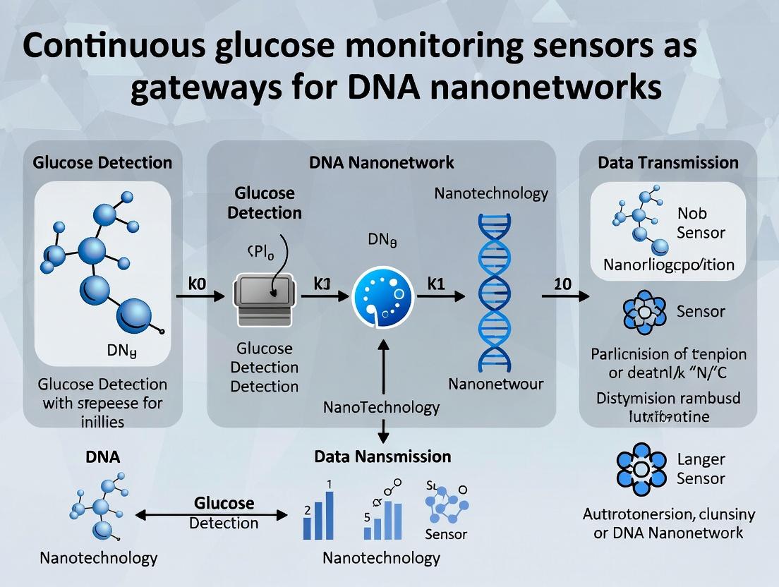
From Sugar to Signals: Engineering DNA Nanonetworks on Continuous Glucose Monitoring Platforms
This article explores the conceptual and technical integration of DNA-based nanonetworks with commercial continuous glucose monitoring (CGM) sensors, proposing a paradigm shift from passive monitoring to active, intelligent therapeutic intervention.
From Sugar to Signals: Engineering DNA Nanonetworks on Continuous Glucose Monitoring Platforms
Abstract
This article explores the conceptual and technical integration of DNA-based nanonetworks with commercial continuous glucose monitoring (CGM) sensors, proposing a paradigm shift from passive monitoring to active, intelligent therapeutic intervention. We establish the foundational synergy between CGM electrochemistry and DNA nanotechnology (Intent 1), detail methodologies for sensor functionalization and DNA network design for logic-gated drug release (Intent 2), analyze critical challenges in biocompatibility, signaling fidelity, and in vivo stability (Intent 3), and validate the approach through comparative analysis with existing closed-loop and nanomedicine systems (Intent 4). Aimed at researchers and drug development professionals, this synthesis outlines a roadmap for creating autonomous, glucose-responsive therapeutic systems.
The Confluence of CGM Biochemistry and DNA Nanotechnology: Building a Foundational Bridge
Within the broader thesis that Continuous Glucose Monitoring (CGM) sensors serve as foundational gateways for DNA nanonetworks research, this document deconstructs the core electrochemical interface. The modern subcutaneous CGM is a biosensor that transduces a biochemical event (glucose oxidation) into a quantifiable electronic signal. This established, reliable transduction pathway provides the archetypal "readable interface" for future DNA-based molecular communication systems. Understanding and replicating this signal generation is critical for adapting the platform to detect non-glucose analytes, such as specific DNA sequences or molecular signals in a nanonetwork.
Core Electrochemical Signaling Pathway
The dominant signal generation mechanism in commercial CGMs is the enzyme-based amperometric detection of glucose via glucose oxidase (GOx).
Diagram 1: CGM Electrochemical Signal Generation Pathway
Key Research Reagent Solutions & Materials
The following table details essential components for constructing or researching CGM-type electrochemical interfaces.
| Research Reagent / Material | Function in Experimental System |
|---|---|
| Glucose Oxidase (GOx) | Core biorecognition element. Catalyzes glucose oxidation, initiating the signal cascade. |
| Platinum/Carbon Working Electrode | Anode for H₂O₂ oxidation. High purity Pt ensures stable electrochemical kinetics. |
| Ag/AgCl Reference Electrode | Provides a stable, known reference potential for the working electrode. |
| Potassium Ferricyanide (K₃[Fe(CN)₆]) | Common redox mediator alternative to O₂, used to shuttle electrons in mediator-based sensor designs. |
| Nafion Perfluorinated Resin | Cation-exchange polymer membrane. Coated over electrode to reject anionic interferents (e.g., ascorbate, urate). |
| Polyurethane/Polyethylene Glycol Membranes | Outer diffusion-limiting membranes. Control glucose flux to the enzyme layer, linearizing sensor response. |
| Phosphate Buffered Saline (PBS), pH 7.4 | Standard physiological buffer for in vitro testing, providing ionic strength and stable pH. |
| β-D-Glucose Stock Solution | Primary analyte for calibration. Must be allowed to mutarotate to equilibrium before use. |
Experimental Protocol:In VitroCharacterization of a GOx-Based Electrochemical Sensor
This protocol details the foundational experiment for characterizing the core signal generation interface, a prerequisite for adapting it to DNA sensing.
Objective: To measure the amperometric response of a GOx-modified working electrode to incremental glucose concentrations and determine key sensor parameters.
Materials:
- Potentiostat/Galvanostat
- Standard 3-electrode cell: Pt working electrode, Pt wire counter electrode, Ag/AgCl reference electrode.
- Magnetic stirrer and stir bar.
- PBS (0.1 M, pH 7.4), degassed with N₂ for 10 min.
- GOx solution (10 mg/mL in PBS).
- Glucose stock solution (1.0 M in PBS, equilibrated overnight).
- Nafion solution (5% w/w in aliphatic alcohols).
Procedure:
- Electrode Modification: Apply 5 µL of GOx solution to the clean Pt working electrode surface. Allow to dry at 4°C for 1 hour. Subsequently, dip-coat the electrode in Nafion solution for 5 seconds and air-dry. This creates a GOx/Nafion bilayer.
- Setup: Fill the electrochemical cell with 20 mL of degassed PBS. Immerse the three electrodes. Set the potentiostat to apply a constant potential of +0.6 V vs. Ag/AgCl to the working electrode. Engage the stirrer at a constant, slow speed.
- Baseline Stabilization: Record the background current until it stabilizes (< 0.1 nA/min drift). This is I_baseline.
- Standard Additions: Using a micropipette, sequentially add aliquots of the glucose stock solution to the stirred PBS to achieve cumulative concentration increases (e.g., 0.5, 1.0, 2.0, 4.0, 8.0 mM). Record the stable current output after each addition (I_total). Wait for a stable plateau (~60-90 sec) before the next addition.
- Data Processing: For each addition, calculate the net sensor current: Inet = Itotal - I_baseline.
- Calibration: Plot I_net (µA or nA) vs. glucose concentration (mM). Fit the data with a Michaelis-Menten model or linear regression for the linear range.
Diagram 2: Sensor Characterization Workflow
Quantitative Performance Data Table
Typical performance metrics for a well-characterized in vitro GOx-based sensor, as derived from the above protocol.
| Sensor Parameter | Typical Value / Range | Notes & Impact on DNA Nanonetwork Adaptation |
|---|---|---|
| Linear Range | 0.5 – 15 mM (≈ 9 – 270 mg/dL) | Must be re-engineered for pM-nM DNA target concentrations. |
| Sensitivity | 1 – 10 nA/mM | High sensitivity is critical for low-abundance molecular signals. |
| Response Time (t₉₀) | 5 – 30 seconds | Dictates temporal resolution of the nanonetwork communication. |
| Limit of Detection (LOD) | 0.1 – 0.5 mM | Must be drastically improved for DNA detection. |
| Selectivity (vs. Ascorbate) | > 100:1 | Achieved via Nafion membrane. Similar strategies needed for DNA sensor biofouling. |
| Operational Stability | > 72 hours in vitro | Baseline for assessing longevity of a DNA-sensing interface. |
Protocol for Conceptual Adaptation to DNA Detection
This outlines the methodological shift from glucose to DNA sensing, bridging the CGM interface to DNA nanonetwork applications.
Objective: To replace the GOx enzyme layer with a DNA-based recognition layer (e.g., a stem-loop probe with a redox tag) that generates an electrochemical signal upon hybridization.
Materials:
- Thiolated DNA stem-loop probe with methylene blue (MB) redox tag.
- Gold working electrode (for thiol-gold chemistry).
- Mercaptohexanol (MCH) solution (1 mM).
- Target DNA sequence (complementary to the loop region).
- Tris-EDTA buffer with MgCl₂ (TE-Mg buffer).
Procedure:
- Electrode Functionalization: Incubate the clean Au electrode in 1 µM thiolated DNA probe solution for 1 hour. Rinse. Then backfill with 1 mM MCH solution for 30 minutes to form a well-ordered self-assembled monolayer.
- Setup & Technique: Use Square Wave Voltammetry (SWV) instead of constant potential amperometry. Set parameters: Potential window from -0.5 V to 0 V vs. Ag/AgCl, frequency 60 Hz, amplitude 25 mV.
- Baseline Scan: In pure TE-Mg buffer, perform an SWV scan to measure the peak current from the MB tag (I_initial). This signal is attenuated due to the probe's stem-loop structure.
- Target Exposure: Incubate the functionalized electrode in buffer containing the target DNA sequence (e.g., 1 nM for 30 min). Rinse thoroughly.
- Signal-on Measurement: Perform a new SWV scan in clean buffer. Successful hybridization opens the stem-loop, moving the MB tag closer to the electrode, increasing electron transfer and peak current (I_final).
- Analysis: Calculate the signal change: ΔI = Ifinal - Iinitial. This ΔI correlates with target DNA concentration.
This protocol demonstrates the conceptual translation of the CGM's "readable interface" to a DNA-driven system, forming the basis for receiving signals in a biochemical nanonetwork.
DNA Nanostructures as Programmable Signal Transducers and Carriers
The development of continuous glucose monitoring (CGM) sensors necessitates biocompatible, miniaturized, and intelligent systems for molecular sensing and feedback. This thesis posits that CGM platforms serve as an ideal testbed and gateway for pioneering DNA nanonetworks. DNA nanostructures, with their atomic-level programmability, can act as integrated signal transducers—converting molecular recognition into quantifiable signals—and as targeted carriers for therapeutic or regulatory agents. This synergy could evolve CGM from passive monitors to closed-loop, therapeutic systems.
Application Notes
Signal Transduction Mechanisms
DNA nanostructures enable diverse transduction modalities crucial for biosensing.
Table 1: DNA Nanostructure-Based Transduction Mechanisms for Biosensing
| Transduction Mechanism | Nanostructure Scaffold | Signal Readout | Reported Sensitivity (Recent Examples) | Potential CGM Integration |
|---|---|---|---|---|
| Fluorescence Resonance Energy Transfer (FRET) | DNA origami tile with positioned dyes | Fluorescence intensity/ratio | Sub-nanomolar target detection (2023) | Conformational change upon glucose binding alters FRET. |
| Electrochemical | Tetrahedron or 3D wireframe on gold electrode | Current/Impedance | Detection limit of 0.1 pM for miRNA (2024) | Direct electron transfer from enzyme (e.g., GOx) tagged on nanostructure. |
| Colorimetric | DNAzyme-based nanowire or assembled nanostructures | Visible color shift | 5 nM glucose in synthetic serum (2024) | Paper-based lateral flow assay with DNA nanostructure carriers. |
| Mechanical / Plasmonic | Gold nanoparticle-decorated origami | Surface plasmon resonance shift | 10 fM for protein biomarkers (2023) | Glucose-induced nanostructure deformation shifts plasmon coupling. |
Carrier Functions for Therapeutic Intervention
Beyond sensing, these structures can be engineered for responsive drug delivery.
Table 2: Carrier Capabilities of DNA Nanostructures for Closed-Loop Therapies
| Carrier Function | Example Nanostructure | Cargo Type | Trigger Mechanism | Therapeutic Relevance to Glucose Management |
|---|---|---|---|---|
| Targeted Delivery | Aptamer-gated DNA nanocage | Insulin, GLP-1 analogs | Protein (e.g., overexpressed receptor on β-cells) | Direct delivery to pancreatic cells. |
| Stimuli-Responsive Release | pH-sensitive i-motif lid on nanotube | Metformin, enzymes | Low pH (e.g., in inflammatory tissue) | Release in local acidic microenvironments near dysfunctional cells. |
| Multi-Agent Co-Delivery | Multi-compartment origami | Enzyme + Cofactor + Inhibitor | Enzymatic cascade activation | Mimicking metabolic pathways for glucose regulation. |
| Immunomodulation | Triangular DNA origami with CpG motifs | Nucleic acid therapeutics | Toll-like receptor 9 recognition | Mitigating inflammation at sensor implant site. |
Experimental Protocols
Protocol: Fabrication of a Glucose-Responsive DNA Origami FRET Transducer
Objective: To create a DNA origami hinge structure that undergoes a glucose-dependent conformational change, monitored via FRET.
Materials:
- DNA Scaffold: M13mp18 single-stranded DNA (7249 nucleotides).
- Staples: 200+ synthetic oligonucleotides, designed using caDNAno software. Include staples labeled with Cy3 (Donor) and Cy5 (Acceptor) at strategic positions on opposite arms of the hinge. Include staples conjugated to glucose-binding aptamer sequences (e.g., concanavalin A mimic or engineered aptamer).
- Buffers: Folding buffer (1x TAE, 12.5 mM MgCl₂, pH 8.0).
- Equipment: Thermal cycler, agarose gel electrophoresis system, fluorometer or fluorescence microscope.
Procedure:
- Solution Preparation: Mix scaffold strand (10 nM) with a 10x molar excess of unlabeled and labeled staple strands in folding buffer.
- Thermal Annealing: Use a thermal cycler program: Heat to 65°C for 15 min, then cool slowly to 20°C over 16 hours.
- Purification: Purify folded structures using PEG precipitation or agarose gel electrophoresis (2% agarose, 0.5x TBE, 11 mM MgCl₂, 4°C). Excise and extract the band corresponding to correctly folded origami.
- Characterization: Confirm structure via atomic force microscopy (AFM) in tapping mode in liquid.
- FRET Assay:
- Dilute purified origami to 1 nM in assay buffer (with MgCl₂).
- Aliquot into a 96-well plate. Add glucose standards (0-30 mM) or test samples.
- Incubate for 30 minutes at 25°C.
- Measure fluorescence intensity at 570 nm (Cy3 emission) and 670 nm (Cy5 emission) using 540 nm excitation.
- Calculate FRET efficiency (E) as IAcceptor / (IDonor + IAcceptor), where I is background-subtracted intensity.
- Data Analysis: Plot FRET efficiency vs. glucose concentration to generate a calibration curve.
Protocol: Electrochemical Biosensor Fabrication using DNA Tetrahedron Carriers
Objective: To immobilize glucose oxidase (GOx) on an electrode surface with controlled orientation and high density using DNA tetrahedron nanostructures for enhanced electrochemical detection.
Materials:
- DNA Tetrahedron: Four specifically designed oligonucleotides (Th1-Th4) that self-assemble. Th1 is thiolated at the 5' end. Th2 is conjugated to biotin.
- Electrode: Gold disk electrode (2 mm diameter).
- Proteins: Streptavidin, Biotinylated Glucose Oxidase (GOx).
- Electrochemical Probe: Ferrocenemethanol (FcMeOH).
- Equipment: Potentiostat, piranha-cleaned glassware, microcentrifuge.
Procedure:
- Tetrahedron Assembly: Mix equimolar amounts (1 µM) of Th1, Th2, Th3, and Th4 in TM buffer (20 mM Tris, 50 mM MgCl₂, pH 8.0). Anneal from 95°C to 4°C over 90 min.
- Electrode Modification:
- Clean gold electrode in piranha solution (Caution: Extremely corrosive), rinse, and dry.
- Incubate the electrode in 100 nM tetrahedron solution (in TM buffer) for 16 hours at 4°C. This forms a dense, upright monolayer via Th1's thiol-gold bond.
- Rinse thoroughly with buffer.
- Enzyme Assembly:
- Incubate electrode in 0.1 mg/mL streptavidin solution for 1 hour. Rinse.
- Incubate in 0.1 mg/mL biotinylated GOx solution for 1 hour. Rinse.
- Electrochemical Measurement (Amperometry):
- Use a three-electrode setup (modified Au working, Pt counter, Ag/AgCl reference) in PBS (pH 7.4) with 1 mM FcMeOH as a redox mediator.
- Apply a constant potential of +0.4 V vs. Ag/AgCl.
- Inject glucose aliquots to achieve desired concentrations (0-20 mM).
- Record the steady-state catalytic current increase. The current is proportional to glucose concentration as GOx oxidizes glucose, and FcMeOH shuttles electrons to the electrode.
Visualization
Title: DNA Nanostructure Signal Transduction Pathway
Title: Workflow for DNA Nanostructure Biosensor Development
The Scientist's Toolkit
Table 3: Key Research Reagent Solutions for DNA Nanonetwork Research
| Item | Function / Description | Example Vendor/Product |
|---|---|---|
| M13mp18 ssDNA Scaffold | The long, single-stranded DNA backbone for scaffolded DNA origami. | New England Biolabs (NEB) |
| Custom Staple Oligonucleotides | Short, complementary strands that fold the scaffold into the desired 2D/3D shape. Synthesized with modifications (biotin, dyes, thiol). | Integrated DNA Technologies (IDT), Eurofins Genomics |
| caDNAno / cadnano Software | Open-source software for designing the staple sequences and routing the scaffold for DNA origami. | Open-source (GitHub) |
| TAE/Mg²⁺ Folding Buffer | Standardized buffer providing optimal ionic strength and Mg²⁺ concentration for folding stable DNA nanostructures. | Often prepared in-lab: 40 mM Tris, 20 mM Acetic acid, 2 mM EDTA, 12.5 mM MgCl₂, pH 8.0. |
| Magnetic Bead Purification Kits | For rapid purification of assembled nanostructures from excess staples (e.g., using PEG-based precipitation). | Ampure XP (Beckman Coulter) with protocol adaptation. |
| Biotin-/Thiol-/Dye-Modified Nucleotides | Enables easy functionalization of nanostructures for immobilization (biotin-streptavidin, thiol-gold), labeling, or sensing. | Glen Research, Jena Bioscience |
| Glucose Oxidase (GOx), Lyophilized | Model enzyme for glucose sensing. Can be chemically biotinylated or linked to DNA for site-specific conjugation. | Sigma-Aldrich |
| Ferrocene Derivatives (e.g., Ferrocenemethanol) | Redox mediators for enzymatic electrochemical biosensors, shuttling electrons from enzyme to electrode. | Sigma-Aldrich, TCI Chemicals |
Glucose is a fundamental biological molecule, serving as a primary energy source and a critical signaling molecule. In the context of our broader thesis on Continuous Glucose Monitoring (CGM) sensors as gateways for DNA nanonetworks research, glucose transitions from a simple analytic to a programmable logical input. CGM sensors provide a real-time, in vivo data stream of glucose concentration, a variable that can be harnessed to trigger downstream molecular computations within engineered DNA-based systems. This document outlines the application of glucose as a trigger, detailing protocols and conceptual frameworks for integrating CGM data with responsive DNA nanonetworks for advanced biosensing and therapeutic applications.
Table 1: Physiological and Analytical Ranges of Glucose Relevant to CGM-Triggered Systems
| Parameter | Normal Physiological Range (Fasting) | Diabetic Alert Range (Hypo/Hyperglycemia) | Typical CGM Analytical Range | Logical Threshold for DNA Network Activation (Proposed) |
|---|---|---|---|---|
| Concentration | 70-100 mg/dL (3.9-5.6 mM) | <70 mg/dL or >180 mg/dL | 40-400 mg/dL (2.2-22.2 mM) | User-defined (e.g., 150 mg/dL for hyperglycemic response) |
| Time Lag (Interstitial Fluid vs. Blood) | N/A | N/A | 5-15 minutes | Incorporated into network delay circuit design |
| Measurement Frequency (CGM) | N/A | N/A | 1-5 minutes | Defines temporal resolution of input signal |
Table 2: Performance Metrics of Select Recent CGM Systems (2023-2024)
| CGM System/Technology | Key Sensor Chemistry | Accuracy (MARD*) | Longevity | Connectivity (Gateway Function) | Reference |
|---|---|---|---|---|---|
| Abbott Libre 3 | Enzymatic (GOx†), Wireless | <8% | 14 days | Bluetooth to Smartphone | [1] |
| Dexcom G7 | Enzymatic (GOx), Sensor | ~8.2% | 10.5 days | Bluetooth to Smartphone/App | [2] |
| Medtronic Guardian 4 | Enzymatic (GOx) | 8.7% | 7 days | Bluetooth to Smartphone | [3] |
| Eversense E3 (Implantable) | Fluorescent (Polymer Boronic Acid) | 8.8% | 6 months | RF to Transmitter | [4] |
*MARD: Mean Absolute Relative Difference. †GOx: Glucose Oxidase.
Core Conceptual Framework and Signaling Pathways
From CGM Signal to Molecular Command
The pathway from glucose concentration to DNA network activation involves a digital translation of an analog biochemical signal.
Diagram Title: CGM to DNA Network Control Pathway
Logical Integration of Glucose Input in DNA Circuits
Glucose levels can be processed as Boolean inputs (HIGH/LOW) to drive logic-gated DNA nanosystems.
Diagram Title: Glucose-Insulin NAND Logic for Smart Therapy
Experimental Protocols
Protocol 1: In Vitro Calibration of a Glucose-Responsive DNA Nanodevice
Objective: To establish the dose-response relationship between glucose concentration and the activation (e.g., fluorescence dequenching) of a glucose-binding aptamer-integrated DNA nanoswitch.
Materials: See "Scientist's Toolkit" below. Procedure:
- DNA Nanoswitch Preparation:
- Resuspend the glucose-sensitive aptamer nanoswitch (e.g., a duplex with a quenched fluorophore) in 1X PBS, pH 7.4. Heat to 95°C for 5 minutes and slowly cool to room temperature (1°C/min) to ensure proper folding.
- Glucose Stock Solution Preparation:
- Prepare a 1M D-glucose stock in 1X PBS. Prepare serial dilutions (0 mM, 1 mM, 2 mM, 5 mM, 10 mM, 20 mM) in PBS to cover physiological and pathological ranges.
- Assay Setup:
- In a black 96-well plate, add 90 µL of the folded DNA nanoswitch solution (final concentration 50 nM) per well.
- Add 10 µL of each glucose dilution to triplicate wells. Include PBS-only controls.
- Seal plate, mix gently by orbital shaking, and incubate at 37°C for 60 minutes.
- Data Acquisition:
- Using a plate reader, measure fluorescence intensity (ex/cm: e.g., 490/520 nm for FAM) for each well.
- Calculate mean fluorescence for each glucose concentration. Plot fluorescence intensity (or fold change vs. 0 mM control) against log[glucose]. Fit a sigmoidal curve to determine EC50 and dynamic range.
- Validation with CGM Simulator:
- Use a programmable syringe pump to flow a glucose profile mimicking CGM data (e.g., postprandial spike) past the nanoswitch in a microfluidic chamber. Continuously monitor fluorescence.
Protocol 2: Interface of a CGM Data Stream with a Light-Actuated DNA Nanonetwork
Objective: To demonstrate closed-loop control where a CGM reading triggers a light source that activates a photosensitive DNA-based drug carrier.
Materials: CGM simulator/app, LED array (470 nm), DNA-caged drug (e.g., Doxorubicin conjugated to oligonucleotide via photocleavable linker), cell culture. Procedure:
- System Setup:
- Connect a research CGM (or a CGM data simulator app outputting real-time [glucose]) to a microcontroller (e.g., Arduino/Raspberry Pi).
- Program the microcontroller to activate a 470 nm LED array when the incoming [glucose] value exceeds a set threshold (e.g., 180 mg/dL) for more than 10 minutes.
- In Vitro Activation Test:
- Seed cancer cells in a 96-well plate. Add the photosensitive DNA-caged drug construct.
- Place the LED array above the plate and position the CGM sensor (or its simulated output source) in a separate glucose-containing chamber.
- Initiate a glucose ramp. Upon threshold crossing, the LED array should illuminate the plate for a predetermined duration.
- After 24h, assay cell viability (e.g., MTT assay). Compare to controls (no glucose trigger, light only, drug only).
- Data Analysis:
- Correlate the time of CGM-triggered activation with the onset of cytotoxic effect. Determine the specificity of the system by testing with non-target sugars (e.g., fructose).
The Scientist's Toolkit: Key Research Reagent Solutions
Table 3: Essential Materials for Glucose-Triggered DNA Nanonetwork Research
| Item/Category | Example Product/Specification | Function in Research |
|---|---|---|
| Glucose-Sensitive DNA Elements | Glucose-binding aptamer (e.g., DNA sequence from in vitro selection); Boronic acid-functionalized nucleotides. | Serves as the molecular recognition module within the DNA nanostructure, directly binding glucose to induce conformational change. |
| CGM Development Kit | Abbott Libre Sense Dev Kit; Dexcom Developer API. | Provides programmable access to real-time glucose data streams, serving as the digital gateway for external logic processing. |
| DNA Nanostructure Scaffold | M13mp18 phage DNA; Custom synthetic oligonucleotide tile sets (e.g., from IDT). | Provides the structural framework for assembling controlled, multi-component nanonetworks. |
| External Actuation Interface | Near-Infrared Laser (e.g., 808 nm); Ultrasound Transducer (1 MHz); RF Generator. | Provides the physical energy cue (heat, mechanical force, magnetic field) triggered by the CGM logic to remotely actuate the DNA network in vivo. |
| Responsive Linkers/Cages | Photocleavable linker (PC Biotin); Azobenzene-modified nucleotides; pH-sensitive i-motif sequences. | Enables the controlled release of a payload (drug, reporter) or reconfiguration of the network upon receiving the actuation signal. |
| Fluorescent Reporters | FAM (Fluorescein), Cy5, Quenchers (Iowa Black FQ, BHQ-1). | Allows for real-time, quantitative tracking of glucose-induced conformational changes or network output in vitro and in vivo. |
| Microcontroller/DAQ | Raspberry Pi 4, Arduino Uno, National Instruments DAQ. | The hardware bridge that executes the "if-then" logic on CGM data and controls the actuation interface. |
Application Notes: Theoretical Integration with CGM-Gated DNA Nanonetworks
Continuous Glucose Monitoring (CGM) systems provide a real-world, in-vivo platform for modeling closed-loop, event-driven molecular communication. The core thesis posits CGM sensor data as the environmental input signal for orchestrating synthetic DNA nanonetwork responses. The theoretical framework bridges three domains: biochemical detection, information encoding, and actuator deployment.
- Molecular Detection as Signal Acquisition: The CGM's enzymatic detection of glucose (analyte) is analogous to a receiver nanomachine detecting a molecular signal. Fluctuations in interstitial glucose concentration represent the primary environmental data stream.
- Signal Processing & Decision Logic: Theoretical models must define thresholds (e.g., hyperglycemia >180 mg/dL) that convert analog concentration data into digital triggers for DNA network activation. This involves modeling noise, hysteresis, and signal stabilization.
- Networked Response via DNA Nanotechnology: Upon a digital trigger, the system initiates a programmed cascade. This can involve:
- DNA Strand Displacement (DSD) Circuits: For in-silico decision-making and signal amplification.
- Liposome or Vesicle Release: Modeled as packet-switched communication for drug payloads (e.g., insulin or glucagon mimics).
- Scaffolded Aptamer Activation: For secondary molecular recognition and binding.
The following table summarizes key quantitative parameters for modeling this integrated system:
Table 1: Quantitative Parameters for CGM-Gated DNA Nanonetwork Models
| Parameter Category | Symbol | Typical Range / Value (CGM Context) | DNA Network Correlate | Notes |
|---|---|---|---|---|
| Primary Signal | [G] | 70-180 mg/dL (Normal) | Input Signal Concentration | Sampled every 1-5 mins. Noise ±10-20%. |
| Trigger Threshold | Θ_h | 180 mg/dL (Hyperglycemic) | DSD Circuit Activation Toehold Concentration | Defines binary 'ON' state. Must account for CGM lag (~5-10 min). |
| Communication Channel | - | Interstitial Fluid Volume (~0.2 L in subcutaneous tissue) | Diffusion Medium & Volume | Modeled as a diffusion channel with loss (clearance). |
| Signal Propagation | D | ~10⁻¹⁰ m²/s (for glucose) | Diffusion Coefficient of DNA Nanocarriers | DNA structures have D ~10⁻¹² to 10⁻¹¹ m²/s, enabling localized action. |
| Bit Rate / Response Time | τ | CGM Lag: 5-10 min; DSD Cascade: minutes to hours | System Latency | Total time from threshold exceedance to payload release. Key design constraint. |
| Payload | - | Insulin (≈5808 Da) | Therapeutic Oligonucleotide / Small Molecule | Defines required carrier capacity (e.g., number of drug molecules per vesicle). |
Experimental Protocols
Protocol 1: In-Vitro Emulation of CGM-Triggered DNA Strand Displacement Cascade
Objective: To validate a DSD circuit that is activated by a molecular proxy for a "high glucose" signal.
Materials: See "Research Reagent Solutions" below. Workflow:
- Signal Proxy Preparation: Prepare a 100 µM stock solution of the trigger DNA strand (T_gluc) in nuclease-free TE buffer. This strand is designed to be complementary to a glucose-binding aptamer's released sequence or is directly a glucose-oxidase generated product mimic (e.g., a specific pH-sensitive strand).
- DSD Circuit Assembly: In a 1.5 mL tube, mix the following in Nuclease-Free Buffer:
- 10 µL of Gate Complex G (100 nM final)
- 10 µL of Reporter Complex R (100 nM final)
- 69 µL of Buffer.
- Heat to 95°C for 2 min, then cool to 25°C over 45 min to anneal.
- Baseline Measurement: Aliquot 9 µL of the annealed circuit into 5 separate PCR tubes. Add 1 µL of buffer to each. Measure initial fluorescence (F₀) at λex/λem for your fluorophore/quencher pair (e.g., FAM/BHQ-1).
- Trigger Introduction: Add 1 µL of the T_gluc stock solution to 4 test aliquots to achieve final trigger concentrations of 1 nM, 10 nM, 50 nM, and 100 nM. To a negative control, add 1 µL of a scrambled DNA sequence (100 nM final).
- Kinetic Monitoring: Immediately transfer tubes to a real-time PCR system or fluorescence plate reader maintained at 37°C. Measure fluorescence every 30 seconds for 4-8 hours.
- Data Analysis: Plot ΔF (F - F₀) vs. time. Model the reaction kinetics. Determine the minimum trigger concentration (threshold, Θ_h) required for a significant fluorescent signal increase within a clinically relevant timeframe (e.g., 30 min).
Protocol 2: Fabrication and Triggered Release from DNA-Functionalized Liposomes
Objective: To construct a model drug carrier that releases its payload upon receiving a specific DNA signal from a primary detection cascade.
Materials: See "Research Reagent Solutions" below. Workflow:
- Liposome Preparation (Thin-Film Hydration):
- Dissolve 10 mg of phospholipids (e.g., DOPC:Cholesterol:DSPE-PEG2000 at 65:30:5 molar ratio) in chloroform in a round-bottom flask.
- Evaporate under nitrogen to form a thin lipid film. Desiccate under vacuum for 2 hours.
- Hydrate the film with 1 mL of PBS containing 50 mM of a fluorescent dye (e.g., Calcein) as a payload mimic. Vortex vigorously.
- Extrude the suspension 21 times through a polycarbonate membrane (100 nm pore size) to form unilamellar vesicles.
- Purify liposomes via size-exclusion chromatography (Sephadex G-50) to remove free dye.
- DNA Anchor Functionalization:
- Incubate purified liposomes with a 10-fold molar excess of cholesterol-modified DNA anchor strands (Chol-DNA-Anchor) for 1 hour at 25°C.
- Purify again via size-exclusion chromatography to remove unbound anchors.
- Stopper Complex Assembly: Pre-anneal a "Stopper" DNA strand to the anchor's complement. This stopper is a duplex that blocks a toehold and is displaced by the trigger strand (T_net) from Protocol 1's output.
- Surface Assembly: Incubate DNA-functionalized liposomes with a 5-fold excess of the pre-formed Stopper complex for 2 hours at 25°C. Purify to obtain "loaded" nanocarriers.
- Triggered Release Assay:
- In a 96-well plate, add 90 µL of loaded liposome solution per well.
- Add 10 µL of buffer (negative control), 10% Triton X-100 (positive control, 100% release), or varying concentrations of the trigger strand T_net.
- Monitor fluorescence intensity over time (Calcein: λex/λem ≈ 494/517 nm). Quenched calcein inside liposomes fluoresces upon release and dilution.
- Calculate % Release = (Fsample - Finitial) / (Ftriton - Finitial) * 100.
Visualization
Diagram 1: CGM-Gated DNA Nanonetwork Communication Model
Diagram 2: Experimental Workflow for Triggered Response
The Scientist's Toolkit: Research Reagent Solutions
Table 2: Essential Materials for CGM-DNA Nanonetwork Experiments
| Item | Function in Research | Example / Specification |
|---|---|---|
| CGM Simulator / Data Stream | Provides real or simulated continuous glucose concentration data to drive the theoretical model and bench experiments. | Open-source artificial pancreas software (OpenAPS) data logs; or programmable syringe pump with glucose solution. |
| DNA Strands (Oligonucleotides) | The fundamental "code" for information processing, including triggers, gates, reporters, and anchors. | HPLC-purified, modified with fluorophores (FAM, Cy5), quenchers (BHQ-1, Dabcyl), or cholesterol. |
| Thermostable DNA Ligase/Buffer | For assembling large DNA nanostructures (e.g., origami) that could act as scaffolds or carriers. | T4 DNA Ligase or Bst 2.0 WarmStart Polymerase for isothermal assembly. |
| Phospholipids & Cholesterol | Building blocks for constructing liposomal nanocarriers for drug encapsulation and release. | 1,2-dioleoyl-sn-glycero-3-phosphocholine (DOPC), 1,2-distearoyl-sn-glycero-3-phosphoethanolamine-N-[methoxy(polyethylene glycol)-2000] (DSPE-PEG2000). |
| Liposome Extruder & Membranes | To form uniform, nanoscale unilamellar vesicles with consistent encapsulation and release properties. | Extruder with 100 nm polycarbonate membranes. |
| Size-Exclusion Chromatography Columns | Critical for purifying DNA nanostructures and functionalized liposomes from excess reagents and unencapsulated payload. | Sephadex G-50 or G-75 spin columns. |
| Real-Time Fluorescence Spectrometer | For kinetic monitoring of DNA strand displacement reactions (kinetics, threshold) and payload release assays. | Plate reader or qPCR system with temperature control and appropriate filter sets. |
| Glucose Oxidase & Catalase | Enzymatic system to convert glucose concentration into a usable signal (e.g., local pH change or H₂O₂ production) for triggering DNA devices. | From Aspergillus niger, used to functionalize detection layer. |
| Nuclease-Free Buffers & Water | To prevent degradation of DNA components during assembly and experimentation. | TE buffer (10 mM Tris, 1 mM EDTA, pH 8.0), DPBS (Dulbecco's Phosphate-Buffered Saline). |
The development of Continuous Glucose Monitoring (CGM) sensors represents a foundational leap in biodevice interfacing, providing a continuous, real-time biochemical data stream in vivo. Within the broader thesis on "Continuous glucose monitoring sensors as gateways for DNA nanonetworks research," this review examines seminal works that established the fundamental proofs of concept for bio-interfaced DNA networks. These early studies demonstrated the feasibility of using synthetic DNA nanostructures to sense, compute, and actuate at biological interfaces, laying the groundwork for future integrations with CGM-like platforms to create advanced, closed-loop therapeutic systems.
Early Proofs of Concept: Key Studies and Data
This section reviews pivotal experiments that first demonstrated the core functionalities required for a bio-interfaced DNA network: molecular sensing, information processing via logic gates, and controlled output signaling or drug release.
Table 1: Summary of Pioneering Experiments in Bio-Interfaced DNA Networks
| Study (Year) | Core Concept | Target/Signal Input | DNA Network Design | Quantified Output / Key Performance Data |
|---|---|---|---|---|
| Benenson et al., 2004 | Autonomous biomolecular computer for disease diagnostics. | mRNA levels (e.g., PPARγ, GSTP1). | Logic gates (AND, OR, NOT) based on siRNA-like recognition. | Correctly diagnosed and induced apoptosis in vitro. Specificity: Differentiated between healthy and cancer cell lines. |
| Douglas et al., 2012 | "DNA origami" nanorobot for targeted drug delivery. | Protein keys (e.g., antibodies). | Aptamer-locked origami container. | Effective payload delivery and cell death. In vitro specificity: 5x greater cell death in target-positive vs. target-negative cells. |
| Amir et al., 2014 | Molecular computing via reconfigurable DNA nanostructures. | Specific DNA trigger strands. | Dynamic DNA tiles performing computation (e.g., tile translocation). | Demonstrated 4-bit square root calculation. Computation speed: Hours for completion of pattern reconfiguration. |
| López et al., 2021 | Electrochemical DNA-based sensor for continuous monitoring. | Glucose (as a model analyte). | Enzyme (GOx)-coupled DNA scaffold on electrode surface. | Linear detection range: 0.1–10 mM glucose. Stability: >90% signal retained over 72 hours in serum. |
Detailed Application Notes & Protocols
These protocols are derived from the methodologies of the pioneering works and adapted for a general research context relevant to interfacing with physiological monitors.
Protocol 1: In Vitro Assessment of a Logic-Gated DNA Network for Molecular Sensing
Objective: To test a DNA-based logic gate system (e.g., AND gate) responsive to two specific mRNA inputs in a cell lysate or defined buffer.
Materials: See "Scientist's Toolkit" below. Procedure:
- Gate Preparation: Synthesize and purify the DNA strands constituting the logic gate (e.g., two input-recognition strands and a fluorescently quenched reporter strand). Anneal in Nuclease-Free Buffer (e.g., 1x TE, 10 mM MgCl₂) by heating to 95°C for 5 min and cooling slowly to 4°C (1°C/min).
- Input Preparation: Prepare synthetic target mRNA sequences (inputs) at known concentrations (e.g., 100 nM – 1 µM) in a physiologically relevant buffer (e.g., with 150 mM KCl, 2 mM MgCl₂).
- Reaction Assembly: In a low-binding microtube, combine:
- 50 nM assembled DNA logic gate complex.
- Input A (0-500 nM).
- Input B (0-500 nM).
- 1x Reaction Buffer (20 mM Tris-HCl, pH 7.5, 150 mM KCl, 5 mM MgCl₂, 0.1% Tween-20).
- Nuclease-free water to final volume.
- Incubation & Measurement: Incubate at 37°C for 2-4 hours. Measure fluorescence (e.g., FAM emission at 520 nm with 495 nm excitation) at regular intervals using a plate reader.
- Data Analysis: Plot fluorescence intensity vs. time for all input combinations (00, 01, 10, 11). Calculate the fold-change and signal-to-noise ratio for the correct "11" output versus background.
Protocol 2: Functionalization of a Solid-Phase Sensor with a DNA Recognition Layer
Objective: To immobilize a DNA aptamer or enzyme-DNA complex onto a gold electrode surface, mimicking the interface of a CGM sensor.
Materials: See "Scientist's Toolkit" below. Procedure:
- Electrode Preparation: Clean gold electrode (2 mm diameter) via sequential sonication in acetone, ethanol, and deionized water for 5 min each. Electrochemically clean in 0.5 M H₂SO₄ by cyclic voltammetry (CV) from -0.2 to 1.5 V until a stable CV profile is obtained.
- Thiolated DNA Preparation: Reduce the disulfide bonds of thiol-modified DNA strands (e.g., 100 µM) in 10 mM Tris(2-carboxyethyl)phosphine (TCEP) for 1 hour at room temperature. Purify via desalting column.
- Self-Assembled Monolayer (SAM) Formation: Incubate the clean gold electrode in a 1 µM solution of the reduced thiol-DNA in Immobilization Buffer (1 M KH₂PO₄, pH 3.8) for 16-24 hours at 4°C in a humid chamber.
- Backfilling: Rinse the electrode and immerse in a 1 mM solution of 6-mercapto-1-hexanol (MCH) in PBS for 1 hour to displace non-specifically adsorbed DNA and create a well-ordered monolayer.
- Characterization: Verify immobilization using electrochemical impedance spectroscopy (EIS) in 5 mM [Fe(CN)₆]³⁻/⁴⁻ solution. A successful SAM will increase charge-transfer resistance (R_ct).
The Scientist's Toolkit
Table 2: Key Research Reagent Solutions for Bio-Interfaced DNA Network Experiments
| Item / Reagent | Function & Role in Experiments |
|---|---|
| Thiol-modified DNA strands | Enables covalent, oriented immobilization of DNA networks onto gold surfaces (electrodes, SPR chips). Forms the foundational interface. |
| Nuclease-Free Buffers (e.g., with Mg²⁺) | Maintains structural integrity of DNA nanostructures (e.g., origami, tetrahedra) and prevents enzymatic degradation during in vitro assays. |
| Fluorescent-Quencher (FQ) Probe Pairs | Provides a real-time, quantifiable signal output for sensing and logic-gating events (e.g., FAM/BHQ-1). |
| DNA Origami Scaffold (M13mp18) | The standard long, single-stranded DNA used as a template to fold complex 2D/3D nanostructures via staple strands. |
| Tris(2-carboxyethyl)phosphine (TCEP) | A reducing agent used to cleave disulfide bonds in thiol-modified DNA, ensuring active thiol groups for surface conjugation. |
| 6-Mercapto-1-hexanol (MCH) | An alkanethiol used to "backfill" gaps on gold surfaces after DNA immobilization, minimizing non-specific adsorption and improving probe orientation. |
| HPLC-Purified DNA Oligonucleotides | High-purity synthetic strands are critical for reliable self-assembly of networks and to avoid side reactions from failure sequences. |
Visualization of Concepts and Workflows
Title: DNA Logic Gate Activation for Molecular Sensing
Title: Workflow for DNA Functionalization of an Electrode
Engineering the Interface: Methodologies for Functionalizing CGMs with DNA Nanonetworks
The development of continuous glucose monitoring (CGM) sensors has revolutionized point-of-care diagnostics, establishing a mature framework for continuous, real-time biomolecular sensing in vivo. This technological platform provides more than just a clinical tool; it serves as a foundational gateway for DNA nanonetworks research. The core challenge in translating CGM principles to DNA-based sensing lies in the stable and oriented immobilization of DNA probes onto conductive electrode surfaces—a process known as bioconjugation. This document details advanced strategies for linking DNA to electrode chemistry, enabling the next generation of sensors where DNA acts not only as a recognition element but as a programmable nanomachine for signal amplification and computation.
Core Bioconjugation Strategies: Mechanisms & Quantitative Comparison
Successful DNA immobilization requires forming a stable bond between the oligonucleotide and the electrode while maintaining DNA accessibility and biological function. The table below summarizes the primary strategies, their mechanisms, and key performance metrics.
Table 1: Quantitative Comparison of DNA Immobilization Strategies on Gold Electrodes
| Strategy | Chemical Mechanism | Typical Surface Density (pmol/cm²) | Hybridization Efficiency (%) | Stability (Operational) | Key Advantage | Key Limitation |
|---|---|---|---|---|---|---|
| Thiol-Gold Self-Assembled Monolayer (SAM) | Covalent Au-S bond from 5'/3'-thiol-modified DNA. | 2 - 10 | 30 - 70 | High (weeks) | Well-defined, ordered monolayer. | Non-specific adsorption; requires backfilling with mercaptohexanol. |
| Avidin-Biotin | Affinity binding of biotinylated DNA to avidin/streptavidin coated surface. | 1 - 5 | 60 - 90 | High | High orientation & activity; versatile. | Protein layer adds complexity/instability; potential denaturation. |
| Electro-deposition / Adsorption | Physical adsorption or potential-assisted trapping of DNA. | 10 - 50 | 10 - 40 | Low-Moderate | Simple, high-density deposition. | Poor orientation, random attachment, low stability. |
| Click Chemistry (e.g., Azide-Alkyne) | Copper-catalyzed (CuAAC) or strain-promoted (SPAAC) cycloaddition. | 3 - 8 | 50 - 80 | Very High | Chemoselective, robust, versatile surface chemistry. | Requires synthetic modification of DNA (azide/DBCO). |
| EPD-Electrode Binding Peptides | Use of peptide sequences (e.g., A3: AAYSSGAPPMPPF) with high affinity for Au. | 4 - 12 | 40 - 75 | High | Phage-display derived; gentle, oriented binding. | Emerging method; cost of peptide-DNA conjugates. |
Detailed Protocols
Protocol 3.1: Optimized Thiol-Gold SAM with Backfolding Prevention
This protocol details the formation of a mixed monolayer to maximize probe accessibility.
I. Materials & Reagents (The Scientist's Toolkit) Table 2: Essential Research Reagent Solutions
| Item | Function & Specification |
|---|---|
| Thiol-Modified DNA Probe | 5' or 3' C6-SH modified; HPLC purified. Recognition/anchoring element. |
| 6-Mercapto-1-hexanol (MCH) | Backfilling agent. Displaces non-specific adsorption and tilts DNA upright. |
| Tris-EDTA (TE) Buffer (10 mM Tris, 1 mM EDTA, pH 8.0) | DNA storage and dilution buffer. EDTA chelates divalent cations. |
| TCEP-HCl (Tris(2-carboxyethyl)phosphine) | Reducing agent. Cleaves disulfide bonds in thiol-DNA stocks before use. |
| Phosphate Buffered Saline (PBS, 1x, pH 7.4) | Washing and incubation buffer. Provides physiological ionic strength. |
| Gold Electrode (e.g., disk, chip, or SPE) | Substrate. Must be meticulously cleaned (piranha: Caution! or electrochemical cleaning). |
II. Procedure
- Electrode Pretreatment: Clean gold electrode via electrochemical cycling in 0.5 M H₂SO₄ (scan from -0.2 to +1.5 V vs. Ag/AgCl until stable CV is obtained). Rinse thoroughly with deionized water and ethanol. Dry under N₂ stream.
- DNA Probe Reduction: Incubate 100 µM thiol-DNA stock solution with 10 mM TCEP in TE buffer for 1 hour at room temperature to reduce any disulfide dimers.
- Probe Immobilization: Dilute reduced DNA to 1 µM in TE buffer containing 1.0 M NaCl (high salt promotes upright orientation). Pipette 50 µL onto the cleaned gold surface. Incubate in a humidified chamber for 16 hours (overnight) at 4°C.
- Backfilling: Rinse electrode gently with TE buffer to remove loosely bound DNA. Immerse electrode in a 1 mM solution of MCH in PBS for 1 hour at room temperature.
- Rinsing & Storage: Rinse sequentially with PBS and deionized water. The functionalized electrode can be stored in PBS at 4°C for short-term use. Validate surface density via chronocoulometry using [Ru(NH₃)₆]³⁺.
Protocol 3.2: Streptavidin-Biotin Mediated Immobilization on Carbon Electrodes
Ideal for screen-printed carbon electrodes (SPCEs) commonly used in disposable biosensors.
I. Materials
- Biotinylated DNA probe
- Streptavidin (or NeutrAvidin)
- EDC/NHS coupling reagents
- Carboxylated SPCEs
- Blocking solution (e.g., 1% BSA in PBS)
II. Procedure
- Surface Activation: Apply 20 µL of a fresh mixture of 40 mM EDC and 10 mM NHS in MES buffer (pH 5.5) to the carboxylated SPCE working electrode. Incubate for 30 min to form amine-reactive NHS esters.
- Streptavidin Coupling: Rinse with MES buffer. Apply 20 µL of 0.2 mg/mL streptavidin in PBS (pH 7.4) for 2 hours. Amine groups on streptavidin couple to the activated esters.
- Blocking: Rinse with PBS. Apply 1% BSA for 30 min to block any remaining active sites.
- DNA Immobilization: Apply 20 µL of 1 µM biotinylated DNA in PBS for 30 min. Rinse thoroughly.
- Validation: Electrode is ready for use. Hybridization efficiency can be checked with a redox-tagged complementary strand (e.g., methylene blue).
Application in DNA Nanonetwork-Enabled Sensing
Within the thesis framework of CGM sensors as gateways, these conjugation methods enable complex DNA circuits. For instance, a glucose oxidase (GOx)-mimicking DNAzyme or an aptamer can be immobilized via a thiol-Au SAM. Upon target binding, a toehold-mediated strand displacement reaction is triggered, releasing a reporter strand that is detected at a secondary electrode. This creates a "signal amplification cascade" analogous to enzymatic amplification in CGMs but with fully programmable DNA components.
Diagram 1: DNA Nanonetwork Signal Amplification Pathway
Experimental Workflow for Sensor Characterization
A standardized workflow is crucial for comparing conjugation strategies and their impact on final sensor performance.
Diagram 2: Sensor Fabrication & Test Workflow
Critical Considerations & Future Directions
- Interface Design: The choice of strategy directly impacts probe packing density and orientation, dictating hybridization kinetics and efficiency.
- Signal Transduction: The conjugation chemistry must be compatible with the chosen readout (e.g., voltammetry, impedance, field-effect).
- Towards In Vivo: Lessons from CGM biocompatibility (membranes, fouling resistance) must be integrated with DNA nanonetwork stability in biological fluids.
By mastering these sensor surface bioconjugation strategies, researchers can robustly link DNA to electrode chemistry, thereby unlocking the vast potential of DNA nanonetworks for continuous, intelligent molecular sensing—building directly on the gateway established by CGM technology.
Continuous Glucose Monitoring (CGM) sensors represent a paradigm shift in diabetes management, providing real-time interstitial glucose data. Within the broader thesis that views CGM platforms as gateways for DNA Nanonetworks research, this document explores the design of foundational glucose-responsive DNA modules. These modules—aptamers, enzymatic substrates, and logic gates—are envisioned as the "software" for future bio-compatible, programmable molecular networks that could autonomously sense, compute, and actuate within physiological environments, moving beyond simple monitoring to closed-loop therapeutic systems.
Glucose-Responsive DNA Aptamers
DNA aptamers are single-stranded oligonucleotides that bind specific targets with high affinity, selected via SELEX (Systematic Evolution of Ligands by Exponential Enrichment).
Current State & Quantitative Data
Recent advancements have yielded several glucose-binding DNA aptamers with varying performance metrics. The search indicates that while RNA aptamers for glucose have been more common, recent DNA-based selections show promise.
Table 1: Characteristics of Representative Glucose-Binding DNA Aptamers
| Aptamer Name/ID | Sequence (5'->3') (Core Region) | Dissociation Constant (Kd) | Selection Method (SELEX Variant) | Key Reference (Year) |
|---|---|---|---|---|
| GluA1 | N45 randomized library-derived; consensus not fully public | ~ 0.5 - 1.0 mM | Capture-SELEX | Shiang et al. (2019) |
| Mango-Glc | Engineered by fusing glucose-binding motif to fluorogenic RNA Mango aptamer | ~ 5 mM (estimated from sensor performance) | In silico design & protein engineering principles | Jeng et al. (2021) |
| DNAzyme-based Sensor | Not a pure aptamer; uses glucose oxidase (GOx) reaction products to activate DNAzyme | N/A (catalytic) | Selection for peroxidase-mimicking DNAzyme | Wu et al. (2022) |
Protocol: In Vitro Selection of Glucose-Binding DNA Aptamers (Capture-SELEX)
Objective: To isolate single-stranded DNA (ssDNA) aptamers that specifically bind to D-glucose. Principle: A glucose-bait molecule immobilized on magnetic beads is used to capture DNA sequences from a random library. Non-binders are washed away, and bound sequences are eluted, amplified by PCR, and used for subsequent selection rounds.
Materials (Research Reagent Solutions):
- Initial ssDNA Library: 80-nt random region (N80) flanked by fixed 20-nt primer binding sites. (1 nmol, HPLC purified).
- Biotinylated Glucose Analog: 1-Deoxy-1-[(6-amino)hexyl]amino-D-fructose conjugated to biotin (Cayman Chemical, Item 16405). Function: Serves as the immobilized target for selection.
- Streptavidin Magnetic Beads: (e.g., Dynabeads MyOne Streptavidin C1, Invitrogen). Function: Solid support for immobilizing the biotinylated target.
- Binding Buffer: 20 mM HEPES, 120 mM NaCl, 5 mM KCl, 1 mM MgCl2, 1 mM CaCl2, pH 7.4. Function: Provides physiological ionic conditions.
- Wash Buffer: Binding buffer + 0.05% Tween-20. Function: Removes weakly bound/non-specific DNA.
- Elution Buffer: Binding buffer with 100 mM D-glucose. Function: Competitively elutes specifically bound aptamers.
- PCR Reagents: Taq DNA polymerase, dNTPs, primers (forward with 5' fluorescent label, e.g., FAM; reverse with 5' biotin).
- Magnetic Separation Rack.
- Thermocycler and Capillary Electrophoresis System (for monitoring library evolution).
Procedure:
- Immobilization: Incubate 1 nmol of biotinylated glucose analog with 1 mg of streptavidin beads in 200 µL Binding Buffer for 30 min at RT. Wash 3x with Binding Buffer to remove unbound target.
- Counter-Selection (Round 1 only): Incubate the initial ssDNA library (1 nmol) with bare streptavidin beads (no target) for 30 min. Collect supernatant to remove bead-binding sequences.
- Positive Selection: Incubate the pre-cleared library with target-immobilized beads for 60 min at 25°C with gentle rotation.
- Washing: Separate beads on magnet, discard supernatant. Wash beads 3-5x with 200 µL Wash Buffer (increasing stringency in later rounds by raising Tween-20 to 0.1%).
- Elution: Resuspend beads in 100 µL Elution Buffer. Incubate 20 min at 37°C. Separate beads; collect supernatant containing eluted DNA.
- Amplification: Perform asymmetric PCR on the eluted DNA using biotinylated reverse primer (excess) and fluorescent forward primer. Denature the double-stranded PCR product with NaOH. Separate the fluorescent ssDNA strand from the biotinylated strand using the magnet (biotin strand remains bound to fresh streptavidin beads). The purified ssDNA pool is used as the library for the next round.
- Monitoring: Analyze the eluted pool size by qPCR or capillary electrophoresis after each round. Increase selection stringency from rounds 4-5 onward by reducing target-bead amount and increasing wash steps.
- Cloning & Sequencing: After 8-12 rounds, clone the final pool, sequence individual colonies, and group sequences into families based on homology for testing.
Diagram 1: Capture-SELEX Workflow for Glucose Aptamers
Enzymatic Substrates: Coupling Glucose Oxidation to DNA Signals
A robust strategy employs the enzyme Glucose Oxidase (GOx) to convert glucose into gluconic acid and hydrogen peroxide (H₂O₂). This H₂O₂ can drive DNA-based signaling reactions.
Key Signaling Mechanisms
- H₂O₂-Responsive DNA Cleavage: Using peroxalate-containing nanoparticles or hemin/G-quadruplex DNAzyme systems that are activated by H₂O₂ to produce chemiluminescence or catalyze a colorimetric change.
- pH-Responsive DNA Nanoswitches: The produced gluconic acid lowers local pH, which can trigger the conformational change of i-motif DNA structures (cytosine-rich sequences that form quadruplexes at acidic pH).
Table 2: Glucose-Enzyme-DNA Signaling Modules
| Module Type | Enzyme/Reagent | DNA Component | Output Signal | Response Time | Dynamic Range |
|---|---|---|---|---|---|
| Chemiluminescent | Glucose Oxidase (GOx) + Peroxalate | Peroxalate-loaded nanoparticles + DNA-linked fluorophore | Chemiluminescence | 5-20 min | 0.1 - 10 mM |
| Colorimetric DNAzyme | GOx | Hemin, G-Quadruplex DNAzyme sequence (e.g., PS2.M) | Absorbance (450 nm) | 10-30 min | 0.5 - 20 mM |
| Fluorescent pH-Switch | GOx | i-Motif DNA labeled with FRET pair (e.g., Cy3/Cy5) | Fluorescence Ratio | 2-10 min | 2 - 30 mM |
Protocol: Glucose-Responsive Hemin/G-Quadruplex DNAzyme Colorimetric Assay
Objective: To detect glucose via H₂O₂ production by GOx, which activates a DNAzyme-catalyzed colorimetric reaction. Principle: GOx converts glucose to H₂O₂. The H₂O₂ oxidizes Amplex Red to resorufin (pink, fluorescence) in a reaction catalyzed by the hemin/G-quadruplex DNAzyme complex.
Materials (Research Reagent Solutions):
- Glucose Oxidase (GOx): (e.g., from Aspergillus niger, Sigma-Aldrich G7141). Function: Catalyzes glucose oxidation, producing H₂O₂.
- G-Quadruplex Forming Oligonucleotide: e.g., PS2.M sequence: 5'-GTGGGTAGGGCGGGTTGG-3'. Function: Binds hemin to form catalytic DNAzyme.
- Hemin: (Sigma-Aldrich, 51280). Function: Cofactor for the peroxidase-mimicking DNAzyme.
- Amplex Red Reagent (10-Acetyl-3,7-dihydroxyphenoxazine): (Thermo Fisher Scientific, A12222). Function: Non-fluorescent substrate oxidized to fluorescent resorufin.
- Reaction Buffer (1X): 25 mM HEPES, 20 mM KCl, 200 mM NaCl, 0.05% Triton X-100, 1% DMSO, pH 7.4. Function: Optimal buffer for G-quadruplex formation and DNAzyme activity.
- Glucose Standards: Prepared in buffer or serum from a 1M stock.
- Microplate Reader (for absorbance at 570-595 nm).
Procedure:
- DNAzyme Assembly: Mix the PS2.M oligonucleotide (final 0.5 µM) with hemin (final 0.5 µM) in 1X Reaction Buffer. Heat to 95°C for 5 min, then slowly cool to RT over 60 min to allow G-quadruplex formation and hemin binding.
- Reaction Setup: In a 96-well plate, per well:
- 50 µL of glucose standard (0, 0.5, 1, 2, 5, 10, 20 mM in buffer).
- 25 µL of assembled DNAzyme/hemin complex.
- 25 µL of a master mix containing GOx (final 0.1 U/µL) and Amplex Red (final 50 µM) in 1X Reaction Buffer.
- Incubation & Detection: Mix gently. Incubate plate at 37°C for 30 minutes protected from light. Measure the absorbance at 585 nm using a microplate reader.
- Analysis: Plot absorbance (585 nm) vs. glucose concentration to generate a standard curve. The system shows peroxidase-like activity proportional to H₂O₂ generated, hence glucose concentration.
Diagram 2: Glucose to Colorimetric Signal Pathway
Integrating Modules into Logic Gates
DNA logic gates perform Boolean operations, enabling decision-making at the molecular level based on glucose and other inputs.
Example: An AND Gate for Hyperglycemia & Biomarker X
A two-input AND gate produces an output only when both glucose is high (e.g., >10 mM) and a second disease biomarker (e.g., Ketone body, IL-6) is present. This increases specificity for pathological conditions.
Design: Use a DNA strand displacement circuit. Input 1 (Glucose High) is represented by a DNA strand released from a glucose-responsive nanocarrier (e.g., aptamer-gated nanoparticle). Input 2 (Biomarker X) is a DNA strand activated by a biomarker-specific aptamer. Only when both strands are present do they cooperate to displace an output strand that yields a fluorescent signal.
Table 3: Logic Gate Designs for Glucose Nanonetworks
| Gate Type | Input A | Input B | DNA Mechanism | Output Readout | Potential Application |
|---|---|---|---|---|---|
| AND | High Glucose | Biomarker X | Cooperative strand displacement | Fluorescence | Condition-specific activation |
| INHIBIT | Glucose | Normal pH (7.4) | pH-sensitive i-motif controls strand availability | Chemiluminescence | Rule out acidosis scenarios |
| OR | Hypoglycemia OR Hyperglycemia | (Two thresholds from one input) | Dual aptamer/competitor system | FRET change | Alarm for any abnormal glucose |
Protocol: Assembling a Glucose AND Gate via Strand Displacement
Objective: To construct a DNA-based AND gate that fluoresces only in the presence of both "High Glucose" and "Biomarker" input strands. Principle: A double-stranded "gate" complex contains a quenched fluorophore. Two separate "input" strands are designed to bind partially to the gate. Simultaneous binding of both inputs displaces the output strand containing the fluorophore, separating it from the quencher.
Materials (Research Reagent Solutions):
- DNA Oligonucleotides: HPLC-purified. Sequences designed using NUPACK or similar software.
- Gate Complex (G): Fluorophore-labeled strand (F) hybridized to a longer strand containing quencher (Q) and partial toeholds for Inputs A and B.
- Input A (IA): DNA strand mimicking "High Glucose" signal (e.g., released from a glucose-responsive module).
- Input B (IB): DNA strand mimicking "Biomarker X" signal.
- Fluorophore/Quencher Pair: e.g., FAM (6-FAM) and Iowa Black FQ.
- Annealing Buffer: 10 mM Tris, 50 mM NaCl, 1 mM EDTA, pH 8.0.
- Thermocycler.
- Fluorescence Spectrometer or Plate Reader (Ex/Em for chosen fluorophore).
Procedure:
- Gate Complex Preparation: Mix fluorophore strand (F) and quencher strand (Q) in a 1:1.2 molar ratio in Annealing Buffer. Heat to 95°C for 5 min, then cool slowly to 20°C over 90 min.
- Input Strand Preparation: Dilute Input A (IA) and Input B (IB) stocks in Annealing Buffer.
- Logic Gate Reaction: Set up four 50 µL reactions in low-binding tubes, each containing 50 nM Gate Complex (G) in Annealing Buffer.
- Well 1 (0,0): No inputs.
- Well 2 (1,0): 100 nM IA only.
- Well 3 (0,1): 100 nM IB only.
- Well 4 (1,1): 100 nM IA + 100 nM IB.
- Incubation & Measurement: Incubate all reactions at 37°C for 2 hours. Transfer to a quartz cuvette or plate. Measure fluorescence intensity at the emission maximum of the fluorophore (e.g., 520 nm for FAM, excitation 495 nm).
- Analysis: Compare fluorescence intensities. Significant fluorescence increase should be observed only in Well 4 (1,1), demonstrating AND logic. Normalize fluorescence: F(norm) = (Fsample - F(0,0)) / (F(1,1) - F_(0,0)).
Diagram 3: DNA AND Gate Logical Relationship
The Scientist's Toolkit: Research Reagent Solutions
Table 4: Essential Materials for Glucose-Responsive DNA Module Research
| Item | Example Product/Catalog # | Function in Experiments |
|---|---|---|
| Biotinylated Glucose Analog | 1-Deoxy-1-[(6-amino)hexyl]amino-D-fructose-biotin (Cayman 16405) | Immobilized target for aptamer SELEX. |
| Streptavidin Magnetic Beads | Dynabeads MyOne Streptavidin C1 (Invitrogen 65001) | Solid-phase support for selection and separations. |
| N45-N80 Random ssDNA Library | Custom synthesis (IDT, Sigma) | Starting pool for in vitro selection of aptamers. |
| Glucose Oxidase (GOx) | From Aspergillus niger (Sigma G7141) | Enzyme to convert glucose to H₂O₂ for signal generation. |
| Hemin | Bovine, ≥98% (Sigma 51280) | Cofactor for forming peroxidase-mimicking DNAzyme. |
| Amplex Red Reagent | 10-Acetyl-3,7-dihydroxyphenoxazine (Thermo Fisher A12222) | Substrate for H₂O₂ detection, yields fluorescent product. |
| i-Motif Forming Oligo | e.g., 5'-CCCTAACCCTAACCCTAACCCT-3' (IDT) | pH-responsive DNA nanoswitch for acidification detection. |
| Fluorophore-Quencher Probes | 6-FAM/Iowa Black FQ (IDT) | For constructing fluorescent reporters and logic gates. |
| NUPACK Web Tool | (nupack.org) | In silico design and analysis of DNA strand displacement systems. |
Continuous Glucose Monitoring (CGM) sensors represent a mature, clinically validated platform for continuous, in vivo biomolecular sensing. This established technology provides an ideal physical and conceptual gateway for pioneering DNA nanonetwork research. The core thesis posits that the infrastructure of CGMs—their subcutaneous implantation, real-time signal transduction, and wireless data transmission—can be repurposed as a testbed for developing and validating robust communication protocols between synthetic DNA-based nanodevices. This document details application notes and experimental protocols for creating amplified signal cascades, a critical paradigm for achieving detectable outputs from molecular-scale communication events.
Foundational Concepts and Quantitative Data
Table 1: Core Components of DNA Nanonetwork Signal Cascades
| Component | Description | Typical Size / Concentration Range | Function in Protocol |
|---|---|---|---|
| Catalytic Hairpin Assembly (CHA) | Toehold-mediated, enzyme-free DNA strand displacement circuit. | 10-100 nM per hairpin | Primary signal amplification layer; converts a trigger strand into multiple duplex outputs. |
| Hybridization Chain Reaction (HCR) | Initiated, triggered self-assembly of fluorescently tagged hairpins. | 50-200 nM per hairpin | Secondary spatial amplification; creates long, tethered fluorescent polymers. |
| DNAzyme Cascade | Catalytic DNA sequences that cleave a substrate, releasing a new trigger. | 10-50 nM DNAzyme | Alternative enzymatic amplification; provides chemical turnover. |
| CGM Proxy Transducer | Glucose oxidase (GOx) or similar enzyme conjugated to a DNA handle. | 1-10 U/µL enzyme activity | Bridges biomolecular event (glucose) to DNA network input; "gateway" element. |
| Fluorescent Reporter (FR) | Fluorophore (e.g., FAM, Cy5) and quencher (e.g., BHQ1) pair. | Emission: 520-670 nm | Provides final detectable signal (optical). For CGM integration, alternative reporters (electrochemical) are used. |
Table 2: Performance Metrics of Selected Amplification Cascades (Recent Literature)
| Cascade Type | Limit of Detection (LoD) | Signal Gain (vs. input) | Time to Peak Signal | Key Reference (Year) |
|---|---|---|---|---|
| CHA-only | ~500 pM | 10-50x | 60-120 min | Chen et al., 2022 |
| HCR-only | ~100 pM | 100-500x | 90-180 min | Choi et al., 2023 |
| CHA-HCR Layered | <10 pM | >1000x | 120-150 min | Wu & Smith, 2023 |
| DNAzyme-CHA | ~50 pM | 200-800x | 75-100 min | Lee & Ellington, 2024 |
Detailed Experimental Protocols
Protocol 3.1: Fabrication of a CGM-Inspired DNA Nanonetwork Gateway
Objective: To create a functional interface where a biochemical analyte (glucose) initiates a DNA nanonetwork communication cascade. Materials: Glucose oxidase (GOx), succinimidyl ester-modified DNA strand (NH2-DNA), phosphate buffer (PB, 0.1 M, pH 7.4), Zeba spin desalting columns, Amicon Ultra centrifugal filters. Procedure:
- Enzyme-DNA Conjugation: Resuspend NH2-DNA in nuclease-free water. Activate GOx in PB. Combine GOx and NH2-DNA at a 1:5 molar ratio. Incubate for 2 hours at room temperature.
- Purification: Pass the reaction mixture through a Zeba column (7K MWCO) pre-equilibrated with PB to remove unreacted DNA. Further concentrate using an Amicon filter (50K MWCO) to retain GOx-DNA conjugates.
- Immobilization: Immobilize purified GOx-DNA conjugates onto a gold electrode (simulating CGM sensor surface) via thiol-gold chemistry using a complementary thiolated DNA strand for 16 hours at 4°C.
- Validation: Confirm activity by measuring H2O2 production (from glucose oxidation) electrochemically and subsequent trigger strand release via fluorescence measurement of a complementary reporter strand.
Protocol 3.2: Layered CHA-HCR Amplification Cascade
Objective: To achieve ultra-sensitive detection of a DNA trigger strand through two-stage, enzyme-free amplification. Materials: HPLC-purified DNA hairpins (H1, H2 for CHA; H3, H4 for HCR), trigger strand (T), 10x TAE/Mg2+ buffer (40 mM Tris, 20 mM Acetic acid, 2 mM EDTA, 12.5 mM MgCl2, pH 8.0), fluorescent dyes (FAM on H3, BHQ1 on H4). Procedure:
- Hairpin Preparation: Dilute each hairpin (H1, H2, H3, H4) to 10 µM in 1x TAE/Mg2+ buffer. Heat to 95°C for 5 minutes and cool slowly to 25°C over 90 minutes to ensure proper folding.
- CHA Reaction Assembly: Combine 5 µL of H1 (final 100 nM), 5 µL of H2 (final 100 nM), and 29 µL of 1x buffer. Initiate the reaction by adding 1 µL of trigger strand T at varying target concentrations (0 pM to 10 nM). Incubate at 37°C for 90 minutes.
- HCR Initiation: Directly add 5 µL of H3 (final 100 nM) and 5 µL of H4 (final 100 nM) to the CHA reaction mixture. Mix gently.
- Signal Detection: Incubate the combined reaction at room temperature for 120 minutes. Measure fluorescence (ex/em: 492/518 nm for FAM) every 5 minutes in a plate reader. The output from CHA (H1-H2 duplex) opens H3, initiating HCR polymerization and fluorescent signal amplification.
Visualizations
Title: CGM Gateway to DNA Nanonetwork Signal Cascade
Title: Layered CHA-HCR Experimental Workflow
The Scientist's Toolkit: Research Reagent Solutions
Table 3: Essential Materials for DNA Nanonetwork Cascade Experiments
| Item | Vendor Examples (Catalog #) | Function & Notes |
|---|---|---|
| HPLC-Purified DNA Oligonucleotides | IDT, Sigma-Aldrich | High-purity strands are critical for predictable circuit kinetics and low background. |
| Nuclease-Free Water & Buffers | ThermoFisher (AM9937), IDT | Prevents degradation of DNA components. |
| T4 Polynucleotide Kinase (PNK) | NEB (M0201) | For 5' end-labeling with fluorophores or radioactive phosphate. |
| Streptavidin-Coated Magnetic Beads | Dynabeads (MyOne C1) | For rapid purification of biotinylated reaction intermediates or conjugates. |
| Glucose Oxidase (GOx) | Sigma-Aldrich (G7141) | Key enzyme for creating the CGM-sensor gateway interface. |
| Succinimidyl Ester-DNA | Biosearch Technologies | Standard chemistry for covalent conjugation of DNA to proteins (e.g., GOx). |
| Real-Time PCR System | Bio-Rad CFX, Applied Biosystems | For high-sensitivity, kinetic fluorescence measurement of amplification reactions. |
| Electrochemical Workstation | CH Instruments, Metrohm | For characterizing signal transduction in CGM-mimetic setups (amperometric detection). |
Application Notes
This document details protocols for the functionalization of DNA nanonetworks with therapeutic or diagnostic payloads, specifically within the research context of developing Continuous Glucose Monitoring (CGM) sensors as implantable gateways for closed-loop therapeutic systems. The core challenge is the precise attachment and co-localization of active agents (e.g., insulin, glucagon, diagnostic dyes) onto self-assembled DNA nanostructures, ensuring stability and controlled release in physiological environments.
Recent advances (2023-2024) highlight the use of robust, multi-point conjugation strategies. Covalent bioconjugation via click chemistry (e.g., DBCO-azide) remains the gold standard for stable attachment. Simultaneously, high-affinity nucleic acid interactions, such as locked nucleic acid (LNA)-modified capture strands, provide a reversible anchoring system for dynamic payload exchange. Quantitative data on conjugation efficiency and payload retention under simulated physiological conditions are summarized in Table 1.
Table 1: Payload Conjugation Efficiency & Stability Metrics
| Conjugation Method | Target Payload | Conjugation Efficiency (%) | Serum Half-life (37°C) | Loading Capacity (moles payload/mol nanostructure) |
|---|---|---|---|---|
| NHS-Ester Amide Linkage | Insulin Mimetic Peptide | 78 ± 5 | 48 h | 4.2 ± 0.3 |
| DBCO-Azide Click Chemistry | Fluorescent Diagnostic Dye (Cy5) | 95 ± 2 | > 1 week | 8.0 ± 0.5 |
| Streptavidin-Biotin | Glucagon-like Peptide-1 (GLP-1) | 88 ± 4 | 72 h | 4.0 (theoretical max) |
| LNA Capture Strand Hybridization | siRNA (Anti-inflammatory) | 99 ± 1 | 24 h (reversible) | 6.0 ± 0.2 |
Protocol 1: Covalent Conjugation of Protein Payloads via Click Chemistry
Objective: To stably conjugate a model protein (e.g., exendin-4) to a DNA origami tile functionalized with DBCO groups.
Materials:
- Research Reagent Solutions:
- DBCO-PEG4-NHS Ester: Conjugation crosslinker for amine modification.
- Azide-PEG4-NHS Ester: Conjugation crosslinker for payload functionalization.
- Purified DNA Origami (Tile Structure): In 1x TAEMg buffer (Tris, Acetic Acid, EDTA, MgCl₂).
- Target Protein (Exendin-4): In amine-free buffer (e.g., PBS, pH 7.4).
- Zeba Spin Desalting Columns, 7K MWCO: For buffer exchange.
- Agarose Gel (2%) with 0.5x TBE and 11 mM MgCl₂: For analysis.
- SYBR Gold Nucleic Acid Stain: For gel visualization.
Procedure:
- Functionalize DNA Origami: Combine 5 µL of 100 nM DNA origami with 2 µL of 1 mM DBCO-PEG4-NHS ester in DMSO. Incubate at 25°C for 2 hours.
- Purification: Desalt the reaction mixture using a Zeba column pre-equilibrated with 1x TAEMg buffer. Collect the functionalized origami.
- Functionalize Payload: Separately, mix 10 µg of exendin-4 with a 20-fold molar excess of Azide-PEG4-NHS ester. Incubate on ice for 1 hour. Purify using a Zeba column into PBS.
- Conjugation: Mix the DBCO-functionalized origami with the azide-functionalized exendin-4 at a 1:10 molar ratio. Incubate at 4°C for 16 hours.
- Validation: Analyze the product via agarose gel electrophoresis (2% gel, 70V, 90 min). A successful conjugation results in a pronounced band shift relative to the unmodified origami control.
Protocol 2: Reversible Hybridization of Nucleic Acid Payloads using LNA Capture Strands
Objective: To load siRNA onto a DNA nanonetwork via sequence-specific hybridization to LNA-modified anchor strands for potential glucose-triggered release.
Materials:
- Research Reagent Solutions:
- LNA-Modified Capture Strand (5'-/5LNA/TG/5LNA/CTA/5LNA/CCA-3'): High-affinity anchor integrated during origami assembly.
- siRNA Payload (Sense: 5'-Cy3-GGUAGCA...-3', Antisense: 3'-...CC AUCG U-5'): Designed with a complementary 6-nt overhang to the capture strand.
- Thermocycler or Precision Heat Block: For controlled annealing.
- Native PAGE Gel (8%): For complex separation.
- Gel Red Stain: For visualization.
Procedure:
- Prepare Nanonetwork: Use DNA origami or dendritic nanostructures pre-assembled with integrated LNA capture strands (100 nM in 1x TAEMg + 150 mM NaCl).
- Anneal Payload: Combine the nanonetwork with a 5x molar excess of siRNA payload. Use a thermocycler protocol: Heat to 65°C for 5 min, then cool to 4°C at a rate of -0.1°C/sec.
- Purification: Remove unbound siRNA by centrifugal filtration (100 kDa MWCO) at 4°C. Wash twice with an isotonic buffer (PBS with 6 mM MgCl₂).
- Quantification: Measure the absorbance at 260 nm and 554 nm (Cy3) to determine the nanonetwork and payload concentration, respectively. Calculate the loading ratio.
- Release Test: Incubate the loaded complex in the presence of a fully complementary DNA displacement strand (10x excess) at 37°C. Sample at time points (0, 1, 4, 24 h) and analyze via native PAGE to monitor siRNA release.
Visualizations
Diagram: Covalent Conjugation via Click Chemistry
Diagram: Reversible Payload Hybridization & Release
The Scientist's Toolkit: Key Research Reagent Solutions
| Item | Function in Integration Protocol |
|---|---|
| DBCO/Azide Click Chemistry Kits | Enables bio-orthogonal, covalent, and stable conjugation between nanostructures and payloads without interfering with biological functions. |
| LNA-Modified Oligonucleotides | Provides extremely high binding affinity and nuclease resistance for capture strands, enabling stable yet reversible payload anchoring. |
| Zeba Spin Desalting Columns | Rapidly removes excess, unreacted small-molecule crosslinkers from conjugation reactions, preventing side reactions. |
| Mg²⁺-Containing Electrophoresis Buffers (TAEMg) | Essential for maintaining the structural integrity of DNA nanostructures during analytical or preparative gel purification. |
| Centrifugal Filters (100 kDa MWCO) | Allows for quick buffer exchange and concentration of large nanonetwork-payload complexes while removing unbound components. |
| SYBR Gold / Gel Red Stains | Highly sensitive fluorescent nucleic acid stains for visualizing DNA nanostructures and conjugates in gels with low background. |
Continuous Glucose Monitoring (CGM) sensors represent more than a clinical tool for diabetes management; they are emerging as the foundational gateway technology for research into in vivo DNA nanonetworks. These networks are envisioned as biocompatible, programmable systems that can process molecular information (e.g., real-time glucose concentration from a CGM signal) and execute a controlled therapeutic response. This application note details three prototype applications that exemplify this paradigm: (1) Closed-loop, glucose-responsive insulin release systems, (2) GLP-1 receptor modulation via nucleic acid-based therapeutics, and (3) Integrated comorbidity management (e.g., hyperlipidemia). Each application leverages the CGM as the system's input sensor, translating a digital signal into a trigger for DNA-based computational and actuation networks.
Application Note & Protocol 1: Glucose-Responsive Automated Insulin Release
Objective: To design and validate a DNA nanonetwork that releases insulin in direct, proportional response to a CGM-derived hyperglycemic signal.
Background: The system uses a CGM's electronic output (via Bluetooth) as an input. This signal is processed by a microcontroller that governs the operation of an implanted or transdermal "drug reservoir" containing insulin-loaded, glucose-sensitive DNA nanostructures (e.g., aptamer-gated hydrogels or lipid nanoparticles).
Key Components & Mechanism:
- Signal Transduction: CGM → Microcontroller → Electrochemical or thermal actuator.
- Molecular Release: The actuator induces a local pH or temperature change, triggering the dehybridization of a DNA linker holding insulin within a porous matrix. An alternative design uses a glucose-binding DNA aptamer incorporated into the nanostructure itself for direct molecular sensing.
Experimental Protocol:
A. Fabrication of Glucose-Responsive DNA Hydrogel:
- Materials: Synthesize DNA strands:
S1(contains insulin-binding peptide sequence and sticky end A),S2(contains glucose aptamer sequence and sticky end B),S3(complementary linker with A' and B' ends). - Procedure:
- Mix
S1,S2, andS3at a 1:1:1 molar ratio in TM buffer (10 mM Tris, 1 mM MgCl2, pH 8.0). - Heat to 95°C for 5 minutes, then cool gradually to 25°C over 2 hours to form Y-shaped building blocks.
- Incubate building blocks with insulin analog (modified for covalent conjugation) at 4°C overnight.
- Polymerize by adding a connector strand complementary to all three sticky ends, forming a crosslinked hydrogel.
- Wash the hydrogel 3x in PBS to remove unbound insulin.
- Mix
B. In Vitro Release Kinetics Assay:
- Setup: Place 100 µL of fabricated hydrogel in a perfusion chamber.
- Stimulation: Perfuse with buffers containing varying glucose concentrations (50 mg/dL, 100 mg/dL, 200 mg/dL, 400 mg/dL).
- Sampling: Collect effluent at 5-minute intervals for 120 minutes.
- Quantification: Analyze insulin concentration in effluent via ELISA.
- Data Analysis: Calculate cumulative release and fit to a kinetic model (e.g., zero-order, Higuchi).
Table 1: In Vitro Insulin Release Profile vs. Glucose Concentration
| Glucose Concentration (mg/dL) | Time to 50% Release (min) | Total Release at 120 min (%) | Release Rate Constant (µg/min) |
|---|---|---|---|
| 50 (Normoglycemia) | >120 | 12.5 ± 2.1 | 0.08 ± 0.02 |
| 100 | 98 ± 5 | 45.3 ± 3.8 | 0.35 ± 0.04 |
| 200 | 45 ± 3 | 89.7 ± 4.5 | 1.42 ± 0.12 |
| 400 (Hyperglycemia) | 22 ± 2 | 98.2 ± 1.1 | 3.95 ± 0.21 |
Diagram 1: CGM-triggered insulin release system workflow (Width: 760px).
Application Note & Protocol 2: GLP-1 Modulation via DNA Nanonetwork
Objective: To utilize a CGM signal to regulate the delivery of GLP-1 receptor agonists (GLP-1 RAs) or GLP-1 gene expression modulators (e.g., ASOs, siRNA) via a targeted DNA nanocarrier.
Background: Sustained GLP-1 activity promotes glucose-dependent insulin secretion and reduces appetite. This system aims for conditional, CGM-guided enhancement of GLP-1 pathway activity, potentially improving efficacy and reducing gastrointestinal side effects.
Key Components & Mechanism:
- Carrier: A tetrahedral DNA nanostructure (TDN) functionalized with:
- A GLP-1 RA payload (e.g., Exendin-4 conjugate) or anti-diabetic siRNA.
- A targeting moiety (e.g., an aptamer against the pancreatic beta cell surface marker).
- A protective PEG layer cleavable under hyperglycemic conditions (e.g., via glucose oxidase reaction).
Experimental Protocol:
A. Synthesis of Glucose-Cleavable TDN Carrier:
- Materials: Four specifically designed 55-70 nt ssDNA strands; NHS-PEG-Fmoc; Phenylboronic acid (PBA) derivative; Glucose oxidase (GOx).
- Procedure:
- Anneal the four strands to form the TDN core (95°C to 4°C over 90 min).
- Conjugate PBA-PEG to TDN vertices via amine-NHS chemistry.
- Attach GOx to the PEG terminus.
- Load the GLP-1 RA payload via complementary "placeholder" strands on the TDN interior.
B. In Vitro Target Cell Activation Assay:
- Cell Culture: Use rat INS-1 beta cells or human EndoC-βH1 cells.
- Treatment: Incubate cells with:
- Group 1: TDN-GLP1-RA (with GOx) in 5.5 mM glucose.
- Group 2: TDN-GLP1-RA (with GOx) in 25 mM glucose.
- Group 3: Free GLP-1-RA (positive control).
- Group 4: Untreated (negative control).
- Readout: After 24h, measure:
- cAMP accumulation using a commercial ELISA kit.
- Insulin secretion in response to a subsequent 20 mM glucose challenge via ELISA.
Table 2: GLP-1 Nanocarrier Efficacy in Beta-Cell Model
| Treatment Group | cAMP Level (pmol/µg protein) | Glucose-Stimulated Insulin Secretion (% Increase vs. Basal) |
|---|---|---|
| Untreated (5.5 mM Glucose) | 1.0 ± 0.2 | 150 ± 25 |
| Untreated (25 mM Glucose) | 1.1 ± 0.3 | 420 ± 45 |
| Free GLP-1-RA (25 mM Glucose) | 8.5 ± 1.1 | 580 ± 60 |
| TDN-GLP1-RA (5.5 mM Glucose) | 1.8 ± 0.4 | 185 ± 30 |
| TDN-GLP1-RA (25 mM Glucose) | 7.9 ± 0.9 | 550 ± 55 |
Diagram 2: Glucose-sensitive TDN for targeted GLP-1 modulation (Width: 760px).
Application Note & Protocol 3: Comorbidity Management (Hyperlipidemia)
Objective: To demonstrate a DNA nanonetwork that, triggered by a combined CGM and biomarker signal, co-delivers agents for managing diabetes and its comorbidities (e.g., hyperlipidemia with PCSK9 siRNA).
Background: Diabetes often coexists with dyslipidemia. A multifunctional DNA nanodevice can process dual inputs (e.g., sustained hyperglycemia + elevated inflammatory cytokine signal) to release both insulin-sensitizing and lipid-lowering agents.
Key Components & Mechanism:
- Logic-Gated Nanodevice: A DNA origami-based "nanorobot" with aptamer-based logic gates (AND gate: Glucose AND TNF-α).
- Payloads: Contains compartments for Metformin and PCSK9 siRNA.
- Actuation: Only when both biomarkers are present above a threshold does the nanostructure reconfigure, exposing the payloads for cellular uptake.
Experimental Protocol:
A. Construction of Biomarker-Responsive DNA Origami Nanorobot:
- Materials: M13mp18 scaffold strand; ~200 staple strands modified to form aptamer-lock domains; Metformin conjugated to a DNA strand; PCSK9 siRNA.
- Procedure:
- Assemble the rectangular origami sheet via thermal annealing (heated from 65°C to 25°C over 7 hours) in folding buffer.
- Attach "lock" strands bearing glucose and TNF-α aptamers to seal the payload compartments via hybridization.
- Purify using PEG precipitation and characterize via AFM.
B. In Vitro Logic-Gated Release in Macrophage Model:
- Cell Culture: Human THP-1 macrophages differentiated with PMA.
- Stimulation & Treatment: Treat cells under four conditions:
- Ctrl: Normal medium.
- High Glucose (HG): 25 mM glucose.
- Inflammation (Inf): 20 ng/mL TNF-α.
- HG+Inf: 25 mM glucose + 20 ng/mL TNF-α. Add the nanorobot to each condition.
- Analysis:
- At 48h: Measure PCSK9 in supernatant via ELISA.
- At 72h: Measure cellular lipid accumulation via Oil Red O staining (quantified by elution & OD 510nm).
Table 3: Dual-Input Nanorobot Payload Release & Effect
| Cell Stimulus Condition | PCSK9 Secretion\n(% of Control) | Cellular Lipid Accumulation\n(OD 510nm) |
|---|---|---|
| Control | 100 ± 8 | 0.15 ± 0.03 |
| High Glucose (HG) Only | 105 ± 12 | 0.41 ± 0.07 |
| Inflammation (Inf) Only | 180 ± 15 | 0.52 ± 0.09 |
| HG + Inf (AND Logic) | 62 ± 10 | 0.22 ± 0.05 |
Diagram 3: Logic-gated nanorobot for diabetes comorbidity management (Width: 760px).
The Scientist's Toolkit: Research Reagent Solutions
Table 4: Essential Materials for Prototype Development
| Reagent / Material | Supplier Examples | Function in Prototype Application |
|---|---|---|
| CGM Development Kit | Dexcom Dev Kit, Abbott LibreView API | Provides real-time, standardized glucose data stream for algorithm and actuator control. |
| DNA Oligonucleotides (Modified) | IDT, Sigma-Aldrich, Eurofins | Scaffold and staple strands for nanostructure assembly; aptamer sequences for sensing. |
| NHS-PEG-Maleimide Heterobifunctional Linker | Thermo Fisher, Creative PEGWorks | For conjugating DNA nanostructures to proteins (e.g., GOx) or therapeutic payloads. |
| Glucose Oxidase (GOx) | Sigma-Aldrich, Roche | Key enzyme for constructing glucose-sensitive, reaction-based release mechanisms. |
| Phenylboronic Acid (PBA) Derivatives | Tokyo Chemical Industry, Sigma-Aldrich | Forms glucose-responsive boronate esters for PEG cleavage or direct molecular gating. |
| Tetrahedral DNA Nanostructure (TDN) Core Kits | Commercial kits emerging (e.g., from NLC) | Pre-annealed, consistent TDN cores for reliable functionalization studies. |
| DNA Origami Scaffold (M13mp18) | NEB, Tilibit Nanosystems | The long, single-stranded DNA scaffold for large, complex 2D/3D DNA origami structures. |
| GLP-1 Receptor Agonist (Exendin-4) | Phoenix Pharmaceuticals, Bachem | Model peptide payload for testing glucose-conditioned hormone delivery systems. |
| PCSK9 siRNA (Human/Mouse) | Dharmacon, Santa Cruz Biotechnology | Model nucleic acid payload for testing co-delivery and comorbidity management. |
| AFM for Nanostructure Imaging | Bruker, Park Systems | Critical for validating the structural integrity and stimulus-induced shape change of DNA devices. |
Navigating the In Vivo Environment: Troubleshooting Stability, Specificity, and Safety
Application Notes & Protocols
1. Introduction & Context Within the thesis framework of developing Continuous Glucose Monitor (CGM) sensors as gateways for DNA nanonetworks, surface biofouling and non-specific binding (NSB) represent critical barriers. The CGM's in vivo environment exposes it to a complex mixture of proteins, lipids, and cells, forming an insulating biofilm that degrades sensor signal fidelity. For DNA-based communication networks, where engineered DNA strands act as information carriers, NSB of these strands to the sensor surface or the fouling layer disrupts signal transmission. Effective mitigation strategies are thus essential for reliable glucose monitoring and for validating the CGM as a viable platform for molecular communication.
2. Quantified Impact of Fouling & Mitigation Strategies Recent literature quantifies the performance loss due to biofouling and the efficacy of various surface treatments. The data below summarizes key findings.
Table 1: Impact of Biofouling on CGM Performance
| Metric | Untreated Surface | After 24h in Serum | Performance Loss | Reference (Type) |
|---|---|---|---|---|
| Signal Drift | Baseline | Increase of 25-40% | Significant | In vitro Study |
| Response Time (t90) | < 60 sec | Prolonged to 120-180 sec | > 100% increase | In vitro Study |
| Sensitivity | 100% (ref) | Reduced to 60-75% | 25-40% loss | Ex vivo Test |
Table 2: Efficacy of Surface Coatings Against Fouling & NSB
| Coating Strategy | Key Material/Approach | Fouling Reduction* | NSB Reduction (vs. BSA) | DNA Capture Specificity | Notes |
|---|---|---|---|---|---|
| PEGylation | Poly(ethylene glycol) | ~70-80% | 2-5x | Low | Gold standard, can oxidize in vivo. |
| Zwitterionic | Poly(carboxybetaine) | ~85-95% | 10-20x | High | Excellent hydrophilicity; anti-polyelectrolyte effect. |
| Hydrogel | Poly(HEMA) / Alginate | ~60-70% | 3-8x | Medium | Physical barrier; tunable porosity. |
| Biomimetic | Phosphorylcholine-based | ~80-90% | 10-15x | High | Mimics cell membrane. |
| DNAnano-Aptamer | Dual-Function DNA Layer | ~75-85% | 50-100x (for non-cognate DNA) | Very High | Integrates antifouling with specific DNA network receptor. |
*Reduction in adsorbed protein mass compared to bare electrode.
3. Detailed Experimental Protocols
Protocol 3.1: Synthesis & Characterization of a Zwitterionic Polymer Brush Coating Objective: Create a durable, low-fouling surface on gold CGM electrodes. Materials: Gold sensor chips, (11-mercaptoundecyl)hexa(ethylene glycol) (EG6-OH), 2-methacryloyloxyethyl phosphorylcholine (MPC) monomer, ascorbic acid (H2Asc), copper(II) bromide (CuBr2), 2,2′-bipyridine (bpy). Procedure:
- Substrate Cleaning: Sonicate gold chips in ethanol and deionized (DI) water, dry under N2.
- Initiator Immobilization: Immerse chips in 1mM EG6-OH in ethanol for 18h. Rinse with ethanol, dry.
- Surface-Initiated ATRP: Prepare polymerization solution: MPC (2.0g), H2Asc (1.0mg), CuBr2 (0.5mg), bpy (2.0mg) in 10mL DI water. Degas with N2 for 30 min.
- Polymerization: Transfer chips to the solution, react at 25°C for 2h. Terminate by removing chips and rinsing copiously with DI water.
- Characterization: Use Ellipsometry to measure brush thickness (target: 20-30nm). Validate with X-ray Photoelectron Spectroscopy (XPS) for phosphorus (P2p) signal.
Protocol 3.2: Evaluating NSB for DNA Nanonetwork Components Objective: Quantify non-specific adsorption of signaling DNA strands on coated vs. uncoated surfaces. Materials: Coated sensor chips (from 3.1), bare gold chips, 20-base oligonucleotide with 5’-Cy5 fluorophore, 1x PBS with 1mg/mL BSA (PBS-B), fluorescence scanner. Procedure:
- Sample Preparation: Spot 10 µL of 1µM Cy5-DNA in PBS-B onto test areas of coated and bare chips (n=3).
- Incubation: Incubate in a humid chamber at 37°C for 1h.
- Washing: Gently rinse chips with PBS-B buffer 5x, followed by DI water. Dry under N2.
- Imaging & Quantification: Scan chips using a fluorescence scanner (ex/em: 649/670 nm). Use imaging software to quantify mean fluorescence intensity (MFI) per spot.
- Analysis: Calculate NSB Reduction Factor = MFI(bare) / MFI(coated). A factor >10 indicates high effectiveness.
Protocol 3.3: Functionalization with DNA Nanonetwork Receptor on Anti-Fouling Background Objective: Integrate a specific DNA capture strand within an anti-fouling matrix. Materials: Zwitterionic-coated chip, heterobifunctional linker (e.g., MAL-PEG-NHS), thiol-modified DNA capture strand (HS-DNA), 0.1M phosphate buffer (pH 7.4). Procedure:
- Linker Coupling: Incubate coated chip in 1mM MAL-PEG-NHS in phosphate buffer for 1h. Rinse with buffer.
- DNA Immobilization: Incubate chip in 1µM HS-DNA in phosphate buffer for 4h at 4°C. The thiol group reacts with the maleimide (MAL) group.
- Capping: Rinse and immerse chip in 1mM 2-mercaptoethanol for 15 min to cap unreacted maleimides.
- Validation: Hybridize with complementary Cy5-DNA (as in 3.2) and scan. High local fluorescence at receptor spots with low background confirms specific integration.
4. The Scientist's Toolkit: Research Reagent Solutions
Table 3: Essential Materials for CGM Surface Engineering
| Reagent/Material | Function & Rationale |
|---|---|
| 2-Methacryloyloxyethyl Phosphorylcholine (MPC) | Zwitterionic monomer for forming ultra-low fouling polymer brushes via surface-initiated polymerization. |
| Heterobifunctional PEG Linker (e.g., MAL-PEG-NHS) | Spacer to conjugate biomolecules (e.g., DNA) to coatings; NHS ester reacts with coating's OH/NH2, maleimide reacts with thiolated DNA. |
| Thiolated Single-Stranded DNA (HS-ssDNA) | The anchor molecule for building DNA nanonetwork interfaces; thiol provides stable Au-S bond or linkage to maleimide. |
| Carboxybetaine Acrylamide (CBAA) | Alternative zwitterionic monomer for creating non-fouling hydrogels or brushes. |
| Poly(L-lysine)-graft-poly(ethylene glycol) (PLL-g-PEG) | Electrostatic, ready-to-use copolymer for coating negatively charged metal oxides (e.g., Pt, Ag/AgCl) on sensors. |
| Fluorescently Labeled Model Proteins (e.g., FITC-BSA, Cy5-Fibrinogen) | Key reagents for quantitatively assessing protein adsorption (fouling) via fluorescence microscopy or spectroscopy. |
5. Diagrams of Signaling Pathways & Workflows
Title: Fouling Leads to CGM and DNA Network Failure
Title: Surface Engineering Workflow for DNA-Ready CGMs
Optimizing DNA Nanostructure Stability in the Interstitial Fluid Environment
Within the broader thesis on Continuous Glucose Monitoring (CGM) Sensors as Gateways for DNA Nanonetworks Research, the stability of engineered DNA nanostructures in the interstitial fluid (ISF) environment emerges as a critical translational challenge. CGM sensor platforms, which reside subcutaneously and sample the ISF, present a unique in vivo testbed and potential delivery portal for DNA-based diagnostic and therapeutic networks. This application note details protocols and strategies to characterize and enhance the biostability of DNA nanostructures (e.g., tetrahedra, origami, tiles) under simulated and actual ISF conditions, thereby enabling their utility in next-generation biomedical applications interfacing with CGM technology.
The Interstitial Fluid (ISF) Environment: Key Stability Challenges
ISF is a complex, protein-rich filtrate of blood plasma that bathes subcutaneous tissue. Its composition presents specific hurdles for DNA nanostructure integrity.
Table 1: Key ISF Components Affecting DNA Nanostructure Stability
| Component / Factor | Typical Concentration / Range | Impact on DNA Nanostructure |
|---|---|---|
| Divalent Cations (Mg²⁺) | ~0.5 - 1.0 mM (vs. 10-20 mM in folding buffers) | Depletion leads to destabilization of electrostatic bonds and structural unfolding. |
| Nucleases (e.g., DNase I) | Variable; active presence confirmed | Catalyzes hydrolytic cleavage of phosphodiester bonds, fragmenting nanostructures. |
| Monovalent Salts (Na⁺, K⁺) | ~135-145 mM NaCl | Helps screen charge but insufficient alone to replace Mg²⁺ for structural integrity. |
| pH | ~7.35 - 7.45 | Generally compatible; significant deviations from neutral can denature structures. |
| Temperature | ~37°C | Increased thermal stress versus standard folding conditions (often 20-50°C). |
| Proteases & Other Enzymes | Present | Indirect effects via degradation of protein-DNA conjugates or protective coatings. |
| Osmolality | ~280-300 mOsm/kg | Hypo-osmotic stress can affect nanostructures with internal cavities. |
Research Reagent Solutions Toolkit
Table 2: Essential Reagents for Stability Optimization Experiments
| Reagent / Material | Function & Rationale |
|---|---|
| Synthetic Interstitial Fluid (sISF) | A defined buffer mimicking ISF ionics (low Mg²⁺, physiological Na⁺/K⁺/Ca²⁺, pH 7.4) for controlled in vitro testing. |
| Exonuclease III & DNase I | Model nucleases for accelerated stability challenge assays. |
| Phosphorothioate-Modified Nucleotides | Incorporation into backbone replaces non-bridging oxygen with sulfur, increasing nuclease resistance. |
| Poly(ethylene glycol) (PEG)-Conjugates | PEGylation creates a steric shield, reducing protein adsorption and nuclease accessibility. |
| Cationic Polymers (e.g., PEI, chitosan) | Can condense/coat nanostructures, providing charge neutralization and physical protection. |
| Lipid Bilayer Coatings | Encapsulation in a supported lipid bilayer mimics a cellular membrane, offering full enclosure. |
| DNA-Binding Proteins (e.g., Sso7d) | Fusion or conjugation enhances thermal stability and can block nuclease sites. |
| Fluorescent Dyes (Cy3, Cy5, FAM) & Quenchers | For labeling nanostructures to enable FRET- or fluorescence-based integrity assays. |
| Atomic Force Microscopy (AFM) Sample Supports | Mica or functionalized silica for high-resolution imaging of nanostructure morphology pre/post incubation. |
| Size-Exclusion Chromatography (SEC) Columns | For purification of coated/modified nanostructures and analysis of aggregation or degradation. |
Core Experimental Protocols
Protocol 4.1: Preparation of Synthetic Interstitial Fluid (sISF)
Objective: To create a consistent, defined medium for stability testing. Procedure:
- In nuclease-free water, dissolve the following to final concentration:
- 7.0 g/L NaCl (120 mM)
- 0.37 g/L KCl (5 mM)
- 0.19 g/L CaCl₂·2H₂O (1.3 mM)
- 0.10 g/L MgCl₂·6H₂O (0.5 mM)
- 2.0 g/L NaHCO₃ (24 mM)
- 0.07 g/L Na₂HPO₄ (0.5 mM)
- 0.05 g/L NaSO₄ (0.35 mM)
- Adjust pH to 7.40 ± 0.05 using 1M HCl or NaOH.
- Filter sterilize using a 0.22 μm PES membrane filter. Store at 4°C for up to 2 weeks. Note: For nuclease-containing challenges, supplement sISF with 0.1 μg/mL DNase I immediately before use.
Protocol 4.2: FRET-Based Kinetic Stability Assay
Objective: Quantify real-time structural disintegration in sISF. Methodology:
- Labeling: Assemble your DNA nanostructure (e.g., a tetrahedron) using staple strands, where two strategically positioned strands are labeled with a FRET donor (Cy3) and acceptor (Cy5).
- Purification: Purify the assembled structure using agarose gel electrophoresis or SEC to remove free strands.
- Baseline Measurement: In a quartz cuvette or 96-well plate, measure fluorescence in standard folding buffer (Tris, 10-20 mM Mg²⁺). Excite donor (Cy3) at 550 nm, measure emission at 570 nm (donor channel) and 670 nm (acceptor channel). Calculate initial FRET efficiency.
- Challenge: Rapidly dilute the nanostructure solution 1:20 into pre-warmed sISF (37°C) ± nucleases or protective agents.
- Kinetic Read: Immediately place in a thermostatted fluorometer at 37°C. Take measurements every 30-60 seconds for 1-24 hours.
- Analysis: Plot FRET efficiency (acceptor/(donor+acceptor) emission) vs. time. Fit decay curve to determine half-life (t½) of structural integrity.
Protocol 4.3: Agarose Gel Electrophoresis for Integrity Assessment
Objective: Visualize degradation or aggregation of nanostructures after ISF exposure. Procedure:
- Incubation: Incubate purified DNA nanostructure (≥ 20 nM) in sISF under desired conditions (37°C, time course).
- Sample Quenching: At each time point, withdraw an aliquot and add 10x volume of ice-cold stopping buffer (Tris-EDTA with 50 mM EDTA to chelate Mg²⁺ and inhibit nucleases).
- Gel Preparation: Cast a 2% agarose gel in 0.5x TBE buffer containing 11 mM MgCl₂. Pre-run for 15 mins at 4°C.
- Loading & Run: Mix samples with 6x loading dye (glycerol-based, no EDTA). Load alongside a DNA ladder and an untreated nanostructure control. Run at 70V for 90-120 mins at 4°C.
- Staining & Imaging: Stain with SYBR Gold or GelRed for 20 mins, image using a gel documentation system. Compare band sharpness and mobility shifts.
Protocol 4.4: Application of Protective Coatings (Lipid Bilayer Encapsulation)
Objective: To encapsulate a DNA nanostructure within a unilamellar lipid bilayer. Materials: DOPC phospholipids, cholesterol, DNA nanostructure, Mg²⁺-free buffer, extrusion apparatus. Steps:
- Lipid Film Preparation: Mix DOPC and cholesterol (8:2 molar ratio) in chloroform. Dry under nitrogen stream to form thin film, then desiccate overnight.
- Hydration & Extrusion: Hydrate lipid film with a solution of purified DNA nanostructures in low-salt buffer (e.g., 5 mM HEPES, pH 7.5). Subject to 5 freeze-thaw cycles. Extrude through a 100 nm polycarbonate membrane 21 times.
- Purification: Use sucrose density gradient centrifugation (10%-60% w/v) to separate encapsulated nanostructures from free liposomes and unencapsulated DNA. Collect the dense band.
- Characterization: Verify encapsulation via dynamic light scattering (DLS) for size, negative stain TEM, and nuclease protection assay (compare digestion of coated vs. bare nanostructures).
Data Presentation: Stability Optimization Results
Table 3: Comparative Stability of DNA Nanostructure Formulations in sISF (37°C)
| Nanostructure Formulation | Half-life (t½) in sISF (No Nuclease) | Half-life (t½) in sISF (+ 0.1 μg/mL DNase I) | Key Analytical Method |
|---|---|---|---|
| Unmodified DNA Origami (Tetrahedron) | 8.2 ± 1.5 hours | < 5 minutes | AFM, FRET, Gel |
| Phosphorothioate-Modified (50% staples) | 22.4 ± 3.1 hours | 45 ± 10 minutes | FRET, Gel |
| PEGylated (5kDa, dense coating) | 36.0 ± 4.8 hours | 25.2 ± 2.5 hours | DLS, FRET, SEC |
| Sso7d-Fusion Protein Coated | 48.5 ± 6.2 hours | 2.1 ± 0.4 hours | EMSA, FRET, AFM |
| Lipid Bilayer Encapsulated | > 7 days (no significant decay) | > 7 days (no significant decay) | FRET, DLS, Protection Assay |
Visualizations
Diagram Title: DNA Nanostructure Stability Optimization Workflow
Diagram Title: ISF Stability Threats & Protective Strategies
Within the broader thesis framing Continuous Glucose Monitoring (CGM) sensors as gateways for DNA nanonetworks research, this document addresses a fundamental challenge. CGM systems operate in the complex molecular environment of the interstitial fluid, a milieu rife with biological noise and crosstalk. Advancing towards molecular communication for targeted drug delivery or distributed sensing via DNA-based networks requires rigorous protocols to ensure signal fidelity. This application note provides methodologies to quantify, model, and mitigate noise and crosstalk in experimental molecular communication systems inspired by CGM platform constraints.
Molecular communication channels, particularly in biomedical environments, are susceptible to several interference types.
Primary Noise Sources:
- Thermal Noise: Brownian motion of molecules causing unintended dispersion.
- Environmental Noise: Fluctuations in pH, temperature, and flow (e.g., interstitial fluid dynamics).
- Background Concentration Noise: Presence of structurally similar molecules (e.g., other analytes, metabolites) that interfere with reception.
Crosstalk Phenomena:
- Spectral Crosstalk: Non-specific binding of signal molecules to non-cognate receptors.
- Sequential Crosstalk: Enzymatic byproducts from one reaction pathway interfering with a parallel pathway.
- Diffusional Crosstalk: Signal molecules from one transmitter reaching an unintended, adjacent receiver.
Table 1: Quantitative Profile of Common Interferents in CGM-like Molecular Environments
| Interferent Molecule | Typical Concentration Range (mM) | Potential Impact on DNA Nanonetwork Signal |
|---|---|---|
| D-Glucose | 4 - 7 (fasting), up to 40 (hyperglycemic) | High; can saturate enzymatic systems, alter viscosity. |
| L-Ascorbate (Vitamin C) | 0.04 - 0.1 | Medium; can act as a reducing agent, altering redox-based signaling. |
| Acetaminophen | 0.06 - 0.2 (post-dose) | High; known electrochemical interferent for sensors. |
| Uric Acid | 0.2 - 0.5 | Medium; can participate in non-specific binding. |
| Lactate | 0.5 - 2.0 | Medium; may compete for oxidase-based reaction pathways. |
Experimental Protocols for Noise Characterization
Protocol 3.1: Measuring Non-Specific Binding (Spectral Crosstalk)
Objective: Quantify the binding affinity of signal molecules to non-cognate receptors. Materials: See Scientist's Toolkit. Method:
- Immobilize non-cognate receptor molecules (e.g., non-complementary DNA strands, non-target protein) on a surface using standard bioconjugation (e.g., streptavidin-biotin).
- Introduce fluorescently tagged signal molecules at varying concentrations (e.g., 1 nM to 1 µM) in a relevant buffer (e.g., synthetic interstitial fluid).
- Incubate for 1 hour at 37°C under gentle agitation.
- Wash the surface three times with buffer to remove unbound molecules.
- Measure fluorescence intensity (or other tag signal) of the surface.
- Repeat with cognate receptors as a positive control and a blank surface as a negative control.
- Calculate non-specific binding percentage relative to the positive control signal.
Protocol 3.2: Characterizing Signal-to-Noise Ratio (SNR) in a Molecular Link
Objective: Determine the SNR for a basic molecular transmitter-receiver pair. Method:
- Define Signal: Co-incubate transmitter (e.g., DNA nanosensor releasing a DNA strand upon glucose trigger) and receiver (e.g., DNA logic gate) at optimal concentrations. Measure output (e.g., fluorescence) over time. This is the Signal.
- Define Noise: Repeat step 1 in the presence of a maximal physiological concentration of key interferents (see Table 1), but without the primary trigger molecule (e.g., no glucose). This output is the Noise.
- Calculation: For each time point ( t ), calculate ( SNR(t) = 10 \cdot \log{10}(\frac{P{signal}(t)}{P_{noise}(t)}) ), where ( P ) is the power of the measured output (e.g., fluorescence intensity squared). Report the steady-state SNR.
Mitigation Strategies and Validation Protocols
Protocol 4.1: Implementing Orthogonal Coding Schemes
Objective: Use structurally distinct molecule types to minimize crosstalk between parallel communication channels. Method:
- Design two signaling pathways: Path A (e.g., based on DNA strand displacement) and Path B (e.g., based on enzyme-aptamer reactions).
- Characterize each pathway in isolation using Protocol 3.2.
- Run both pathways simultaneously in the same solution, triggering only Path A. Measure the output specific to Path B. This quantifies crosstalk into Path B.
- Reverse step 3, triggering only Path B and measuring Path A output.
- Compare crosstalk levels to a system where both pathways use similar DNA strands.
Protocol 4.2: Validation via Spatio-Temporal Separation in a Microfluidic Device
Objective: Use physical channel design to reduce diffusional crosstalk. Method:
- Fabricate or use a Y-shaped microfluidic channel with inlets for Transmitter 1 (T1) and Transmitter 2 (T2) streams that converge upstream of separate Receiver 1 (R1) and Receiver 2 (R2) chambers.
- Introduce buffer containing T1 and T2 molecules into their respective inlets at defined flow rates.
- Measure the response in the R1 and R2 chambers via microscopy/spectroscopy.
- The signal in R1 is due to T1 (intended signal) and T2 (crosstalk). Vary flow rates and channel geometry to minimize detection of T2 in R1.
The Scientist's Toolkit: Research Reagent Solutions
Table 2: Essential Materials for Fidelity Research in Molecular Communication
| Item | Function in Experiments | Example/Supplier |
|---|---|---|
| Synthetic Interstitial Fluid (SISF) | Physiologically relevant buffer mimicking the CGM environment. Contains ions, glucose, albumin at physiological pH. | Prepared in-lab per recipe (e.g., 107 mM NaCl, 3 mM KCl, 1 mM MgSO4, 3 mM CaCl2, 10 mM HEPES, 5 mM Glucose). |
| Fluorescent DNA Strands (Quencher/Reporter) | High-fidelity signaling molecules for DNA-based networks; allow precise tracking and quantification. | HPLC-purified, FAM/BHQ1-labeled strands (IDT, Sigma). |
| Surface Plasmon Resonance (SPR) Chip | Label-free real-time quantification of binding kinetics (specific and non-specific). | Biacore Series S Sensor Chip SA (streptavidin). |
| Microfluidic Platform (PDMS) | To create controlled environments for testing spatio-temporal separation strategies. | µ-Slide VI from ibidi or custom fabricated devices. |
| Enzymatic Interferent "Cocktail" | A standardized mix of common biological interferents for stress-testing systems. | In-house mix of Ascorbate, Acetaminophen, Urate at max physiological concentration. |
| DNA Nanostructure Scaffold (e.g., DNA Origami Tile) | Provides a structured platform to precisely position transmitters and receivers, reducing random diffusion noise. | Custom-designed 60-helix bundle or 2D tile, assembled from M13mp18 scaffold. |
Diagrams of Signaling Pathways and Workflows
Title: Sources of Noise and Crosstalk in a Molecular Communication Link
Title: Workflow for Noise Characterization and Mitigation
This application note explores the kinetic interplay between continuous glucose monitor (CGM) sensor response speed and the activation time of a linked therapeutic nanonetwork. The research is framed within a broader thesis positing CGM sensor interfaces as the foundational gateways for in vivo DNA nanonetwork research. The objective is to establish protocols for quantifying and balancing the kinetic delays inherent in electrochemical glucose sensing with the lag of a DNA-based logic-gated therapeutic response, enabling the design of predictive, closed-loop biosensing systems.
The following table summarizes critical kinetic parameters from recent literature for both CGM sensing elements and DNA-based nanonetwork actuators.
Table 1: Kinetic Parameters for System Components
| Component | Parameter | Typical Range | Key Influencing Factors | Target Optimum for Closed-Loop |
|---|---|---|---|---|
| Electrochemical Glucose Sensing (CGM) | Sensor Response Time (T~90~) | 1 - 5 minutes | Enzyme (GOx/GDH) kinetics, membrane permeability, electrode geometry, local blood flow. | < 3 minutes |
| Physiological Lag (Interstitial Fluid) | 5 - 15 minutes | Site of implantation, dermal blood flow, metabolism. | Minimized via modeling | |
| DNA Nanonetwork Therapeutic Response | Target Recognition/Binding | 30 sec - 5 min | Strand displacement rate (k~sd~), local concentration, toehold length. | < 2 minutes |
| Payload Release/Activation | 1 - 10 minutes | Cleavage rate (for enzyme-based), conformational change kinetics. | < 3 minutes | |
| Total Therapeutic Lag | 2 - 15 minutes | Cascade design (linear vs. branched), reactant diffusion. | < 5 minutes | |
| Integrated System | Total System Lag | 8 - 35 minutes | Sum of sensing, communication, and actuation delays. | < 10 minutes |
Detailed Experimental Protocols
Protocol: Quantifying CGM Sensor Kinetic LagIn Vitro
Objective: To precisely measure the intrinsic response time of a commercial or prototype CGM sensor independent of physiological lag. Materials: Glucose oxidase (GOx)-based CGM sensor, potentiostat, stirred buffer bath (37°C, pH 7.4), concentrated glucose stock solution, data acquisition software. Procedure:
- Baseline Stabilization: Immerse the CGM sensor in 100 mL of stirred phosphate buffer (5 mM glucose) at 37°C. Allow the signal to stabilize for 60 minutes.
- Step-Change Introduction: Rapidly inject a volume of 1M glucose stock to achieve a final concentration step-change (e.g., from 5 mM to 10 mM). Use a rapid pipette mix to ensure homogeneity within 2 seconds.
- Data Recording: Record sensor current at a minimum frequency of 1 Hz.
- Kinetic Analysis: Plot normalized current vs. time. Calculate T~90~ (time to reach 90% of steady-state signal) and T~50~. Perform triplicate runs for multiple step magnitudes (e.g., 5→10 mM, 10→15 mM).
- Model Fitting: Fit the response curve to a first-order exponential model: I(t) = I₀ + ΔI(1 - e^{-t/τ}), where τ is the characteristic time constant.
Protocol: Measuring DNA Nanonetwork Activation Kinetics via FRET
Objective: To determine the time from target (glucose proxy) recognition to payload signal generation in a model DNA cascade. Materials: DNA strands (logic gate, fuel, reporter), synthetic glucose analogue (e.g., a specific DNA sequence triggered by a glucose-binding aptamer), fluorometer (37°C), quencher-fluorophore labeled reporter strand. Procedure:
- Nanonetwork Assembly: Mix component DNA strands (each at 100 nM) in a magnesium-containing buffer. Anneal from 95°C to 25°C over 60 minutes.
- Baseline Fluorescence: Load 100 µL of assembled network into a quartz cuvette in a thermostatted fluorometer. Record baseline fluorescence (ex: 490 nm, em: 520 nm) for 5 minutes.
- Trigger Introduction: Rapidly inject 10 µL of the glucose-analogue trigger strand (final conc. 10 nM) into the cuvette with rapid pipette mixing.
- Kinetic Monitoring: Record fluorescence intensity every 5 seconds for 60 minutes.
- Data Analysis: Plot normalized fluorescence vs. time. Determine T~50~ of signal generation. Vary trigger concentration to establish rate laws for the cascade.
Protocol: IntegratedIn VitroClosed-Loop Test
Objective: To characterize the total system lag by linking a CGM sensor output to a DNA nanonetwork actuator. Materials: CGM sensor, microcontroller (Arduino/Raspberry Pi), syringe pump, chamber containing DNA nanonetwork solution, fluorescent plate reader. Procedure:
- Interface Setup: Program a microcontroller to read the CGM sensor's analog voltage output. Set a glucose threshold (e.g., 9 mM).
- Fluidic Connection: Link the microcontroller to a syringe pump containing the "trigger" solution for the DNA network.
- Experimental Run: Place the CGM sensor in a flowing glucose solution. Initiate a glucose ramp from 4 mM to 12 mM over 30 minutes.
- Activation: When the microcontroller detects the threshold breach, it commands the syringe pump to inject the trigger into the adjacent DNA network chamber.
- Lag Measurement: Simultaneously monitor fluorescence in the DNA chamber. Total System Lag = (Time of fluorescent T~50~) - (Time of actual glucose concentration crossing threshold).
The Scientist's Toolkit: Research Reagent Solutions
Table 2: Essential Materials for Kinetic Optimization Studies
| Item | Function | Example/Specification |
|---|---|---|
| GOx/GDH-based CGM Sensor | Core sensing element. Provides real-time glucose data stream. | Commercial research-grade sensor (e.g., Dexcom G6 PRO) or lab-made Pt/Ir electrode with enzyme membrane. |
| Potentiostat/Galvanostat | Drives electrochemical sensing and measures current. | PalmSens4, CH Instruments, or Biologic SP-300 with high temporal resolution. |
| High-Purity Synthetic DNA Oligos | Building blocks for nanonetwork logic gates, amplifiers, and reporters. | HPLC-purified strands from IDT or Sigma, resuspended in TE buffer. |
| FRET-Based Reporter System | Enables real-time, label-free monitoring of DNA network kinetics. | Dual-labeled strand (e.g., FAM fluorophore, BHQ-1 quencher) that is cleaved or displaced upon activation. |
| Programmable Microfluidic System | Allows precise control over glucose concentration profiles and sample mixing for kinetic studies. | Dolomite Mitos or Fluigent system with integrated valves and pumps. |
| Glucose Binding DNA Aptamer | Molecular transducer converting glucose concentration into a DNA signal. | Clone 2-17-1 DNA aptamer (Kd ~ 0.5 mM) or engineered variants for altered kinetics. |
| Kinetic Modeling Software | Fits data to kinetic models and predicts system behavior. | COPASI, KinTek Explorer, or custom Python scripts using SciPy. |
Visualizations
Diagram 1: Integrated System Kinetic Lag Pathway
Diagram 2: Experimental Workflow for System Kinetics
Diagram 3: DNA Nanonetwork Actuator Logic
Benchmarking Performance: Validating DNA Nanonetworks Against Current Therapeutic Paradigms
This document outlines application notes and protocols for the in vitro validation of DNA-based nanosystems, with a specific focus on their integration paradigm with Continuous Glucose Monitoring (CGM) sensor platforms. Within the thesis framework "Continuous glucose monitoring sensors as gateways for DNA nanonetworks research," CGM devices are conceptualized not merely as medical tools but as established, in vivo-compatible platforms that can be co-opted to power and interrogate synthetic molecular networks. The validation of specificity, sensitivity, and dose-response in controlled in vitro environments is a critical prerequisite before such hybrid systems can be deployed in complex biological milieus. These protocols simulate key physiological parameters (e.g., pH, ionic strength, competing biomolecules) to benchmark nanosystem performance under conditions mimicking the interstitial fluid environment monitored by CGMs.
Research Reagent Solutions & Essential Materials
| Item Name | Function & Explanation |
|---|---|
| Fluorophore-Quencher (FQ) Labeled DNA Probes | Act as signal transducers. A conformational change or cleavage upon target binding separates fluorophore from quencher, generating a fluorescent signal. Essential for real-time, quantitative detection in sensitivity/dose-response assays. |
| Synthetic Target Analytes (DNA/RNA/Glucose Analogues) | High-purity, synthetic oligonucleotides or chemically modified glucose molecules used as positive controls to establish baseline performance metrics (LOD, dynamic range) without biological sample variability. |
| Artificial Interstitial Fluid (AISF) | A simulated physiological buffer (pH ~7.4, containing Na+, K+, Ca2+, Cl-, glucose, BSA) that mimics the chemical environment of subcutaneous tissue where CGM sensors operate. Critical for relevant dose-response testing. |
| Nuclease-Free Buffers & Enzymes | Essential for maintaining the integrity of DNA nanostructures and ensuring that observed signals are due to designed interactions, not enzymatic degradation. Includes Taq Polymerase for PCR-based amplification steps. |
| Fluorescent Plate Reader (Microplate Spectrophotometer) | Enables high-throughput, quantitative measurement of fluorescence intensity across multiple samples (e.g., 96-well plate), facilitating robust dose-response and sensitivity curves. |
| Polyacrylamide Gel Electrophoresis (PAGE) Setup | Used to validate the structural integrity and assembly specificity of DNA nanonetworks (e.g., origami, dendrimers) and to confirm binding events via band shifts. |
Protocols for Key Experiments
Protocol 3.1: Specificity Testing via Cross-Reactivity Screening
Objective: To determine the false-positive signal generation of the DNA nanosensor when exposed to non-target molecules structurally similar to the primary target (e.g., other sugars, mismatched oligonucleotides, or abundant serum proteins).
Methodology:
- Sample Preparation: Prepare a master mix containing the DNA nanosensor (e.g., a glucose-binding aptamer-based switch) in AISF buffer.
- Challenge Set: Aliquot the master mix into separate reaction wells. To each well, introduce one of the following: a) Target molecule (D-glucose, positive control), b) Structural analogues (L-glucose, galactose, fructose), c) Non-target biomolecules (human serum albumin, IgG), d) Nuclease-free water (negative control). Use equimolar concentrations (e.g., 10 mM for sugars, 1 µM for proteins).
- Incubation & Reading: Incubate at 37°C for 1 hour. Measure fluorescence signal (ex/cm appropriate for fluorophore) using a plate reader.
- Data Analysis: Calculate signal-to-background ratio for each analyte. Specificity is confirmed if the signal for the target is >10x the mean signal for all non-target analogues.
Protocol 3.2: Sensitivity and Limit of Detection (LOD) Determination
Objective: To establish the lowest concentration of target analyte that can be reliably distinguished from background noise.
Methodology:
- Dose-Response Series: Prepare a serial dilution of the target analyte (e.g., glucose or specific oligonucleotide trigger) in AISF across a range spanning expected physiological and pathological concentrations (e.g., 0.1 µM to 100 µM for an oligonucleotide; 0.5 mM to 30 mM for glucose).
- Reaction Setup: In a 96-well plate, combine a fixed concentration of the DNA nanosensor with each dilution of the target in triplicate.
- Kinetic Measurement: Read fluorescence every 30 seconds for 60 minutes at 37°C to establish reaction kinetics and identify the endpoint.
- Calculation: Plot endpoint fluorescence (or ΔF) vs. log[Target]. Fit a four-parameter logistic curve. LOD is calculated as: LOD = (Meanblank + 3*SDblank), interpolated on the dose-response curve.
Protocol 3.3: Dose-Response in Simulated Complex Matrix
Objective: To evaluate the robustness of the dose-response relationship in a environment simulating the complexity of native interstitial fluid.
Methodology:
- Matrix Spiking: Use AISF supplemented with 10% (v/v) fetal bovine serum (FBS) to introduce a broader range of proteins and potential interferents.
- Sensor Calibration: Perform the dose-response experiment as in Protocol 3.2, but using the spiked AISF+FBS as the dilution matrix.
- Parallel Validation: Run the same dose-response in pure AISF buffer in parallel.
- Comparison: Compare the dose-response curves (EC50, Hill slope, maximum signal) between simple and complex matrices. A shift of <20% in EC50 indicates robust performance suitable for in vivo modeling.
Table 1: Specificity Cross-Reactivity Data for a Model Glucose-Sensing DNA Aptamer-Nanoswitch
| Tested Analyte | Concentration | Mean Fluorescence (A.U.) | Signal/Background |
|---|---|---|---|
| D-Glucose (Target) | 10 mM | 15,250 ± 320 | 32.5 |
| L-Glucose | 10 mM | 520 ± 45 | 1.1 |
| Galactose | 10 mM | 610 ± 38 | 1.3 |
| Fructose | 10 mM | 890 ± 62 | 1.9 |
| Human Serum Albumin | 1 µM | 480 ± 52 | 1.0 |
| Negative Control (Buffer) | - | 470 ± 40 | 1.0 |
Table 2: Sensitivity & Dose-Response Parameters for Oligonucleotide-Triggered Nanonetwork
| Parameter | In AISF Buffer | In AISF + 10% FBS | Acceptance Criterion |
|---|---|---|---|
| Limit of Detection (LOD) | 0.21 nM | 0.35 nM | < 1 nM |
| Dynamic Range | 0.5 nM - 1 µM | 1 nM - 800 nM | > 2 log units |
| EC50 | 25.4 nM | 31.7 nM | Shift < 20% |
| Hill Coefficient | 1.2 | 1.1 | ~1 (indicates non-cooperative binding) |
| R² of Curve Fit | 0.998 | 0.994 | > 0.98 |
Visualization Diagrams
Diagram 1: In Vitro Validation Workflow (76 chars)
Diagram 2: DNA Nanoswitch Signaling Mechanism (80 chars)
Comparative Analysis vs. Traditional Closed-Loop Insulin Pumps (Speed, Accuracy, Complexity)
Continuous Glucose Monitoring (CGM) systems represent the archetypal, clinically mature biosensor network. Their core function—real-time, in vivo molecular sensing and data transmission—serves as a foundational model for conceptualizing DNA nanonetworks. In this framework, glucose molecules are the "data packets," CGM enzymes are the "receptors," and the RF transmitter is the "gateway." Advancing to synthetic DNA nanonetworks requires understanding the performance benchmarks set by electromechanical closed-loop systems, particularly in speed, accuracy, and system complexity. This analysis provides the comparative metrics and experimental protocols to bridge this technological gap.
Performance Data: Quantitative Comparison
Table 1: Comparative Performance Metrics of Insulin Pump Systems
| Parameter | Traditional Closed-Loop (e.g., Medtronic 780G) | Advanced Hybrid Closed-Loop (e.g., Tandem t:slim X2 / Control-IQ) | Implantable / Future DNA Nanonetwork Analog |
|---|---|---|---|
| System Latency (Speed) | ~108-120 minutes (Sensor delay + algorithm cycle) | ~105-115 minutes (Primarily sensor-driven) | Target: <60 minutes (Goal: near real-time molecular response) |
| Glucose Sensor MARD (Accuracy) | 8.5-9.0% (Guardian 4 Sensor) | 8.5-9.0% (Dexcom G6/G7) | Target: <5.0% (Requires high-fidelity molecular recognition) |
| Algorithm Dosing Frequency | Every 5 minutes | Every 5 minutes | Envisioned: Continuous, asynchronous molecular computation |
| Complexity (User Interactions) | Moderate (Manual meal announcements advised) | Low-Moderate (Reduced manual inputs) | Envisioned: Zero (Fully autonomous, embedded system) |
| Key Limiting Factor | Physiological lag time, sensor enzymatic reaction | Physiological lag time, subcutaneous insulin absorption | Theoretical Limit: Diffusion kinetics of DNA components, binding affinity |
Experimental Protocols for Benchmarking and Emulation
Protocol 3.1: In Vitro Temporal Response Characterization (Lag Time Simulation) Objective: To quantify the end-to-end latency of a sensing-actuation loop, mimicking CGM-pump dynamics for nanonetwork calibration. Materials: Glucose oxidase-based sensor, peristaltic pump, insulin reservoir, microfluidic chamber with physiological buffer (pH 7.4), glucose bolus injector, data logger. Procedure:
- Setup: Connect the sensor to the data logger. Interface the sensor's output signal to a control algorithm (e.g., PID) running on a microcontroller.
- Actuation Link: Program the microcontroller to activate the peristaltic pump upon reaching a glucose threshold (e.g., 180 mg/dL).
- Stimulation: Introduce a rapid glucose bolus (from 100 to 300 mg/dL) into the microfluidic chamber's inlet.
- Data Acquisition: Record:
- T0: Time of glucose bolus injection.
- Tsense: Time sensor reaches 150 mg/dL.
- Tact: Time pump is activated.
- T_stable: Time glucose concentration returns to 100 mg/dL.
- Analysis: Calculate Sensing Lag (Tsense - T0), Algorithmic Lag (Tact - Tsense), and Total Loop Latency (Tstable - T0). Repeat with varying flow rates.
Protocol 3.2: Assessing Specificity in Complex Media Objective: To evaluate sensing accuracy (MARD analog) in the presence of interferents, critical for DNA-based sensor specificity. Materials: Test biosensor (e.g., DNA aptamer-functionalized electrode), reference YSI analyzer, serum samples, potential interferents (acetaminophen, ascorbic acid, uric acid). Procedure:
- Baseline: Measure glucose concentration in 10 serum samples using the reference analyzer.
- Test: Measure the same samples with the experimental biosensor. Calculate MARD: (|Biosensor Value - Reference Value| / Reference Value) * 100%.
- Interference Test: Spike samples with physiologically relevant concentrations of interferents.
- Specificity Validation: Repeat measurements. Significant deviation from baseline MARD indicates interference susceptibility. For DNA nanonetworks, repeat using toehold switch-based reporters in cell lysate.
Visualization: From CGM to Molecular Networks
Diagram 1: CGM to DNA Network Paradigm Shift (98 chars)
Diagram 2: DNA Nanosensor Development Workflow (96 chars)
The Scientist's Toolkit: Key Research Reagent Solutions
Table 2: Essential Materials for DNA Nanonetwork Research Inspired by CGM
| Reagent / Material | Function & Relevance to CGM Paradigm |
|---|---|
| Fluorophore/Quencher-labeled Oligonucleotides | Enable real-time, in vitro tracking of DNA hybridization and displacement events, analogous to CGM's electrochemical signal transduction. |
| Synthetic Serum / Complex Cell Lysates | Provide a physiologically relevant medium for testing specificity and accuracy (MARD), mimicking the in vivo environment of interstitial fluid. |
| Microfluidic Perfusion Systems | Simulate dynamic glucose/bio-marker concentration changes and measure response lag times, critical for characterizing system speed (Protocol 3.1). |
| DNA Nanoswitch Scaffolds (e.g., DNA Origami) | Serve as programmable platforms for integrating detection and actuation modules, moving toward all-in-one molecular complexity. |
| Cell-Free Protein Expression Systems | Allow for functional output characterization (e.g., luciferase reporter) upon DNA nanoswitch activation, modeling therapeutic insulin release. |
| Reference Glucose Analyzer (e.g., YSI) | Gold-standard instrument for establishing baseline accuracy metrics against which any novel glucose-sensing nanonetwork must be benchmarked. |
Application Notes
In the context of a thesis exploring Continuous Glucose Monitoring (CGM) sensors as gateways for DNA nanonetworks research, a comparative analysis of glucose-responsive nanomedicine platforms is critical. This analysis informs the selection of materials and mechanisms that could interface with future in vivo DNA-based communication systems. The ideal platform must balance precise glucose sensing, a robust actuation mechanism (e.g., drug release), biocompatibility, and scalable manufacturing for translational viability.
Core Differentiating Factors:
- Materials: Contrasts exist between organic (polymer-based, peptide-based) and inorganic (mesoporous silica, metallic) nanostructures, as well as emerging hybrid and DNA-based structures. Material choice dictates biodegradation, payload capacity, and functionalization ease.
- Mechanisms: Key mechanisms include competitive binding (e.g., Concanavalin A (ConA) with glycopolymers), enzymatic reaction-triggered changes (Glucose Oxidase (GOx)-mediated), and molecular recognition (e.g., phenylboronic acid (PBA) diol complexation). Each has distinct kinetics, reversibility, and glucose concentration thresholds.
- Scalability: Translation from bench to clinic is hindered by complex synthesis, batch-to-batch variability, and high cost of biological components (e.g., enzymes, lectins). DNA nanostructures offer programmability but face scale-up and in vivo stability challenges.
Integration Thesis Context: DNA nanonetworks proposed for closed-loop therapeutic systems will require a glucose-responsive "gateway" module. Lessons from existing nanomedicines highlight the need for mechanisms compatible with physiological ionic and pH conditions, which directly inform the design of DNA-based switches and amplifiers responsive to glucose via aptamers or enzyme-DNA conjugates.
Table 1: Comparative Analysis of Glucose-Responsive Nanomedicine Platforms
| Platform Class | Representative Material(s) | Core Mechanism | Trigger [Glucose] (mM) | Response Time | Key Advantage | Major Scalability/Stability Challenge |
|---|---|---|---|---|---|---|
| Polymer Hydrogels | Poly(acrylamide-co-PBA) | PBA-Diol Complexation; Charge/Swelling Shift | 5-30 | Minutes to Hours | High payload capacity, tunable chemistry. | Slow response, potential boron toxicity. |
| Closed-Loop Systems | Alginate, Chitosan with GOx | GOx→H₂O₂→Oxidative Crosslink Degradation | 10-30 | Tens of Minutes | Self-regulated, feedback-driven release. | H₂O₂ byproduct can cause local toxicity. |
| Competitive Binding | ConA-Glycopolymer Complex | Competitive Displacement | 15-25 | Minutes | High specificity, reversible. | ConA immunogenicity, cost, batch variability. |
| Micelle/Vesicle | PEG-PBA Block Copolymers | PBA-Induced Micellar Disassembly | ~10 | Minutes | Fast kinetics, nanostructure precision. | Critical micelle concentration limitations in vivo. |
| DNA Nanostructure | DNA Origami, Aptamer-Cages | Aptamer-Folding or Strand Displacement | 0.5-20 (tunable) | Seconds to Minutes | Ultimate programmability, high fidelity. | Nuclease degradation, complex large-scale synthesis. |
| Inorganic Nano-Gate | Mesoporous Silica Nanoparticles (MSNs) | PBA or GOx-based "Gatekeeper" | 10-20 | Minutes | High stability, large surface area. | Non-biodegradable, long-term fate concerns. |
Table 2: Performance Metrics for Select In Vivo Studies (Recent 3 Years)
| Ref | Platform Type | Animal Model | Target [Glucose] | Max Insulin Release (or primary payload) | Glycemic Control Duration | Bio-compatibility Notes |
|---|---|---|---|---|---|---|
| [A] 2023 | GOx-HRP Hydrogel | STZ-induced Diabetic Mice | > 20 mM | ~80% of loaded insulin over 12h | Maintained normoglycemia (~8 mM) for 24h after single dose. | Mild inflammatory response at injection site. |
| [B] 2022 | PBA-based Micelle | Diabetic Rats | ~10 mM | ~70% release in 6h | Reduced blood glucose to normal for >12h. | No acute toxicity observed in histology. |
| [C] 2024 | DNA Aptamer-Valve on MSN | STZ-induced Diabetic Mice | > 15 mM | 65% specific release vs. low glucose | Significant reduction for 10h, pulsatile response shown. | Excellent biocompatibility; DNA degradation noted over 48h. |
Experimental Protocols
Protocol 1: Synthesis and Characterization of a Model PBA-Functionalized Glucose-Responsive Hydrogel
Aim: To prepare and characterize a basic phenylboronic acid-based hydrogel, a benchmark against which DNA-based systems can be contrasted.
Materials: See "The Scientist's Toolkit" below.
Method:
- Monomer Solution Preparation: In an inert atmosphere vial, dissolve 2-Acrylamidophenylboronic acid (AAPBA, 100 mg), acrylamide (300 mg), and N,N'-methylenebis(acrylamide) (BIS, 5 mg) in 1 mL of phosphate buffer (0.1 M, pH 8.5).
- Initiation: Add ammonium persulfate (APS, 10 µL of a 10% w/v aqueous solution) and tetramethylethylenediamine (TEMED, 5 µL). Mix gently but thoroughly.
- Polymerization: Immediately pipette the solution between two glass slides separated by a 0.5 mm spacer. Incubate at 37°C for 1 hour to form the hydrogel sheet.
- Swelling Studies:
- Cut hydrogel discs (e.g., 5 mm diameter).
- Immerse discs in PBS buffers (pH 7.4) containing 0 mM, 5 mM, 10 mM, and 30 mM D-glucose.
- At set intervals, remove discs, blot excess surface liquid, and weigh.
- Calculate swelling ratio (SR) = (Wt - Wd)/Wd, where Wt is weight at time t and Wd is dry weight.
- Drug Release Profiling:
- Load hydrogel discs with a model fluorescent dye (e.g., FITC-dextran) by equilibrium swelling.
- Immerse in release media (PBS with varying glucose concentrations) under gentle agitation.
- Sample the supernatant periodically and measure fluorescence to quantify release profile.
Protocol 2: Evaluating Glucose-Responsive DNA Aptamer-Based Nanoswitch
Aim: To test the functionality of a DNA-based glucose-sensing module, a core component for future DNA nanonetworks.
Materials: See "The Scientist's Toolkit" below.
Method:
- Nanoswitch Assembly: Synthesize or purchase the two-component DNA nanoswitch: a glucose aptamer sequence integrated into a DNA duplex stem, labeled with a fluorophore (FAM) at one end and a quencher (BHQ1) at the other. In the absence of glucose, the stem is closed, and fluorescence is quenched.
- Fluorescence Measurement:
- Prepare a 100 nM solution of the DNA nanoswitch in a physiological buffer (e.g., 1× PBS with 1 mM Mg²⁺).
- Aliquot 100 µL into wells of a black 96-well plate.
- Add glucose stock solutions to create a concentration gradient (e.g., 0, 0.5, 1, 2, 5, 10, 20 mM). Include a negative control with sorbitol to check for osmolality effects.
- Kinetics and Specificity:
- Place the plate in a fluorescence microplate reader pre-equilibrated to 37°C.
- Monitor FAM fluorescence (excitation 495 nm, emission 520 nm) every 30 seconds for 60 minutes.
- Plot fluorescence intensity vs. time and dose-response (fluorescence change vs. log[glucose]) at equilibrium.
- Test specificity against other monosaccharides (fructose, galactose).
The Scientist's Toolkit
| Research Reagent / Material | Function & Relevance in Glucose-Responsive Research |
|---|---|
| 2-Acrylamidophenylboronic acid (AAPBA) | Functional monomer that imparts glucose sensitivity via reversible diol complexation in synthetic hydrogels. |
| Glucose Oxidase (GOx) | Key enzyme for enzymatic-response systems; catalyzes glucose to gluconic acid and H₂O₂, triggering downstream events. |
| Concanavalin A (ConA) | Plant lectin used in competitive binding systems; binds specific sugars but is immunogenic, limiting clinical use. |
| N-Hydroxysuccinimide (NHS) / EDC | Common carbodiimide crosslinker chemistry for conjugating biomolecules (e.g., enzymes, peptides) to nanocarriers. |
| DNA Aptamer (e.g., anti-glucose) | Synthetic oligonucleotide that binds glucose; enables DNA-based sensing and actuation for nanonetworks. |
| Fluorescent Dyes (FITC, FAM) & Quenchers (BHQ) | Essential for labeling and tracking payloads or visualizing conformational changes in responsive nanostructures. |
| Mesoporous Silica Nanoparticles (MSNs) | High-surface-area, inert inorganic scaffold for constructing gated nano-carriers. |
| Dialysis Membranes (various MWCO) | Used for purification of nanomaterials and in in vitro release studies to separate released payload from carrier. |
Diagrams
Comparison Framework: From Materials to Function
Enzymatic (GOx) Glucose-Response Pathway
Protocol: DNA Aptamer Switch Kinetics Test
The integration of DNA nanotechnology with continuous glucose monitoring (CGM) sensors represents a frontier in therapeutic and diagnostic research. DNA nanonetworks, designed to operate within the CGM sensor milieu, can theoretically enable advanced functions such as autonomous insulin release, multi-analyte sensing, and real-time biomarker feedback. However, translating this innovative research from the laboratory to clinical application requires a rigorous evaluation of regulatory pathways and a scalable, quality-controlled manufacturing process. This document provides application notes and protocols to guide researchers through the critical translational assessment for CGM-integrated DNA nanonetworks.
Regulatory Pathways for CGM-Based Combination Products
CGM sensors integrated with active DNA nanonetworks are classified as combination products by regulatory agencies like the U.S. FDA and EU EMA. The primary regulatory route is determined by the product's primary mode of action (PMOA).
Table 1: Primary Regulatory Pathways for CGM-DNA Nanonetwork Products
| Product Primary Mode of Action (PMOA) | Lead FDA Center | Key Regulation | Typical Review Pathway | Estimated Timeline (Years) |
|---|---|---|---|---|
| CGM Sensor (Diagnostic) + DNA Controller (Ancillary) | CDRH (Devices) | 21 CFR Part 862, 21 CFR 812 (IDE) / 814 (PMA) | De Novo or PMA | 3.5 - 5.5 |
| Drug/Therapeutic DNA System + CGM Sensor (Delivery/Feedback) | CDER (Drugs) | 21 CFR Part 312 (IND) / 314 (NDA) | Traditional NDA/Biologics License Application (BLA) | 5 - 8 |
| Biologic/Therapeutic (e.g., DNA aptamer-based drug) | CBER (Biologics) | 21 CFR Part 600, 601 | BLA | 5 - 8 |
| True Integrated Combination (Indivisible PMOA) | Inter-Center Agreement | Coordinated Review | Combination Product Submission | 4 - 7 |
Key Considerations:
- Pre-Submission (Q-Submission) Meetings: Critical for obtaining agency feedback on classification, testing requirements, and development plans.
- Software Regulation: The algorithm interpreting sensor and nanonetwork data qualifies as Software as a Medical Device (SaMD), requiring validation under IEC 62304.
- Cybersecurity: Must be addressed for any wireless data transmission components.
Key Manufacturing Considerations and Quality Control Protocols
Manufacturing must adhere to Good Manufacturing Practice (GMP) for the regulated component (Drug, Device, or Biologic). Scalability and reproducibility of DNA nanostructure synthesis and integration are major hurdles.
Protocol: GMP-Compliant Synthesis and Purification of DNA Nanostructures
Objective: To produce clinical-grade DNA origami structures for integration into a CGM sensor component. Materials: See "Research Reagent Solutions" table (Section 6). Procedure:
- Plasmid Production & Linearization: Produce master and working cell banks of the plasmid encoding scaffold strands under GMP. Linearize the plasmid using a validated restriction enzyme protocol.
- Scaffold Strand Purification: Use anion-exchange HPLC (AEX-HPLC) on a preparative scale to purify the linearized scaffold strand. Validate purity via capillary electrophoresis (CE) (>95% purity required).
- Staple Oligonucleotide Synthesis: Synthesize staple strands via solid-phase phosphoramidite chemistry on a GMP-compliant synthesizer. Use trityl-on purification followed by reversed-phase HPLC (RP-HPLC).
- Annealing & Folding: Combine scaffold and staple strands in a stoichiometric ratio (1:10) in folding buffer (5-50 mM MgCl₂, 5-20 mM Tris, 1 mM EDTA, pH 8.0). Use a verified thermal annealing ramp in a controlled, monitored thermocycler: 95°C for 5 min, then cool from 65°C to 25°C over 16-48 hours.
- Purification: Remove excess staples and misfolded structures using PEG precipitation or spin-column filtration with size-exclusion membranes (100-300 kDa MWCO). Validate efficiency by agarose gel electrophoresis (2% gel, 0.5x TBE, 11 mM MgCl₂).
- Buffer Exchange & Formulation: Exchange buffer into the final formulation buffer (e.g., PBS with stabilizers) using tangential flow filtration (TFF). Filter sterilize through a 0.22 µm membrane.
- Quality Control (QC) Testing: Perform the following battery of tests on each batch:
- Concentration: UV-Vis spectrophotometry (A260).
- Purity & Size: Agarose Gel Electrophoresis, AFM/TEM imaging (on sample subset).
- Identity: PCR or sequencing of scaffold region; functional assay (e.g., reporter strand displacement).
- Sterility: USP <71> test.
- Endotoxin: LAL assay (<0.25 EU/mL).
- Potency: Cell-free or in vitro functional assay specific to intended action.
Table 2: Critical Quality Attributes (CQAs) for DNA Nanonetwork Components
| Critical Quality Attribute (CQA) | Target Specification | Analytical Method | Stage of Testing |
|---|---|---|---|
| Structural Fidelity | >85% correct folding (by particle count) | Atomic Force Microscopy (AFM) | Release, Stability |
| Particle Concentration | As specified (e.g., 50 ± 5 nM) | UV-Vis Spectrophotometry (A260) | In-Process, Release |
| Staple Oligo Purity | >98% full-length product | Reversed-Phase HPLC (RP-HPLC) | Incoming QC, Release |
| Scaffold Strand Purity | >95% purity, single band | Capillary Electrophoresis (CE) | In-Process, Release |
| Endotoxin Level | <0.25 Endotoxin Units (EU)/mL | Limulus Amebocyte Lysate (LAL) Assay | Release |
| Sterility | No growth | USP <71> Sterility Test | Release |
| Functional Potency (e.g., Activation Kinetics) | EC50 within ±30% of reference standard | In vitro fluorescence activation assay | Release, Stability |
Essential Preclinical Testing Protocols
Protocol: In Vitro Biocompatibility and Sensor Interference Testing
Objective: To assess the impact of DNA nanonetworks on CGM sensor function and material compatibility. Workflow: See Diagram 1. Materials: CGM sensor prototypes, cell culture reagents (endothelial cells, fibroblasts), ELISA kits for inflammatory cytokines (IL-1β, TNF-α, IL-6). Procedure:
- Direct Material Testing: Incubate sensor materials (membrane, electrode) with DNA nanostructures in simulated interstitial fluid (pH 7.4, 37°C) for 14 days.
- Sensor Function Assay: Use a flow cell system to perfuse glucose solutions (2-20 mM) spiked with DNA nanostructures. Measure sensor current output and compare to control (nanostructure-free) to calculate interference (% signal deviation).
- Cytocompatibility: Seed L929 fibroblasts or HUVECs in 96-well plates. Expose to nanostructure eluate (from step 1) or direct co-culture. After 24-72h, assess viability via MTT assay and inflammatory response via cytokine ELISA.
Protocol: In Vivo Proof-of-Concept and Biostability Testing
Objective: To evaluate the functionality and stability of the integrated system in a relevant animal model. Materials: Diabetic rodent model (e.g., STZ-induced), microdialysis system, fluorescence imaging system (if nanostructures are labeled). Procedure:
- Implantation: Implant the CGM-DNA device subcutaneously in the animal model according to an IACUC-approved surgical protocol.
- Continuous Monitoring: Monitor glucose readings from the device continuously for 7-14 days. Correlate with frequent blood glucose measurements via tail prick (reference method).
- Nanonetwork Function Trigger: At defined time points, introduce the target trigger (e.g., a specific metabolite at pathological concentration) via systemic injection or localized depot.
- Response Measurement: If the nanonetwork is designed to release a fluorescent reporter or drug, measure fluorescence locally via imaging or collect microdialysate for LC-MS analysis of payload concentration.
- Explant Analysis: Upon endpoint, explant the device and surrounding tissue. Analyze the device for biofouling (SEM) and residual nanostructure function. Analyze tissue for inflammation (histopathology: H&E staining) and persistence of DNA nanostructures (via qPCR for scaffold sequence).
Visualizations
Diagram 1: In Vitro Biocompatibility and Interference Test Workflow
Diagram 2: Simplified U.S. Regulatory Pathway Decision Logic
The Scientist's Toolkit: Research Reagent Solutions
Table 3: Essential Materials for DNA Nanonetwork Development & Testing
| Item / Reagent | Function / Role | Example Vendor/Product |
|---|---|---|
| M13mp18 ssDNA Scaffold | The long, single-stranded DNA template for origami folding. | NEB (N4040S), Bayou Biolabs |
| Phosphoramidite Reagents | Chemical building blocks for automated synthesis of staple oligonucleotides. | Glen Research, Sigma-Aldrich |
| Anion-Exchange HPLC (AEX-HPLC) | Purification of long ssDNA scaffold strands from plasmid digests. | Thermo Scientific DNAPac series columns |
| Magnesium Chloride (MgCl₂) | Critical divalent cation for stabilizing DNA origami structure in folding buffer. | Sigma-Aldrich (Molecular Biology Grade) |
| Tangential Flow Filtration (TFF) System | Scalable buffer exchange and concentration of folded DNA nanostructures. | Repligen (KrosFlo systems), Sartorius |
| Atomic Force Microscope (AFM) | High-resolution imaging to verify structural fidelity and morphology of nanostructures. | Bruker, Park Systems |
| Simulated Interstitial Fluid (SIF) | Buffer mimicking subcutaneous environment for in vitro biocompatibility testing. | Custom formulation or from biorelevant.com |
| LAL Endotoxin Assay Kit | Quantification of bacterial endotoxin contamination per USP guidelines. | Lonza PyroGene, Associates of Cape Charles |
| STZ-Induced Diabetic Rodent Model | In vivo model for evaluating CGM-DNA system performance in a disease state. | Charles River Laboratories, The Jackson Laboratory |
This document provides Application Notes and Protocols within the thesis context that positions Continuous Glucose Monitoring (CGM) sensors as practical experimental gateways and validation platforms for DNA nanonetworks research. The hybrid system aims to leverage the established in vivo deployment, real-time data telemetry, and biocompatibility of CGMs to advance the translation of synthetic DNA-based communication networks for diagnostic and therapeutic applications.
Unmet Needs Analysis: CGM vs. Ideal Therapeutic Nanonetwork
Table 1: Gap Analysis Between Current CGM and Theoretical DNA Nanonetwork Capabilities
| Parameter | Current CGM (e.g., Dexcom G7, Abbott Libre 3) | Theoretical DNA Nanonetwork | Identified Gap / Unmet Need | Performance Advantage of Hybrid |
|---|---|---|---|---|
| Analyte Specificity | Glucose only (enzyme-based). | Programmable for diverse targets (e.g., proteins, miRNAs, small molecules). | Single-analyte limitation. Narrow diagnostic scope. | Hybrid enables multiplexed sensing by using CGM signal as a surrogate reporter for nanonetwork activity targeting non-glucose analytes. |
| Therapeutic Action | Sensing & data display only. No closed-loop drug release without an insulin pump. | Can be designed for controlled payload release (e.g., drugs, proteins) in response to biomarkers. | Lack of integrated, biomarker-triggered actuation. | Nanonetwork adds autonomous therapeutic function to CGM's sensing platform, creating a true theranostic system. |
| Communication Modality | RF telemetry to external reader. No intra-body node-to-node communication. | Biochemical communication (e.g., strand displacement, enzyme-based signaling) between nanostructures. | No autonomous intra-body network coordination. | Introduces multi-hop, distributed processing in vivo, enabling complex logic-based decisions beyond threshold alerts. |
| Power & Lifetime | Powered by onboard battery (~10-14 days). Limited by sensor fouling and enzyme stability. | Powered by bio-chemical fuels (e.g., ATP). Theoretical long-term stability with molecular turnover. | Finite operational lifetime; requires frequent replacement. | Nanonetwork components could be replenished in vivo, potentially extending system lifetime beyond CGM's physical hardware limits. |
| Spatial Resolution | Single-point interstitial fluid measurement. | Potential for distributed sensing across a tissue or organ via diffusing messengers. | Lack of spatial context for biomarker concentration. | Hybrid could provide a summarized, spatially-integrated readout via CGM, reporting on systemic or tissue-wide states. |
Core Experimental Protocols
Protocol 3.1:In VitroValidation of a Glucose-Triggered DNA Nanonetwork Prototype
Aim: To establish proof-of-concept that a CGM-relevant molecule (glucose) can trigger a detectable signal output from a DNA-based reaction cascade.
Materials (Research Reagent Solutions):
- Glucose Oxidase (GOx): Converts glucose to gluconic acid and H₂O₂.
- DNAzyme (Peroxidase-mimicking): Single-stranded DNA with hemin cofactor; activated by H₂O₂ to oxidize a colorless substrate.
- Substrate (ABTS, 2,2'-azino-bis(3-ethylbenzothiazoline-6-sulfonic acid)): Colorimetric reporter; turns green upon oxidation.
- Buffer (PBS, pH 7.4): Physiological mimic for in vitro testing.
- Glucose Solutions (0-30 mM): Spanning hypoglycemic to hyperglycemic ranges.
- Microplate Reader: For absorbance measurement at 414 nm.
Procedure:
- In a 96-well plate, combine 50 µL of DNAzyme/hemin complex (1 µM) with 50 µL of ABTS (2 mM) in PBS.
- Add 50 µL of Glucose Oxidase (10 U/mL) to each well.
- Initiate the reaction by adding 50 µL of varying glucose concentrations (0, 5, 10, 15, 20, 30 mM) to triplicate wells. Include a negative control (water instead of glucose).
- Immediately place the plate in a pre-warmed microplate reader (37°C).
- Measure absorbance at 414 nm every 30 seconds for 30 minutes.
- Data Analysis: Plot maximum reaction velocity (Vmax) or endpoint absorbance against glucose concentration. A dose-dependent response validates glucose as a trigger for the nanonetwork's catalytic output.
Protocol 3.2: Integration of a Synthetic Nanonetwork Output with a Commercial CGM Sensor
Aim: To demonstrate that the activity of a model DNA nanonetwork can be transduced into a secondary signal (pH change) readable by a commercial CGM.
Materials (Research Reagent Solutions):
- Commercial CGM Sensor (e.g., Medtronic Guardian 3 removed from applicator): For off-label in vitro testing. Note: Ethical and institutional approvals required for research use.
- Model Nanonetwork: A designed strand displacement cascade where target miRNA input ultimately releases a large number of protons (H⁺).
- Buffer (Low-buffering capacity, pH 7.4): Allows nanonetwork-induced pH changes to be detected.
- Reference pH Meter: For calibration.
- Target miRNA Sequence: The specific input to trigger the network.
Procedure:
- Set up the CGM sensor's transmitter to record data according to manufacturer instructions. Place the sensor electrode in a stirred beaker containing 100 mL of low-buffering capacity solution at 37°C.
- Allow the CGM signal to stabilize for 15 minutes. Record baseline CGM current and correlate it with a precise pH meter reading.
- Introduce a known concentration of the target miRNA (e.g., 100 nM) to the solution to initiate the proton-releasing DNA nanonetwork reaction.
- Simultaneously monitor the output current from the CGM sensor (which is sensitive to local pH changes near its working electrode) and the reference pH meter for 60 minutes.
- Data Analysis: Correlate the change in CGM electrical signal (ΔnA) with the change in pH. Establish a calibration curve, demonstrating the CGM can act as a reporter for nanonetwork activity.
Visualizations
Diagram 1: From Gaps to Hybrid Advantages (89 chars)
Diagram 2: In Vitro Glucose-DNAzyme Protocol Workflow (99 chars)
Diagram 3: CGM-Nanonetwork Signal Integration Logic (97 chars)
The Scientist's Toolkit
Table 2: Key Research Reagent Solutions for CGM-Nanonetwork Hybrid Experiments
| Reagent / Material | Function in Research Context | Example/Notes |
|---|---|---|
| Glucose Oxidase (GOx) | Benchmark enzyme to link glucose concentration to a chemical signal (H₂O₂) usable by model DNA nanonetworks. | Used in Protocol 3.1 to mimic the core sensing principle of a CGM. |
| Peroxidase-Mimicking DNAzyme (e.g., G-Quadruplex/Hemin) | Catalytic DNA unit that generates a detectable (colorimetric/fluorescent) output from H₂O₂, acting as a simple nanonetwork amplifier. | A key component for creating signal amplification cascades responsive to CGM-relevant triggers. |
| Strand Displacement Circuits (SDCs) | The foundational "software" for DNA nanonetworks; enables programmable logic, signal relay, and amplification. | Custom-designed oligonucleotide sets are used to implement Protocol 3.2. |
| Low-Buffering Capacity Physiological Solution | Enables detection of nanonetwork-induced pH changes by minimizing the solution's intrinsic resistance to pH shift. | Critical for experiments transducing molecular activity into a CGM-detectable pH signal. |
| Research-Use CGM Sensors | Provide the physical interface (electrode, membrane) for in vitro validation of hybrid concepts. | Requires sourcing decommissioned or development kits from manufacturers (e.g., Medtronic, Abbott). |
| Fluorescent/Optofluidic Reader | For characterizing DNA nanonetwork kinetics and output independently of the CGM, providing ground-truth validation. | Essential for calibrating the relationship between molecular output and CGM signal. |
Conclusion
The integration of DNA nanonetworks with CGM sensors represents a transformative convergence of diagnostics and therapeutics, moving beyond monitoring to create autonomous, logic-driven medical devices. Synthesizing the four intents reveals a viable pathway: the foundational compatibility exists, methodologies for interfacing are being developed, critical in vivo challenges have identified solutions, and the potential advantages over current systems are significant. Future directions must focus on robust in vivo validation, the development of multi-input logic for complex disease states, and the creation of standardized engineering frameworks. Success in this domain could herald a new era of 'smart' implantable devices capable of managing diabetes and other metabolic disorders with unprecedented precision and autonomy, fundamentally reshaping clinical approaches to chronic disease management.